The Cajal Club is pleased to announce the call for nominations for the 35th annual Krieg Cortical Kudos. These awards recognize outstanding neuroscientists and doctoral students who are investigating the “cerebral cortex” and/or its connections. The awards are funded by generous donations from Dr Wendell J. Krieg, the first President of the Cajal Club, and his wife, Roberta Krieg. The officers and board members of the Cajal Club will act as the Award Panel for the selection of winners. The following categories of prizes will be awarded at the Cajal Club Social at the annual meeting of the Society for Neuroscience in Chicago in November 2021. 1. The Cortical Discoverer Prize will be awarded to a senior neuroscientist who has substantially contributed to our knowledge of the cerebral cortex and/or its connections. The award includes receipt of the Cajal medal, an inscribed certificate, and $5000 plus $250 in support of...
In this review of neuroanatomical studies of the genetically epilepsy-prone rat (GEPR), three main topics will be covered. First, the number of GABAergic neurons and total neurons in the inferior colliculus of GEPRs will be compared to those of the nonepileptic Sprague-Dawley rat. Next, the number of small neurons in the inferior colliculus will be described in both developmental and genetic analyses of GEPRs and their backcrosses. Last, results from two types of studies on the propagation pathways for audiogenic seizures in GEPRs will be shown. Together, these studies demonstrate a unique GABAergic, small neuron defect in the inferior colliculus of GEPRs that may playa vital role in the initiation and spread of seizure activity during audiogenic seizures. (C) 2015 Elsevier Inc. All rights reserved.
Numerous animal models of epileptogenesis demonstrate neuroplastic changes in the hippocampus. These changes occur not only for the mature neurons and glia, but also for the newly generated granule cells in the dentate gyrus. One of these changes, the sprouting of mossy fiber axons, is derived predominantly from newborn granule cells in adult rats with pilocarpine-induced temporal lobe epilepsy. Newborn granule cells also mainly contribute to another neuroplastic change, hilar basal dendrites (HBDs), which are synaptically targeted by mossy fibers in the hilus. Both sprouted mossy fibers and HBDs contribute to recurrent excitatory circuitry that is hypothesized to be involved in increased seizure susceptibility and the development of spontaneous recurrent seizures (SRS) that occur following the initial pilocarpine-induced status epilepticus. Considering the putative role of these neuroplastic changes in epileptogenesis, a critical question is whether similar anatomic phenomena occur after epileptogenic insults to the immature brain, where the proportion of recently born granule cells is higher due to ongoing maturation. The current study aimed to determine if such neuroplastic changes could be observed in a standardized model of neonatal seizure-inducing hypoxia that results in development of SRS. We used immunoelectron microscopy for the immature neuronal marker doublecortin to label newborn neurons and their HBDs following neonatal hypoxia. Our goal was to determine whether synapses form on HBDs from neurons born after neonatal hypoxia. Our results show a robust synapse formation on HBDs from animals that experienced neonatal hypoxia, regardless of whether the animals experienced tonic-clonic seizures during the hypoxic event. In both cases, the axon terminals that synapse onto HBDs were identified as mossy fiber terminals, based on the appearance of dense core vesicles. No such synapses were observed on HBDs from newborn granule cells obtained from sham animals analyzed at the same time points. This aberrant circuit formation may provide an anatomic substrate for increased seizure susceptibility and the development of epilepsy.
Abstract Granule cells of the normal adult rodent dentate gyrus generally have the typical morphology of bipolar cells. Their apical dendrites arise from one pole and arborize into the molecular layer, while the axon originates from the base of the granule cell body and extends into the hilus subjacent to the granule cell layer.1 Two exceptions to this rule have been observed. Sometimes recurrent basal dendrites arise from the base of granule cell bodies and then curve back through the granule cell layer in the direction of the molecular layer, where they join apical dendrites.2–4 Despite this unusual origination, dendrites of dentate granule cells in rodents arborize exclusively in the molecular layer. The other exception is the rare instance of an axon originating from the granule cell's apical dendrite or the apical pole of its cell body.4 In this instance, the axon descends into the hilus without giving rise to collaterals. Both of these morphologies suggest that rat granule cells are more heterogeneous than was previously indicated. Dentate granule cells from humans and nonhuman primates differ from granule cells from rodents; primate granule cells commonly have basal dendrites. Seress and Mrzljak5 were the first to show that primate granule cells display basal dendrites in normal brain. Other studies confirmed this observation and showed that many granule cells in monkey have basal dendrites that enter the hilus.6 These basal dendrites have large, complex spines and smaller “stubby” spines. About 10% of granule cells in the monkey dentate gyrus exhibit basal dendrites. Pertinent to this review is the finding that greater numbers of granule cells with hilar basal dendrites are found in the temporal lobes of epileptic humans compared to normal human control tissues.7,8 The remainder of this chapter will focus on the seizure-induced formation of hilar basal dendrites in rodents and the potential significance of hilar basal dendrites in epileptogenesis.
Neurogenesis in the hippocampal dentate gyrus persists throughout the lifespan of mammals, however, the rate of neurogenesis decreases as the animal ages. Although seizures increase neurogenesis in young adult brains, this relationship has not been shown in aged animals. Using doublecortin (DCX) immunocytochemistry, the number of DCX-labeled cells in the dentate gyrus from aged rats (23 months of age) was assessed 30 days following pilocarpine-induced seizures and was compared to the number obtained from age-matched control rats. DCX-labeled cells were located in the subgranular zone, at the border between the hilus and the granule cell layer, and within the granule cell layer in both epileptic and control aged brains. When comparing the aged epileptic rats to age-matched controls, there was a significant increase in the number of DCX-labeled cells that was almost four and a half-fold. Therefore, aged rats also display an increase in adult neurogenesis following seizures.
It is known that the incidence of epilepsy increases with age, but only a few studies have investigated the consequences and mechanisms of seizure and epilepsy in aged animals. Astrocytic changes are known to directly influence neuronal excitability and seizure susceptibility. However, information regarding alterations to astrocytes after seizures in aged animals is lacking in the literature. In the present study, the density and morphology of astrocytes expressing GFAP were investigated in the hippocampus of aged rats that experienced status epilepticus induced by pilocarpine. One month after seizures, astrocytes in aged rats have increased volume and present activated morphology. Despite these morphological changes, the density of astrocytes was not altered in the hippocampus of aged rats after seizures.
Author(s): Bentivoglio, Marina; Jones, Edward G; Mazzarello, Paolo; Ribak, Charles E; Shepherd, Gordon M; Swanson, Larry W
Seizure‐induced hilar basal dendrites on dentate granule cells are observed in several rodent models of temporal lobe epilepsy. Ultrastructural evidence showed that basal dendrites receive predominantly excitatory synapses, including many from mossy fibers. Such highly interconnected granule cells with basal dendrites are suggested to enhance hyperexcitability within the dentate network. For an expanded treatment of this topic see Jasper’s Basic Mechanisms of the Epilepsies, Fourth Edition (Noebels JL, Avoli M, Rogawski MA, Olsen RW, Delgado‐Escueta AV, eds) published by Oxford University Press (available on the National Library of Medicine Bookshelf [NCBI] at http://www.ncbi.nlm.nih.gov/books).
Although pyramidal cells are the main excitatory neurons in the cerebral cortex, it has recently been reported that they can evoke inhibitory postsynaptic currents in neighboring pyramidal neurons. These inhibitory effects were proposed to be mediated by putative axo-axonic excitatory synapses between the axon terminals of pyramidal cells and perisomatic inhibitory axon terminals [Ren M, Yoshimura Y, Takada N, Horibe S, Komatsu Y (2007) Science 316:758-761]. However, the existence of this type of axo-axonic synapse was not found using serial section electron microscopy. Instead, we observed that inhibitory axon terminals synapsing on pyramidal cell bodies were frequently apposed by terminals that established excitatory synapses with neighbouring dendrites. We propose that a spillover of glutamate from these excitatory synapses can activate the adjacent inhibitory axo-somatic terminals.
 Seress Laszlo合作论文数23Lee A. Shapiro合作论文数Department of Anatomy & Neurobiology, University of California at Irvine23
 Xiaoxin Yan (严小新)合作论文数Xiangya School of Basic Medical Sciences, Central South University19
 Khashayar Dashtipour合作论文数College of Medicine, University of California at Irvine14
 Igor Spigelman合作论文数School of Dentistry, University of California, Los Angeles;IMCI Pharmaceuticals12
 J. Victor Nadler合作论文数Medical Center, Duke University;Department of Pharmacology and Cancer Biology, School of Medicine, Duke University10
 Andre Obenaus合作论文数Department of Radiation Medicine, Loma Linda University10Gary M. Peterson合作论文数7
 Paul Buckmaster合作论文数Comparative Medicine Department, Stanford University7
 Tallie Z. Baram合作论文数Department of Pediatrics, School of Medicine, University of California-Irvine6
 James E. Vaughn合作论文数TRADOC Studies Directorate;City Of Hope National Medical Center5
 Roberts, Rosalinda C.合作论文数Department of Psychiatry and Behavioral Neurobiology, Heersink School of Medicine, The University of Alabama at Birmingham5
 Gary Lynch合作论文数Department of Anatomy and Neurobiology, School of Medicine, University of California, Irvine;Department of Psychiatry & Human Behavior, School of Medicine, University of California, Irvine;Center for the Neurobiologyof Learning and Memory, University of California, Irvine5Ph Tran合作论文数4
 Christine M. Gall合作论文数Department of Anatomy & Neurobiology, School of Medicine, University of California, Irvine;Department of Neurobiology and Behavior, School of Biological Sciences, University of California, Irvine4
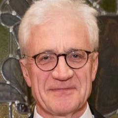 Wolfgang Oertel合作论文数Hertie Foundation, Philipps Universität of Marburg4
 Roland Giolli合作论文数4Kihachi Saito合作论文数4M.J. Korn合作论文数4
 Richard T. Robertson合作论文数Chao Family Comprehensive Cancer Center, University of California, Irvine;School of Biological Sciences, University of California, Irvine;Department of Anatomy & Neurobiology, School of Medicine, University of California, Irvine4L. A. Shapiro合作论文数3JH FALLON合作论文数3Kwan L. Ng合作论文数3Qunyong Zhou合作论文数University of California Irvine3
 Zsolt Toth合作论文数Research Group on Laser Physics, the Hungarian Academy of Sciences3Alex P. Yong合作论文数3Eugene Roberts合作论文数3Maxine M. Okazaki合作论文数3Eric Bednarski合作论文数3Marco S. Navetta合作论文数3
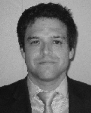 Zachary D. Perez合作论文数3JE VAUGHN合作论文数3
 Pooja Upadhyaya合作论文数2Alan M Wong合作论文数2Tt Dinh合作论文数2Rosalinda C. Roberts合作论文数2Gm Peterson合作论文数2Lulu Wang合作论文数2Xiaoning Bi合作论文数2
 Nicholas C Brecha合作论文数David Geffen School of Medicine, University of California, Los Angeles2
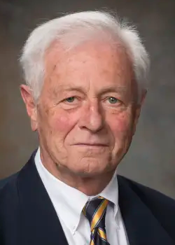 Csaba Leranth合作论文数Department of Neuroscience, Yale School of Medicine, Yale University2Lauterborn Julie C合作论文数Department of Anatomy and Neurobiology, University of California2Farias P A合作论文数2
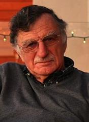 Edward G. Jones合作论文数Center for Neuroscience, University of California, Davis2
 Larry Swanson合作论文数Departments of Biological Sciences and Psychology, College of Letters, Arts and Sciences, University of Southern California2
 Jacqueline F. Mcginty合作论文数Department of Neurosciences, College of Medicine, Medical University of South Carolina;Neuroscience Institute, Medical University of South Carolina2
 Marina Bentivoglio合作论文数Department of Neurosciences,Biomedicineand Movement Sciences, University of Verona2
 Sofia Diaz Cintra合作论文数Instituto De Neurología, National Autonomous University of Mexico2
 Mark G. Frei合作论文数2




