Complications following lifesaving liver transplantation can be devastating and must be managed properly to optimize the patient and allograft survival. There are non-immune, non-infectious complications which present a severe risk to survival of both the patient and the allograft. These include primary graft non-function (PNF) and hepatic artery thrombosis (HAT). Other complications manifest less urgently but continue to represent potentially lethal consequences to both the patient and the hepatic allograft. These include vena cava outflow disruptions, portal venous outflow derangements, and portal vein thrombosis (PVT). Successful management of these complications is optimized with a multidisciplinary approach to the care of liver transplant recipients. We describe their definition, epidemiology, pathophysiology, related factors, presentation, operative and non-operative management, outcomes, and future directions of these potentially catastrophic complications.
Liver transplantation continues to rapidly evolve, and in 2020, 8906 orthotopic liver transplants were performed in the United States. As a technically complex surgery with multiple vascular anastomoses, stenosis and thrombosis of the venous anastomoses are among the recognized vascular complications. While rare, venous complications may be challenging to manage and can threaten the graft and the patient. In the last 20 years, endovascular approaches have been increasingly utilized to treat post-transplant venous complications. Herein, the evaluation and interventional treatment of post-transplant venous outflow complications, portal vein stenosis, portal vein thrombosis, and recurrent portal hypertension with transjugular intrahepatic portosystemic shunt (TIPS) are reviewed.
The incidence of end-stage renal disease has continued to increase. Similarly, the number of patients living with a functioning renal allograft has also increased. Transplantation has improved with advances in surgical techniques, immunosuppression, and better control of comorbid conditions. Transplantation is transformative and offers the greatest potential for restoring a healthy, productive, and durable life to appropriately selected patients. This article describes factors to address in selection of renal transplant candidates and discusses commonly encountered perioperative events. Paramount to selecting appropriate candidates is the collaboration between a multidisciplinary team focused on a systematic process guided by protocols and common practices.
Acute esophageal necrosis (AEN) is rare and characterized endoscopically by distal esophageal ulceration, blackening, and necrosis. It typically arises in patients with multiple comorbidities who have significant systemic disease and frailty. Specific precipitating events are variable. Evidence suggests a multifactorial etiology likely involving esophageal ischemia in the setting of corrosive injury from gastric contents and impaired tissue repair mechanisms. In the transplant setting, immunosuppression likely plays a substantial role. We report a case of AEN in a 70-year-old man following a renal transplant.
PURPOSE:When donor specific HLA antibodies (DSA) are identified, the predictive value of whether a certain strength of reactivity (mean fluorescence intensity, MFI) leads to a positive crossmatch is uncertain. To determine this, we compared the DSA MFI results we generated locally for nationally distributed proficiency samples against the percentage of other laboratories reporting a positive crossmatch.METHOD:DSA MFI from single antigen beads reported by our laboratory for nationally-distributed proficiency testing survey samples was compared against the aggregate percentage of participating laboratories reporting the crossmatch positive using direct, antiglobulin-enhanced microcytotoxic (CDC-AHG), or flow cytometric methods from 2011 to 2015.RESULTS:180 surveys were analyzed. Positive CDC-AHG and flow cytometric crossmatches were associated with MFI greater than 8554 and 2748 respectively for HLA class I, and 6919 and 3707 respectively for class II. Institutional MFI less than 3000 had high positive predictive values (0.98, 0.85, 0.81) for negative direct, AHG, and flow crossmatches, while MFI greater than 8000 had high negative predictive values for a positive direct, AHG, and flow crossmatches (1.00, 1.00, 0.97).CONCLUSION:Review of locally-generated MFI results as part of participating in proficiency testing allow for predictability of crossmatch results against other laboratories, providing a replicable model for other participating centers.
IRD organs are classified by the Public Health Service to be at above-average risk for harboring human immunodeficiency virus, hepatitis C, and hepatitis B. Traditionally underutilized, there exists even greater reluctance for their use in pediatric patients. We performed a retrospective analysis via the United Network for Organ Sharing database of all pediatric renal and hepatic transplants performed from 2004 to 2008 in the United States. Primary outcomes were patient and graft survival. Proportional hazards regression was performed to control for potentially confounding factors. Waitlist time, organ acceptance rates, and infectious transmissions were analyzed. There were 1830 SRD renal, 92 IRD renal, 1695 SRD hepatic, and 59 IRD hepatic transplants. There were no statistically significant differences in allograft or patient survival in either group. Acceptance rates of IRD organs were lower for kidney (1.5% IRD vs 4.82% SRD) and liver (1.99% IRD vs 4.51% SRD). One transmission of a bloodborne pathogen involving a pediatric recipient out of 7797 unique transplants was reported from 2008 to 2015. IRD organs appear to have equivalent outcomes. Increasing their utilization may improve access to transplant while decreasing wait times and circumventing waitlist morbidity and mortality.
Intensive care units are breeding grounds for resistant organisms, necessitating early and aggressive anti-microbial therapy. Cefepime is a fourth generation cephalosporin known for its extended spectrum of activity against Gram-positive and Gram-negative bacteria, more so than any other drug in its class. Widespread utility can also be attributed to the fact it is an antipseudomonal agent; cefepime is carefully reserved to treat severe infections and cases with known multidrug resistance. A key characteristic that serves to differentiate cefepime among other cephalosporins is its ability to resist degradation by beta-lactamases; making cefepime a front-line agent for treating Enterobacteriaceae. Cefepime is minimally metabolized by the liver and excreted nearly unchanged by the kidneys, making it imperative to renally dose. Illustrating its potency, the dosage must be cut in half as creatinine clearance approaches 11 mL/min. Frequent adverse effects of cefepime include: positive Coombs test result without hemolysis (16%), rash (4%), elevated alanine aminotransferase (3%), fever/headache/pain (1%). Post marketing reports suggests that there is evidence for potential neurotoxicity, namely encephalopathy. Further reports have elucidated that the majority of severe encephalopathy cases have been in patients who necessitated renal adjustment of their doses (Jallon, et al., 2000). However, the encephalopathy is reversible and symptomatology appears to resolve within 24-48 h of discontinuation, this underscores two things; first, that renal failure is a significant risk factor for cefepime-induced reversible encephalopathy and second, the importance of reducing dosages in renally impaired patients (Garces, et al., 2008). Despite case reports, the incidence and natural disease course of this side effect is under appreciated. Here we describe our experience in two separate patients, with resumption of cefepime therapy in one.
Using kidneys from deceased donors whose demise was secondary to ethylene glycol (EG) toxicity requires considerable thought and planning. The exact impact that kidneys from these donors could have is unclear. The shortage of viable organs and growing wait list mortality should lead us to consider these allografts as potential life-saving transplants. Because it is crucial for the transplant community to use every available allograft, we need to develop processes that optimize each possible scenario. This article is a discussion of the viability of kidneys from a donor with EG-induced brain death and a proposed algorithm for encouraging the use of renal allografts after EG toxicity.
The growing disparity between organ supply and demand has become the greatest hurdle facing transplant professionals and life-saving transplants. Because the organ shortage has become the rate-limiting step to effective transplants, it is critical for the transplant community to identify viable mechanisms to expand the donor pool and use every available allograft. Although using kidneys from deceased donors whose demise was secondary to ethylene glycol (EG) toxicity requires great deliberation and precise timing as described by Barbas et al [5], using hepatic allografts in this setting involves far less risk. The following is a discussion of a 61-year-old male who was diagnosed with end-stage liver disease secondary to non-alcoholic steatohepatitis and ultimately underwent a life-saving transplant with a liver recovered from a donor with EG-induced brain death and allocated nationally due to trepidation by local and regional centers to use the liver from a donor after EG toxicity.
Introduction: Hemolytic uremic syndrome (HUS) following kidney transplantation is a devastating complication that may result in substantial morbidity and allograft loss. While calcineurin inhibitor-induced HUS has been well described, anti-donor specific antibody production may be an alternative pathway in the pathogenesis of HUS. We present a case of HUS following kidney transplantation, and will present evidence that donor specific antibodies (DSA) may be a factor in platelet activation and end-organ injury resulting in post-transplant HUS. Case description: A 66-year-old female with diabetic nephropathy underwent a deceased donor renal transplant with a 5 HLA mismatched kidney. Her immediate course was uneventful with normalization of her creatinine (Cr). She was re-admitted with rising Cr, oliguria, proteinuria, anemia and thrombocytopenia. A peripheral smear revealed schistocytes, haptoglobin levels were depleted and an allograft biopsy was performed that was suggestive of thrombotic microangiopathy (TMA) with equivocal findings for AMR. Her ADAMTS 13 activity was 103% (normal). She concurrently developed a substantial de novo DSA burden. She underwent therapeutic plasma-pheresis, conversion from tacrolimus to cyclosporine, and received rituximab therapy. She had a complete clinical resolution and remains off of dialysis. Her laboratory markers improved and her antibody titers decreased. Discussion: Post-transplant HUS requires immediate recognition and treatment. This clinical course suggests DSA may be involved in an alternative mechanism of platelet activation leading to HUS and renal insult. Review of the literature suggests this is a rare cause of HUS and we postulate may be under-diagnosed in the transplant population and requires further study. The donor allograft was recovered from a 48 year-old female donor after cardiac death with a terminal creatinine (Cr) of 0.5, and a 5 HLA mismatch. Total cold and warm ischemia times were 14 hours 9 minutes and 56 minutes, respectively. The left renal allograft was placed in the right retroperitoneal space with a peritoneal window. Given a PRA of 55%, she received thymoglobulin induction intra-operatively, and was admitted to the surgical intensive care unit in stable condition. She required vasopressor support for the first 36 hours to maintain an adequate mean arterial pressure and was oliguric. On postoperative day two, she started ambulating and was making urine suggestive of graft function. She was transferred out of the intensive care unit on postoperative day three. She developed anemia and thrombocytopenia (platelets to 96) during the immediate post-transplant period likely secondary to Thymoglobulin induction, and ultimately required transfusion of one unit of packed red blood cells on postoperative day five for a hemoglobin of 6.8. She was transitioned to Tacrolimus at the conclusion of the Thymoglobulin induction (total dose 5 mg/kg), and was discharged home on postoperative day seven after successfull Journal of Immunobiology Jo ur na l of Immunoology
Contemporary Surgical Management of Liver, Biliary Tract, and Pancreatic Disease, pp. 127-144 (2013) No AccessTECHNICAL MANAGEMENT OF ASCITESCarlos E. Marroquin and Bridget M. MarroquinCarlos E. MarroquinUniversity of Rochester, Rochester, New York, USA and Bridget M. MarroquinUniversity of Rochester, Rochester, New York, USAhttps://doi.org/10.1142/9789814293068_0009Cited by:0 (Source: Crossref) PreviousNext AboutSectionsPDF/EPUB ToolsAdd to favoritesDownload CitationsTrack CitationsRecommend to Library ShareShare onFacebookTwitterLinked InRedditEmail Abstract: The following sections are included: INTRODUCTION PARACENTESIS TRANSJUGULAR PORTOSYSTEMIC SHUNT SURGICAL SHUNTS MESOCAVAL SHUNT PORTACAVAL SHUNTS PERITONEOVENOUS SHUNT REFERENCES FiguresReferencesRelatedDetails Recommended Contemporary Surgical Management of Liver, Biliary Tract, and Pancreatic Disease Metrics History PDF download
Contemporary Surgical Management of Liver, Biliary Tract, and Pancreatic Disease, pp. 393-403 (2013) No AccessSURGICAL TECHNIQUES: BILE DUCT RESECTION/RECONSTRUCTIONSarah Evans and Carlos E. MarroquinSarah EvansDuke University, Durham, North Carolina, USA and Carlos E. MarroquinUniversity of Rochester, Rochester, New York, USAhttps://doi.org/10.1142/9789814293068_0033Cited by:0 PreviousNext AboutSectionsPDF/EPUB ToolsAdd to favoritesDownload CitationsTrack CitationsRecommend to Library ShareShare onFacebookTwitterLinked InRedditEmail Abstract: The following sections are included: PATHOPHYSIOLOGY OF BILIARY STRICTURES PREOPERATIVE IMAGING AND STENT PLACEMENT PREOPERATIVE EVALUATION AND PREPARATION CONDUCT OF ABDOMINAL ACCESS AND EXPOSURE BILE DUCT RESECTION Resection of Hilar Bile Duct Tumors End-to-side Roux-en-Y Hepaticojejunostomy for Proximal Obstructions Side-to-Side Roux-en-Y Intrahepatic Hepaticojejunostomy for Biliary Decompression DISTAL OBSTRUCTION End-to-Side Roux-en-Y Choledochojejunostomy or Extrahepatic Hepaticojejunostomy Side-to-Side Roux-en-Y Hepaticojejunostomy or Choledochojejunostomy Cholecystojejunostomy Side-to-Side Choledochoduodenostomy Laparoscopic Bilioenteric Bypass REFERENCES FURTHER READING FiguresReferencesRelatedDetails Contemporary Surgical Management of Liver, Biliary Tract, and Pancreatic DiseaseMetrics History PDF download
Contemporary Surgical Management of Liver, Biliary Tract, and Pancreatic Disease, pp. 237-249 (2013) No AccessSURGICAL TECHNIQUES: LIVER PROCUREMENT AND TRANSPLANTATIONDeepak Vikraman and Carlos E. MarroquinDeepak VikramanDepartment of Surgery, Duke University, Durham, North Carolina, USA and Carlos E. MarroquinDepartment of Surgery, University of Rochester, Rochester, New York, USAhttps://doi.org/10.1142/9789814293068_0017Cited by:0 PreviousNext AboutSectionsPDF/EPUB ToolsAdd to favoritesDownload CitationsTrack CitationsRecommend to Library ShareShare onFacebookTwitterLinked InRedditEmail Abstract: The following sections are included: ORGAN RECOVERY DONATION AFTER CARDIAC DEATH RECIPIENT PROCEDURE REFERENCES FiguresReferencesRelatedDetails Contemporary Surgical Management of Liver, Biliary Tract, and Pancreatic DiseaseMetrics History PDF download
Liver cysts were initially thought to be rare and incidental findings on surgical exploration. However with the increasing use and refinement of modern imaging modalities, the known incidence has increased to between 3% and 5%. This represents an underestimate as most cysts are unrecognized and asymptomatic. The first reported case of a simple nonparasitic liver cysts occurred in 1856 by Bristowe. In contrast, hydatid or parasitic disease has been described since antiquity. Liver cysts are broadly classified as either congenital or acquired and can be subdivided into the following subcategories:
 Paul C. Kuo合作论文数Department of Surgery, University of South Florida;Tampa General Hospital;Muma College of Business, University of South Florida27
 Tuttle-Newhall Janet E合作论文数13B. H. Collins合作论文数8Mark S. Orloff合作论文数8Christopher T Barry合作论文数8Bradley Collins合作论文数7Randeep Kashyap合作论文数7Desai D M合作论文数6Saman Safadjou合作论文数6
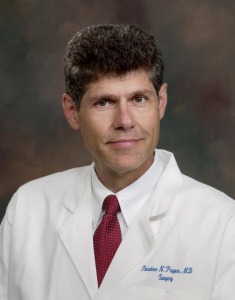 Theodore N. Pappas合作论文数Department of Surgery, School of Medicine, Duke University6Saravanan Ramalingam合作论文数5Gopal A. Ramaraju合作论文数5John E. Scarborough合作论文数5Shaleen Agarwal合作论文数5Benedict Maliakkal合作论文数Albany Medical College5Dev M. Desai合作论文数5
 Bryan Clary合作论文数Department of Surgery, School of Medicine, University of California, San Diego4
 Ricardo Pietrobon合作论文数Durham VA Medical Center;SporeData Inc4D. W. Butterly合作论文数4
 Hongtao Guo合作论文数Department of Surgery, Duke University Medical Center4Erick B. Edwards合作论文数3JE Tuttle-Newhall合作论文数3Kyla M Bennett合作论文数3Matt Harris合作论文数3
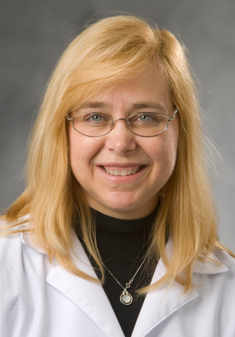 Debra L. Sudan合作论文数Division of Abdominal Transplantation, Department of Surgery, Duke University3Philip Y. Wai合作论文数3
 Jaime A. Pineda合作论文数Cognitive Neuroscience Laboratory, Department of Cognitive Science, University of California San Diego3Barbara Burgess合作论文数2Sr Smith合作论文数2Alastair Smith合作论文数2
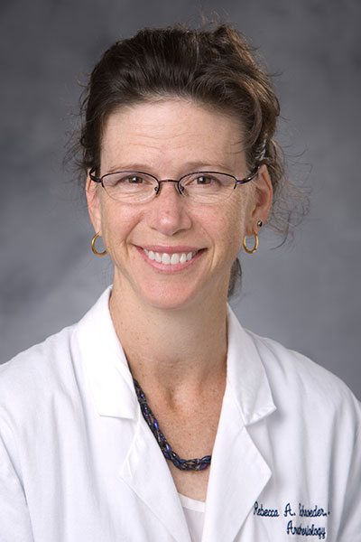 Rebecca A. Schroeder合作论文数Duke Anesthesiology, Duke University School of Medicine2Reddy Srinevas K合作论文数2Majdalany Bill S合作论文数2
 Nicole Yarid合作论文数2Wrenn Sean M合作论文数2Aram F. Hezel合作论文数Department of Medical Oncology;Center for Applied Cancer;Belfer Institute for Innovative Cancer Science;Dana-Farber Cancer Institute;Children ' s Hospital;Stem Cell Program;Department of Genetics;Medical School, Harvard;Harvard Stem Cell Institute;Department of Pathology;Comprehensive Cancer Center;University of Texas Southwestern Medical Center;Massachusetts General Hospital;Belfer Institute for Innovative Cancer Science, University of Texas Southwestern Medical Center2Mark K. Fung合作论文数2
 Charlotte Ryan合作论文数Boston College2
 Andrew S. Barbas合作论文数Medical Center, Duke University2Dongfeng Chen合作论文数2Bridget M. Marroquin合作论文数2Brad Collins合作论文数2
 Alok Khorana合作论文数Taussig Cancer Institute, Cleveland Clinic2W A Schell合作论文数1J.E. Scarborough合作论文数1M S Orloff合作论文数1J Elizabeth Tuttle-Newhall合作论文数1Anant D. Bhave合作论文数1
 Peter Callas合作论文数1




