BACKGROUND:As medical imaging demand grows, there is increasing stress on the currently available workforce to deliver consistent, high-quality imaging studies while ensuring rapid study turnaround times and round-the-clock radiology coverage. Advances in remote access technology facilitating remote scan assistance and control are now commercially available to address these pressing clinical needs. METHODS:This work evaluated an early clinical application of a virtual scanner operations system (syngo Virtual Cockpit (VA13A, Siemens Healthineers, Erlangen, Germany) for remote magnetic resonance imaging (MRI) monitoring and scan control at three geographically distant outpatient sites associated with our primary institution. RESULTS:The system facilitated execution of technically complex oncologic MRI exams at these geographically distant clinics with no measurable impact on acquisition time compared to MR imaging performed at our primary hospital location. Additional operational improvements were realized with the use of the system, including remote staff training, technical assistance, and scanning during staff shortages. This early iteration of remote scanning had some limitations including limited utility for additional assistance in the scanning of those protocols that require complex physical setup. Moreover, connectivity issues were noted to be a limiting factor that contributed to operational delays. It was still necessary to have an onsite MRI technologist at the scanner console to interface with the patient and ensure safe operation. CONCLUSION:Despite these limitations, our initial experience demonstrates that the use of remote MRI scanning support facilitates staffing flexibility while providing expanded patient access to oncology MRI services.
PURPOSE Artificial intelligence (AI) has been used in medicine for decades, but recent advances in machine learning and large language models have rapidly expanded its accessibility and applications in oncology. Although AI offers efficiency in data analysis, synthesis, and communication, important questions remain regarding output reliability, research rigor, data security, and appropriate reliance on automated tools, underscoring the need for thoughtful implementation and training. METHODS To better understand current AI use in academic oncology and perceptions about utility, we conducted a survey of faculty and trainees at a comprehensive cancer center. RESULTS Among 227 respondents (55% women; 64% clinical faculty), 58% reported using AI several times per month or more, whereas 15% had never used it. The most common applications were summarizing academic or research information and generating data visualizations. Attitudes toward AI were generally positive: 74% agreed that AI will improve cancer diagnosis within the next decade. In contrast, views were more cautious regarding end-of-life decision making, with 35% disagreeing that AI would be beneficial in that context. Despite broad interest, a substantial training gap emerged. Nearly 93% of respondents endorsed the need for dedicated AI training, and approximately half reported not knowing where to find reliable learning resources. Lower AI use was associated with female gender and age over 60 years. CONCLUSION AI use among oncology faculty and trainees is common but variable, with differences across demographic and professional groups. These findings highlight the need for structured, accessible training and institutional guidance to promote appropriate, equitable, and high-quality integration of AI into oncology research and clinical care.
Biological aging occurs heterogeneously across individuals and organs. However, current measures of biological age incompletely capture organ-specific differences in health and disease risk. Because chest CT visualizes multiple thoracic organs, it offers an opportunity to quantify structural aging across organ systems. Here, we developed MOSAIC-Age, a framework characterizing eight organ-specific aging clocks on chest CT. The clocks were developed and validated using 9,971 CT scans from CT-RATE and MIDRC, and subsequently locked and applied to two independent prospective cohorts with 35,293 participants from the National Lung Screening Trial and Genetic Epidemiology of COPD study. CT-derived biological age gaps (BAGs) were examined in relation to lifestyle and socioeconomic factors, prevalent comorbidities, incident chronic diseases, and all-cause and cause-specific mortality. Higher BAGs, indicating organs that appeared older on CT than expected for their chronological age, were broadly associated with adverse health characteristics, chronic disease burden, and increased mortality risk. Multiple disease outcomes were associated with aging across several organs, whereas in multivariable analyses including all eight organ-specific BAGs, the remaining associations were more organ specific. A greater number of markedly older-appearing organs and a faster pace of aging were each associated with higher mortality. Together, these findings demonstrate that routine chest CT captures both shared and organ-specific patterns of biological aging and establish CT-derived organ aging as a quantitative imaging biomarker for assessing multi-organ health and long-term disease risk. ### Competing Interest Statement The authors declare the following financial interests/personal relationships which may be considered as potential competing interests: N. I. V. receives consulting fees from Regeneron, Amgen, Xencor, AstraZeneca, Tempus, Pfizer, Summit, OncoHost, Guardant, ImmunityBio, and research funding from EMD Serono, IDEAYA, Amgen, Summit, Regeneron, Sanofi, BMS, and OncoHost, outside the submitted work. M.C.B.G. has received research funding from Siemens Healthcare. T. C. reports speaker fees/honoraria (including travel/meeting expenses) from ASCO Post, AstraZeneca, Bio Ascend, Bristol Myers Squibb, Clinical Care Options, IDEOlogy Health, Medical Educator Consortium, Medscape, OncLive, PeerView, Physicians' Education Resource, Targeted Oncology; advisory role/consulting fees (including travel/meeting expenses) from AstraZeneca, Bristol Myers Squibb, Daiichi Sankyo, Genentech, Johnson & Johnson, Merck, Nuvalent, oNKo-innate, Pfizer, and RAPT Therapeutics; and institutional research funding from AstraZeneca, Bristol Myers Squibb and Merck. X. L. reports receiving consultant and advisory fees from Eli Lilly, AstraZeneca, EMD Serono, Daiichi Sankyo, Spectrum Therapeutics, Boehringer Ingelheim, Hengrui Therapeutics, Novartis, and research funding from Eli Lilly, Boehringer Ingelheim, all outside of the submitted work. M. A. reports research funding from Genentech, Nektar Therapeutics, Merck, GlaxoSmithKline, Novartis, Jounce Therapeutics, Bristol Myers Squibb, Eli Lilly, Adaptimmune, Shattuck Labs, Gilead, Verismo Therapeutics, and Lyell; advisory board roles for GlaxoSmithKline, Shattuck Labs, Bristol Myers Squibb, AstraZeneca, Insightec, Regeneron, Genprex, and Lyell; speaker fees from AstraZeneca, Nektar Therapeutics, SITC, and Regeneron; and participation on a safety review committee for Nanobiotix-MDA Alliance, Henlius, all outside of the submitted work. A.A.S. reports serving on the advisory board of DELFI Diagnostics and as an advisor to Droplet Biosciences, all outside of the submitted work. D.E.G. reports research funding from AstraZeneca, Karyopharm, and Novocure; stock ownership in Gilead and Medtronic; stock options in Early Marker, Inc. and OncoSeer Diagnostics, Inc.; consulting and advisory roles for AbbVie, AstraZeneca, Bayer, Catalyst Pharmaceuticals, EMD Serono, and GSK; service on data and safety monitoring boards for Daiichi Sankyo, Summit Therapeutics, and Taiho Oncology; royalties from Oxford University Press; and roles as co-founder and Chief Medical Officer of OncoSeer Diagnostics, Inc., all outside of the submitted work. J. Y. C. has received travel sponsorship from Accuray and Varian Medical Systems, and grants from Varian Medical Systems, outside the submitted work. D. L. G. reports honoraria for scientific advisory boards from AstraZeneca, Sanofi, Alethia Biotherapeutics, Menarini, Eli Lilly, 4D Pharma and Onconova, and research support from Janssen, Takeda, Astellas, Ribon Therapeutics, NGM Biopharmaceuticals, Boehringer Ingelheim, Mirati Therapeutics and AstraZeneca, all outside of the submitted work. C. C. W. reports research support from Medical Imaging and Data Resource Center from NIBIB/University of Chicago and royalties from Elsevier, outside of the submitted work. J. V. H. reports receiving advisory/consulting fees from AstraZeneca, Boehringer Ingeheim, Catalyst, Genentech, GlaxoSmithKline, Guardant Health, Foundation Medicine, Hengrui Therapeutics, Eli Lilly, Novartis, Spectrum, Sanofi, Takeda Pharmaceuticals, Mirati Therapeutics, Bristiol Myers Squibb, BrightPath Biotherapeutics, Janssen Global Services, Nexus Health Systems, EMD Serono, Pneuma Respiratory, Kairos Venture Investments, Leads Biolabs, RefleXion, and research funding from GlaxoSmithKline, AstraZeneca, Spectrum, all outside of the submitted work. J. Z. reports grants from Merck, Novartis, Johnson and Johnson; and personal fees from BMS, AZ, Novartis, Johnson and Johnson, GenePlus, Hengrui, Innovent, outside the submitted work. J. W. reports research funding from Siemens Healthcare. The remaining authors declare that they have no competing interests. ### Author Declarations I confirm all relevant ethical guidelines have been followed, and any necessary IRB and/or ethics committee approvals have been obtained. Yes The details of the IRB/oversight body that provided approval or exemption for the research described are given below: The Institutional Review Board of The University of Texas MD Anderson Cancer Center gave ethical approval for this work. I confirm that all necessary patient/participant consent has been obtained and the appropriate institutional forms have been archived, and that any patient/participant/sample identifiers included were not known to anyone (e.g., hospital staff, patients or participants themselves) outside the research group so cannot be used to identify individuals. Yes I understand that all clinical trials and any other prospective interventional studies must be registered with an ICMJE-approved registry, such as ClinicalTrials.gov. I confirm that any such study reported in the manuscript has been registered and the trial registration ID is provided (note: if posting a prospective study registered retrospectively, please provide a statement in the trial ID field explaining why the study was not registered in advance). Yes I have followed all appropriate research reporting guidelines, such as any relevant EQUATOR Network research reporting checklist(s) and other pertinent material, if applicable. Yes CT-RATE imaging data are available through the Hugging Face repository under the repository's access conditions for academic, research, and educational use (https://huggingface.co/datasets/ibrahimhamamci/CT-RATE). MIDRC imaging data are available through the MIDRC Data Commons to registered users under the applicable MIDRC data use agreement (https://data.midrc.org/). The NLST imaging data are available through The Cancer Imaging Archive (https://www.cancerimagingarchive.net/collection/nlst/), and NLST clinical data are available through the NCI Cancer Data Access System (https://cdas.cancer.gov/nlst/). The COPDGene phenotype and imaging data used in this study are available through controlled access via dbGaP under study accession numbers phs000179.v7.p2 and phs004023.v1.p2, respectively. The paired CT/PET-CT data are not publicly available owing to patient privacy considerations but are available for research purposes from the corresponding author upon reasonable request. The pretrained organ-clock model weights are also available from the corresponding author upon reasonable request. National Institutes of Health, R01CA262425, R01CA276178 Cancer Prevention and Research Institute of Texas, RP240117 Victory Houston Permanent Health Funds QIAC Partnership in Research Grant Rexanna's Foundation for Fighting Lung Cancer
Objective.High-grade gliomas (HGG) are aggressive brain tumors with poor prognoses despite an intense standard-of-care (SOC) chemoradiation protocol. Radiotherapy (RT) guidelines use static dose maps, which may not fully account for intratumoral heterogeneities and areas of tumor progression over the course of treatment. We address this limitation by guiding radiation delivery using imaging data to identify proliferative areas which are then targeted by a dose boost, and use mathematical modeling to simulate treatment response between the adjusted and the SOC treatment plan.Approach.Quantitative magnetic resonance imaging data for 15 HGG patients were collected at baseline and the third week of treatment to identify regions of high proliferation to guide the adapted radiation dose plan. The resulting dose plan is then adjusted by a board-certified medical physicist to ensure deliverability using clinically available linear accelerators. The plans are then used to virtually 'treat' each patient via our biology-based mathematical model, individually calibrated to each patient's imaging data acquired prior to dose adaptation.Main results.Our adaptation pipeline predicted a median (range) decrease in tumor burden of 31% (12% and 57%) one month post-therapy across the patient cohort compared to the SOC predicted tumor burden (p< 0.05). After adjusting the dose plan for clinical deliverability, it still predicted a decrease in tumor burden of 30% (7% and 62%) which is also significantly different than the SOC protocol (p< 0.05).Significance.Our clinical-computational platform, integrating mathematical modeling, dose painting, and adaptive RT, can identify alternative radiation dose plans hypothesized to yield statistically greater tumor control than the SOC prescriptions.
BACKGROUND:Historically, patients with ≥5 brain metastases (BM) were considered borderline or poor candidates for stereotactic radiosurgery (SRS). With increased survival and expanded treatment options for BM in the modern era, paradigms of BM management warrant reevaluation. Our objective was to determine whether overall survival (OS) differs between patients receiving SRS for 5 to 9 BMs versus ≥10 BMs and to identify factors associated with favorable outcomes. METHODS:We identified patients with ≥5 BMs treated with SRS at a single institution between 2015 and 2022 who had not received prior central nervous system (CNS) radiation. The CNS activity of systemic regimens was classified according to the 2024 NCCN Guidelines. Outcomes included OS, whole-brain radiation therapy-free survival (WBRT-FS), and CNS progression-free survival (CNS-PFS). RESULTS:A total of 557 patients received Gamma knife surgery to ≥5 BMs (range, 5-23). Median OS time was 11.0 months and did not differ for patients with 5 to 9 BMs (n=440) and those with ≥10 BMs (n=117) (P=.52). On multivariable analysis, improved performance status (hazard ratio [HR], 0.956; P<.001) and receipt of CNS-active systemic therapy after SRS (HR, 0.518; P<.001) were associated with improved OS. Lesion number was not associated with OS (HR, 1.012; P=.466). We created a nomogram predicting 6- and 12-month WBRT-FS that performed well on a validation cohort of (area under receiver operating characteristic curve, 0.8). CONCLUSIONS:Lesion number did not influence OS in patients with ≥5 BMs. Good performance status, receipt of CNS-active systemic therapy after SRS, and stable systemic disease predicted improved OS and WBRT-FS. Our publicly available online nomogram can inform patient-specific treatment decisions for individuals with ≥5 BMs (https://medcalc.mdanderson.org/WbrtCalc).
Abstract Background The incidence and implications of heterogeneous intracranial response to immunotherapy in patients with melanoma brain metastases (MBM) are poorly understood, posing a clinical challenge by complicating disease assessment and management. Herein, we compare homogeneous and heterogeneous progression presentations for MBM treated with ipilimumab and nivolumab (ipi-nivo). Methods We retrospectively identified UT MD Anderson patients with newly diagnosed MBM treated with ipi-nivo in 2018-2023, with ≥1 evaluable MBM at baseline and ≥1 MRI brain scan following ipi-nivo initiation. The longest axial diameter (LAD) for each MBM was extracted from segmented volumes in RayStation. Intracranial progressive disease (IPD) was defined according to modified RECIST criteria. IPD patients with ≥1 evaluable MBM achieving complete response or LAD shrinkage by ≥ 1.5 mm at the time of IPD were considered heterogeneous IPD; otherwise, IPD cases were considered homogeneous. Results Out of 65 MBM patients treated with ipi-nivo, 39 (60%) experienced IPD, including 24 (62%) with homogeneous IPD and 15 (38%) with heterogeneous IPD. Compared to heterogeneous IPD, homogeneous IPD was associated with shorter interval from ipi-nivo start to IPD (median 1.4 vs. 6.0 mo, p < 0.01), simultaneous extracranial progression (50% vs. 13%, p = 0.02), and history of systemic therapy prior to MBM diagnosis (63% vs. 13%, p = 0.01). Patients with homogeneous vs. heterogeneous IPD received similar salvage therapy approaches (p = 1.0): stereotactic radiosurgery (63% vs. 67%); whole brain radiotherapy (25% vs. 20%); systemic therapy alone (13% vs. 13%). However, homogeneous IPD patients had worse three-year overall survival, when measured from start of ipi-nivo (22% vs. 67%, p = 0.02) or date of IPD (22% vs. 60%, p = 0.05). Conclusion These findings suggest that patients with heterogeneous IPD represent a clinically distinct subgroup with meaningful differences in long-term outcomes compared to homogeneous IPD. This highlights the need to account for lesion-level response heterogeneity in clinical assessment, risk stratification, and individualized treatment planning.
Immune checkpoint inhibitors (ICIs) benefit only a subset of patients with metastatic non-small cell lung cancer (NSCLC), but current selection relies on tissue PD-L1 immunohistochemistry (IHC), which is invasive and prone to sampling bias. We developed and validated SCENT (Scalable Ensemble Transformer), a CT-based deep learning model for noninvasive prediction of PD-L1 status and immunotherapy outcomes. In this retrospective study, 972 stage IV NSCLC patients treated with ICIs at MD Anderson were analyzed; SCENT was developed and validated in 640 patients with paired CT and PD-L1 IHC, and clinical applicability was assessed in an additional 332 CT-only patients. Generalizability was evaluated in independent cohorts from Mayo Clinic (n = 72) and the phase III LONESTAR trial (n = 116), where paired baseline and 3-month CT enabled longitudinal assessment. SCENT classified PD-L1 status (50% or higher vs lower) in the MD Anderson cohort with AUC 0.84 (95% CI 0.785 to 0.887), specificity 83.9%, and sensitivity 85.3%, outperforming clinical and radiomics models; external validation achieved AUC 0.80 (Mayo) and 0.78 (LONESTAR). SCENT-derived PD-L1 stratified progression-free survival (HR 1.49, p < 0.001) and overall survival (HR 1.40, p = 0.009), comparable to IHC, and provided complementary prognostic value when combined with IHC, with concordant low-low patients showing the poorest survival (OS HR 1.45, p = 0.008). In LONESTAR, serial SCENT-inferred PD-L1 status showed a borderline association with 3-month progression without paired post-treatment tissue confirmation. SCENT is a generalizable CT-based virtual biopsy for baseline PD-L1 prediction and complementary tissue IHC stratification, with longitudinal use requiring prospective validation.
Medical image segmentation continues to advance rapidly, yet rigorous comparison between methods remains challenging due to a lack of standardized and customizable tooling. In this work, we present the current state of the Medical Imaging Segmentation Toolkit (MIST), with a particular focus on its flexible and modular postprocessing framework designed for the BraTS 2025 pre- and post-treatment glioma segmentation challenge. Since its debut in the 2024 BraTS adult glioma post-treatment segmentation challenge, MIST's postprocessing module has been significantly extended to support a wide range of transforms, including removal or replacement of small objects, extraction of the largest connected components, and morphological operations such as hole filling and closing. These transforms can be composed into user-defined strategies, enabling fine-grained control over the final segmentation output. We evaluate three such strategies - ranging from simple small-object removal to more complex, class-specific pipelines - and rank their performance using the BraTS ranking protocol. Our results highlight how MIST facilitates rapid experimentation and targeted refinement, ultimately producing high-quality segmentations for the BraTS 2025 challenge. MIST remains open source and extensible, supporting reproducible and scalable research in medical image segmentation.
Background:The efficacy of ipilimumab and nivolumab (ipi/nivo) for melanoma brain metastases (MBMs) has been previously reported, leading to uncertainty regarding the optimal role of comprehensive stereotactic radiosurgery (cSRS). We therefore conducted a single-institution retrospective study to compare outcomes of upfront versus deferred cSRS for MBM treated with ipi/nivo. Methods:We identified patients who started ipi/nivo for newly diagnosed MBMs between 2018 and 2023, with or without upfront cSRS. Patients with >15 MBMs, leptomeningeal disease, or whole-brain radiotherapy at baseline were excluded. Outcomes were compared using multivariable regression and reported as adjusted hazard ratios (aHRs) with 95% CIs. Results:Of the 132 patients identified, 52.3% received upfront cSRS and 47.7% did not. Patients who received upfront cSRS had larger maximum MBMs (median 2.3 vs 0.7 cm; P < .001), more symptomatic MBMs (59.4% vs 11.1%; P < .001), higher rates of upfront craniotomy (47.8% vs 7.9%; P < .001), and fewer BRAF V600 mutations (34.8% vs 54.0%; P = .035). Upfront cSRS was not associated with longer overall survival (median 47.0 mo vs not reached; aHR = 1.01 [95% CI, 0.60-1.68]; P = .98) but was associated with reduced incidence of intracranial progression (median 37.6 vs 5.5 mo; aHR = 0.40 [95% CI, 0.25-0.64]; P < .001). Conclusions:In this retrospective study, upfront cSRS was more often used in patients with higher-risk MBM and was associated with improved intracranial control, although no significant survival benefit was observed. These findings suggest that starting ipi/nivo alone may be reasonable for lower-risk MBM, but prospective studies are needed to guide optimal integration of cSRS.
Abstract Low-grade gliomas (LGG) typically grow gradually for years with limited clinical symptoms but may later exhibit erratic growth patterns and undergo transformation to high-grade glioma. This presents challenges to disease management creating a need for novel methods to simulate and predict LGG behavior. Towards this goal, we implemented a data assimilation framework utilizing a previously developed biophysical model capable of spatiotemporally forecasting patient-specific tumor dynamics. The study cohort includes nine LGG patients treated with radiation therapy at the MD Anderson Cancer Center. All patients were longitudinally monitored using magnetic resonance imaging (MRI) to assess tumor cellularity (diffusion-weighted MRI) and extent of disease (T1-weighted MRI with & without gadolinium-based contrast, T2-fluid attenuated inversion recovery). MRI was acquired before treatment and approximately at the 4, 5, 6, and 12-month post-treatment visits. Brain and tumor regions were segmented using a semi-automated algorithm. Our biophysical model is a reaction-diffusion equation that explicitly accounts for tumor cell proliferation, invasion, and treatment response. The model focuses on changes in non-enhancing tumor regions, a hallmark of LGG. Total tumor cell count (TTC) derived from spatiotemporal tumor response forecasts was used to quantify tumor burden. The data assimilation framework incorporates MRI data acquired with each subsequent visit then updates its forecast. Predictive accuracy was quantified via the concordance correlation coefficient (CCC) between the observed and predicted TTC for both short (e.g., 1 - 3 months) and longer-term predictions (e.g., 6, 12-months). A Mann-Whitney U test compared short and longer-term prediction performance. The median and interquartile range (IQR) of the model parameters describing tumor cell proliferation and invasion are reported for each follow up visit. The tumors experienced a median volumetric change of -36.4% over 12 months. The model accurately forecasts TTCs at short (CCC: 0.71) and longer interval times (CCC: 0.96) with no statistically significant difference (p-value: 0.34) in performance between groups. The model estimated tumor cell proliferation rate had a median and IQR of 0.10 (0.06) at the 4-month visit, 0.07 (0.04) for the 5-month visit, 0.04 (0.05) at the 6-month visit, and 0.04 (0.04) days-1 at the 12-month visit. Similarly, the model estimated tumor diffusion coefficient had a median and IQR of 0.15 (0.10), 0.16 (0.10), 0.18 (0.06), and 0.16 (0.09) mm2/days for the 4, 5, 6, and 12-month visit. Our preliminary findings demonstrate the model’s ability to predict patient-specific LGG behavior and offers a step towards the development of a personalized decision support tool for managing LGG care. Citation Format: Sophia Ty, Devika Shankar, Bikash Panthi, Mohamad El-Jammal, Victoria White, Holly Langshaw, Eleni Konstantinopoulou, Hannah Green, Ashi Jain Chakresh, Vishantan Kumar, Thomas E. Yankeelov, Caroline Chung, David A. Hormuth. Developing a data assimilation framework to forecast patient-specific tumor burden in low-grade glioma [abstract]. In: Proceedings of the American Association for Cancer Research Annual Meeting 2026; Part 1 (Regular Abstracts); 2026 Apr 17-22; San Diego, CA. Philadelphia (PA): AACR; Cancer Res 2026;86(7 Suppl):Abstract nr 6840.
IDH-wt diffuse gliomas with histologic grade 2–3 features and distinct molecular characteristics are now classified as molecular glioblastoma, yet outcomes and optimal management remain incompletely defined in a contemporary cohort. Adults with histologic grade 2–3 IDH-wt gliomas diagnosed from 1996 to 2019 were retrospectively identified. Overall survival (OS) and progression-free survival (PFS) were estimated using Kaplan-Meier methods, with Cox regression used to assess prognostic factors. To account for noncanonical IDH mutations, subgroup analyses were performed in those age > 55 or with next generation sequence (NGS) IDH testing. A total of 134 patients were included, with a median follow-up of 29.8 months. Median OS was 35 months (95
Artificial intelligence (AI) is increasingly integrated into oncology across clinical care, research workflows, and translational implementation. This chapter provides a practical overview of these domains, emphasizing opportunities and limitations relevant to real-world oncology practice. In clinical care, AI applications include ambient documentation systems, decision support tools, and remote monitoring platforms. These technologies may reduce administrative burden, assist with evidence synthesis and guideline navigation, and enable longitudinal assessment of symptoms and functional status through wearable and patient-reported data. Emerging tools such as large language models also support patient communication through education, translation, and symptom triage. However, across these applications, performance remains dependent on data quality, validation, and appropriate clinical oversight. In research and scholarship, AI is increasingly used to support grant development, study design, literature review, and manuscript preparation. These tools may improve efficiency, enhance clarity, and assist in identifying research gaps or methodological approaches. At the same time, concerns regarding accuracy, hallucinated content, data privacy, and ethical responsibility require careful oversight. The responsibility for hypothesis generation, methodological rigor, and scientific integrity remains with the investigator. From a translational perspective, a persistent gap exists between model development and clinical deployment. Differences in data quality, infrastructure, and patient populations may limit generalizability, while workflow integration, clinician readiness, and bias mitigation remain critical challenges. Collectively, AI has the potential to enhance oncology care and research, but its impact depends on rigorous validation, equitable implementation, and sustained human oversight.
This roadmap provides a comprehensive framework for integrating diffusion-weighted imaging (DWI) into radiation therapy (RT), with an emphasis on its application in magnetic resonance imaging-guided radiotherapy and its potential for driving biological image-guided adaptive radiotherapy (ART). Developed through collaboration among experts in medical physics, magnetic resonance imaging science, and radiation oncology, the paper aims to bridge disciplinary gaps and foster a shared understanding across scientific, technical, and clinical domains. It benchmarks the current state of DWI in RT, identifies critical challenges, and highlights recent advancements in acquisition, reconstruction, biophysical modeling, quality assurance, clinical validation and translation, as well as emerging concepts. By outlining ongoing efforts and forecasting future developments, this roadmap supports the adoption of DWI as a quantitative imaging biomarker for personalized and ART in precision oncology.
e13648 Background: Artificial intelligence (AI) in cancer care offers opportunities to reduce administrative burden, improve efficiency, and support care delivery, yet it prompts questions around safety, equity, and clinical judgment. To assess real-world perspectives, the Association of Cancer Care Centers (ACCC) conducted a national survey examining how oncology professionals engage with AI, their perceived value, and the barriers to adoption. Methods: ACCC, with guidance from an expert committee, created a national online survey of multidisciplinary staff at US cancer programs from May and August 2025. The 26-item survey assessed experiences and perceptions of AI, organizational adoption and governance, and implementation barriers and facilitators. Completed responses were analyzed using descriptive and stratified statistics in Stata 18, with qualitative data examined using rapid inductive thematic analysis. Results: Respondents (N=168) from 36 states included 61% in care delivery and 39% in administrative/operational roles, primarily from community (46%) and NCI-designated or academic (44%) programs. Care delivery staff were less confident than administrative/operations respondents in describing AI use (21% vs 4% “not at all [confident]”), discussing benefits of AI use (18% vs 7%), and critically evaluating AI systems (26% vs 13%). Administrative/operations respondents reported higher confidence contributing to AI implementation (45% vs 32%) and a higher likelihood to adopt/expand AI for treatment planning (52% vs 34%), clinical trial matching (49% vs 32%), and prior authorization (52% vs 35%). Rural respondents were more likely to express concern about declines in patient-provider communication (63% vs 22% suburban and 30% urban), as were those without AI experience (54% vs 24%). Academic respondents rated improving clinical decision-making/diagnostic accuracy, and establishing performance benchmarks and evaluating AI systems, as higher motivators for integration (0.82 vs 0.41; 0.53 vs 0.26), whereas community respondents rated patient engagement higher (0.36 vs 0.13). Respondents with AI experience reported a higher likelihood to adopt/expand AI for chatbots and virtual assistants (52% vs 16%), real-time alerts (35% vs 14%), personalized patient education (46% vs 27%), and individualized patient navigation (38% vs 11%). Qualitative responses reinforced these findings, highlighting stakeholder engagement, evidence of effectiveness, training, integration, and governance as critical for AI implementation. Conclusions: The results reveal broad AI use despite limited AI tools and governance implementation. Respondents were confident acknowledging AI’s limitations but less confident in evaluating its utility, highlighting opportunities for ACCC to support adoption, governance, and AI integration focused on efficiency and patient outcomes.
Abstract Background More than half of patients diagnosed with metastatic melanoma (MM) eventually develop brain metastases, yet these patients are excluded from most clinical trials. The CheckMate 204 study combining ipilimumab (3mg/kg) plus nivo (1 mg/kg) for patients with asymptomatic MBM revealed an intracranial response in 54% of patients. Grade 3/4 treatment-related adverse events (TRAEs) occurred in 55% of patients. RELATIVITY-047, a global, randomized phase II/III study, evaluated nivo/rela (480 mg/160 mg) compared to nivo monotherapy (480 mg) for patients with unresectable, untreated melanoma and demonstrated improved PFS compared with nivo monotherapy. Grade 3/4 TRAEs occurred in 21% of patients in the combination group compared to 11% receiving monotherapy. The nivo/rela combination is FDA approved for the treatment of MM patients but has not been studied in patients with MBM. Methods This phase II, single‑center trial assesses the safety and efficacy of nivo/rela in patients with MBM. Thirty asymptomatic, anti–PD‑1–naïve patients will be enrolled. Patients are treated with nivo/rela (480 mg/160 mg) every 4 weeks for up to 25 cycles, disease progression, or unacceptable toxicity. The primary objective is to evaluate intracranial response (CR + PR) per modified RECIST 1.1 criteria. The Bayesian Optimal Phase 2 design will be used to monitor futility. Longitudinal blood, tissue, and microbiome samples will be collected, along with neurocognitive and quality‑of‑life assessments. Results At this interim analysis, 22 patients have been treated (age 22-90 years, 55% male, median largest baseline brain lesion 1.25 cm (0.5-2.4 cm), 14% received prior targeted therapy). At a median follow-up of 12 months, intracranial response has been seen in 48% of evaluable patients (6 CR, 4 PR). There are no new safety signals, with 1(5%) patient experiencing Grade 3 TRAEs (colitis, arthralgia). This study continues enrollment.
Intracranial metastases (ICM), specifically parenchymal brain metastases, remain a major clinical challenge in solid tumor oncology, despite recent advances in cancer therapies which have led to improvements in survival for these patients. Improving outcomes even further in this patient population will require a multi-disciplinary approach, including pre-clinical and translational studies, clinical trials, and studies of patient reported outcomes and quality of life. At the 2023 and 2024 joint Society for Neuro-Oncology (SNO) and American Society of Clinical Oncology (ASCO) CNS Metastases Conferences, two ICM collaborative group think tanks convened, composed of diverse, multi-disciplinary stakeholders, including basic and translational researchers, clinical trialists, and clinicians from academia and the community setting. Here we summarize the key knowledge gaps and consensus recommendations put forth by these two think tanks. Advances in ICM research and improvements in patient outcomes will require close inter-specialty and inter-institutional collaboration between stakeholders, including pre-clinical and translational researchers, clinical investigators, industry, and regulatory bodies.
Abstract Gamma knife radiotherapy (RT) treatment is often used as a salvage therapy after progression of melanoma brain metastasis (MBM) lesions under immune checkpoint inhibitor (ICI) blockade therapy; however, waiting until progression of nonresponsive lesions can result in significant symptomatic progression, and the toxicity of RT is greater for larger lesions as it is directly proportional to lesion volume. Robust and reliable standards for consistently maximizing the benefit of combination ICI + RT therapy remain elusive, likely in part due to the complexity of mechanistic interactions between each therapy. Addressing this clinical need will require optimizing the effects of their reported synergistic interactions such as tumor microenvironment modulation and immune activation while mitigating inhibitory effects such as immunosuppressive radiation effects. The ability to identify lesions unlikely to respond to ICI shortly after start of treatment would offer immediate clinical benefit by enabling earlier RT delivery, reduce the volume-dependent toxicities of RT, and reduce symptoms from lesion progression. To address the unmet clinical need for methods to optimize outcomes of combination ICI and RT therapy, we have developed a mechanistic mathematical model based on the biological and physical underpinnings of these complex therapies, and their interactions, which is able to describe and quantify interactions between ICI and RT and their interactions on the tumor. This builds upon our previous ICI modeling work by expanding the model based on the assumption that therapeutic outcome is not only a result of direct interaction between the treatment and disease, but is also a result of complex interplay between heterogeneous tumor populations that may be sensitive or resistant to treatment and their unique interactions with the immune system that serve to maintain or disrupt the tumor-immune equilibrium. RT further modulates this tumor-immune dynamic both by killing T cells in the tumor interior and by stimulating release of T cell activating antigens which can alter T cell activation exterior to the tumor, thereby affecting T cell recruitment into the tumor. By capturing how the complex time-dependent dynamics of tumor-immune interaction and recruitment influences – and is influenced by – transient shifts in treatment sensitive and resistant populations, our model aims to describe and quantify long-term tumor dynamics after ICI + RT therapy. This platform provides a consistent framework for systematically evaluating various proposed mechanisms underlying treatment interactions and how these may determine treatment failure or success. By applying the model to a retrospective, in-house generated melanoma brain metastasis data set, we are examining and refining our model structure with a goal of capturing a wide range of observed tumor dynamics using only clinically available data. This may lead to robust, quantitative methods to maximize treatment outcomes based on the unique characteristics of individual tumors. Citation Format: Alexander R. J. Silalahi, Caroline Chung, James W. Welsh, Zhihui Wang, Joseph D. Butner. Mathematical modeling-based optimization of gamma knife surgery after checkpoint blockade in melanoma brain metastases. [abstract]. In: Proceedings of the AACR Special Conference in Cancer Research: Translating Targeted Therapies in Combination with Radiotherapy; 2025 Jan 26-29; San Diego, CA. Philadelphia (PA): AACR; Clin Cancer Res 2025;31(2_Suppl):Abstract nr B019.
High-grade glioma (HGG) exhibits aggressive behavior and high recurrence rates, with limited reliable non-invasive biomarkers to predict outcomes. Radiomic analysis of longitudinal MRI may offer insights into tumor microenvironment changes associated with recurrence. We retrospectively analyzed 46 HGG patients enrolled in an ongoing clinical trial who underwent T1-weighted post-contrast MRI at baseline and midway through radiotherapy. Radiomic features (N=504) were extracted from 4 tumor subregions: enhancing lesion, edema, necrosis, and resection cavity using IBSI-compliant preprocessing and PyRadiomics. We evaluated changes in radiomic features between time points to distinguish non-recurring (n=9) from recurring (n=37) patients. Statistical significance was assessed via Mann-Whitney U tests with false discovery rate (FDR) correction. Discrimination and effect size were quantified by area under the ROC curve (AUC) and Cliff’s delta respectively. Changes in texture features from peritumoral edema showed the strongest association with recurrence. ‘Low Gray Level Emphasis’ demonstrated excellent discrimination (AUC=0.93, adjusted p=0.04, Cliff’s delta=-0.86), suggesting that increased homogeneity of low-intensity regions may represent hypoxic or immune-suppressed zones. Two additional features from regions of edema that indicate spatial clustering of hypointense voxels consistent with infiltrative spread or vasogenic edema: ‘Long Run Low Gray Level Emphasis’ (AUC = 0.88) and ‘Low Gray Level Run Emphasis’ (AUC = 0.87), showed strong uncorrected significance (p < 0.001). The ‘Run Variance’ from the enhancing lesion (AUC = 0.86, p = 0.0008) also correlated with recurrence but wasn’t significant after FDR correction. Radiomic changes in peritumoral edema from mid-treatment MRI may serve as promising biomarkers of HGG recurrence. The ‘Low Gray Level Emphasis’ feature in edema was the most robust, while other high-AUC texture features warrant further validation. These preliminary findings support the potential use of pre- and mid-treatment MRI radiomics in early risk stratification and personalized treatment planning.
 Li Jing合作论文数52
 Normand J. Laperriere合作论文数Department of Radiation Oncology, Temerty Faculty of Medicine, University of Toronto;Division of Hematology/Oncology, Hospital for Sick Children;Princess Margaret Hospital Cancer Centre36
 Barbara-Ann Millar合作论文数Department of Radiation Oncology, Temerty Faculty of Medicine, University of Toronto35
 Mary Frances Mcaleer合作论文数Department of CNS - Rad Onc Clinical, Division of Radiation Oncology, The University of Texas MD Anderson Cancer Center33Amol J. Ghia合作论文数Huntsman Cancer Institute, University of Utah32Yeboa Debra N合作论文数29
 Cynthia Ménard合作论文数Department of Radiation Oncology, University of Montreal Hospital Center;University of Montreal;University of Toronto29
 Paul D. Brown合作论文数Department of Radiation Oncology, Mayo Clinic28
 David A. Jaffray合作论文数Department of Radiation Physics, Division of Radiation Oncology, The University of Texas MD Anderson Cancer Center;Department of Imaging Physics, Division of Diagnostic Imaging, The University of Texas MD Anderson Cancer Center26Chenyang Wang合作论文数24
 Susan L Mcgovern合作论文数24Sherise D. Ferguson合作论文数24Warren Mason合作论文数Department of Medicine, Temerty Faculty of Medicine, University of Toronto22
 Clifton David Fuller合作论文数Department of Radiation Oncology, Division of Radiation Oncology, The University of Texas MD Anderson Cancer Center, Houston21
 Gelareh Zadeh合作论文数Department of Surgery, Temerty Faculty of Medicine, University of Toronto;Toronto Western Hospital, University Health Network21Maguy Farhat合作论文数Dept Radiat Oncol, Univ Texas20
 Hussein A. Tawbi合作论文数Department of Melanoma Medical Oncology, Division of Cancer Medicine, The University of Texas MD Anderson Cancer Center20Subha Perni合作论文数19
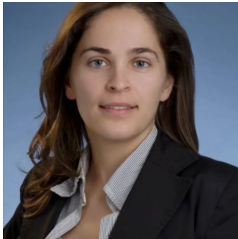 Catherine Coolens合作论文数Department of Medical Biophysics, Temerty Faculty of Medicine, University of Toronto19
 Jeffrey S. Wefel合作论文数Department of Neuro-Oncology, Division of Cancer Medicine, The University of Texas MD Anderson Cancer Center;Department of Radiation Oncology, Division of Radiation Oncology, The University of Texas MD Anderson Cancer Center;Angiochem Corporation;Genentech, Inc;F. Hoffman-La Roche Ltd19
 Sara Loupot Thrower合作论文数Department of Imaging Physics, The University of Texas MD Anderson Cancer CenterUnited States18
 Thomas H Beckham合作论文数18
 David R. Grosshans合作论文数Division of Radiation Oncology, Department of Radiation Oncology, The University of Texas MD Anderson Cancer Center18
 Warren Foltz合作论文数STTARR Innovation Centre/Princess Margaret Hospital17
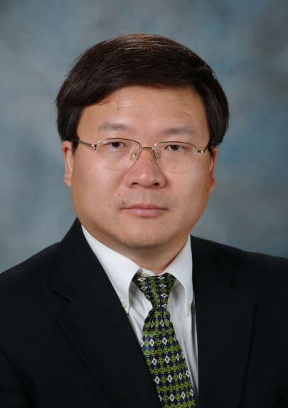 Jihong Wang合作论文数Department of Radiation Physics, Division of Radiation Oncology, MD Anderson Cancer Center, The University of Texas17
 Brian De合作论文数15
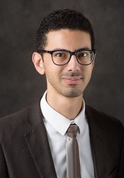 Hesham Elhalawani合作论文数Department of Radiation Oncology, Brigham and Women's Hospital;Harvard Medical School;Dana-Farber Brigham Cancer Center15
 David A Hormuth合作论文数Institute of Imaging Science, Vanderbilt University;Department of Biomedical Engineering, Vanderbilt University;Vanderbilt University, Vanderbilt University15
 Karine Al-Feghali合作论文数15
 Anita Mahajan合作论文数Department of Radiation Oncology, Mayo Clinic;Department of Pediatric and Adolescent Medicine, Mayo Clinic14
 Evanthia Galanis合作论文数Division of Medical Oncology, Department of Oncology, Mayo Clinic;Mayo Clinic Comprehensive Cancer Center, Mayo Clinic14Kristy K. Brock合作论文数14
 Todd A Swanson合作论文数14Bikash Panthi合作论文数13Martin C Tom合作论文数13
 Isabella C. Glitza合作论文数Division of Cancer Medicine, Department of Melanoma Medical Oncology, The University of Texas MD Anderson Cancer Center13
 Mark Bernstein合作论文数Department of Surgery, Temerty Faculty of Medicine, University of Toronto13
 Mark R. Gilbert合作论文数Center for Cancer Research, National Cancer Institute, National Institutes of Health13Erik P. Sulman合作论文数Division of Oncology, Children's Hospital of Philadelphia13
 Samantha M. Buszek合作论文数12
 Jia Wu (吴嘉)合作论文数School of Computer Science and Engineering, Central South University;Monash University12
 Laurence E. Court合作论文数Department of Radiation Physics, Division of Radiation Oncology, The University of Texas MD Anderson Cancer Center12
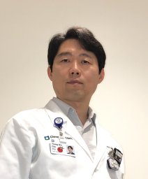 Youngbin Cho合作论文数Princess Margaret Hospital12
 Mairead McNamara合作论文数12
 Amy Heimberger合作论文数Department of Neurosurgery, The University of Texas M. D. Anderson Cancer Center12Holly Langshaw合作论文数11
 Zhihui Wang合作论文数11
 Thomas Yankeelov合作论文数Department of Biomedical Engineering, Cockrell School of Engineering, University of Texas at Austin11Joseph D Butner合作论文数11




