Background/Objectives: In glioma patients, acute neurologic deterioration may reflect nonconvulsive status epilepticus (NCSE), peri-ictal pseudoprogression, treatment-related change, or true progression. As these entities share imaging features, misdiagnosis can delay therapy. This review synthesizes evidence relevant to a proposed electroclinical-imaging framework for distinguishing seizure-related deterioration from treatment-related change and true progression. Methods: This narrative review synthesized English-language literature on tumor-related epilepsy, NCSE, peri-ictal imaging abnormalities, pseudoprogression, radiation necrosis, Response Assessment in Neuro-Oncology (RANO) criteria, and advanced imaging in adult diffuse glioma. Results: Seizure recurrence after glioma treatment may indicate tumor activity, but it may also reflect radiation injury, postoperative gliosis, edema, metabolic disturbance, medication effects, or transient peri-ictal change. NCSE is particularly important because it may present with aphasia, confusion, impaired consciousness, behavioral change, or deficits without convulsions, making electroencephalography central for unexplained deterioration. Seizure activity can also produce transient magnetic resonance imaging (MRI) or positron emission tomography (PET) abnormalities, including enhancement, fluid-attenuated inversion recovery (FLAIR) hyperintensity, diffusion restriction, and perfusion changes that mimic recurrence. Conclusions: This review proposes a structured electroclinical-imaging framework incorporating early EEG, seizure history, treatment timing, corticosteroid and antiseizure response, multimodal imaging, and longitudinal reassessment for prospective evaluation in treated glioma patients with new neurologic decline.
Outpatient neurosurgical oncology has expanded with advances in anesthesia, imaging, and minimally invasive techniques, enabling safe same-day discharge for selected patients undergoing procedures such as stereotactic biopsy and craniotomy. In this review, we find that across multiple international series, same-day discharge rates in several studies ranging from 85 to 95%, with low complication (3–6%) and readmission rates when structured pathways, including standardized selection criteria, enhanced recovery protocols, and routine 4-h postoperative CT imaging, are used. Studies on economic analyses demonstrate substantial cost savings driven by reduced inpatient bed utilization, with no increase in adverse events. Key challenges identified include medicolegal concerns amongst physicians, patient education, and limitations in organization adoption. Telemedicine and remote monitoring are increasingly incorporated to streamline preoperative evaluation and postoperative follow-up, improving access and continuity of care. Emerging technologies such as laser interstitial thermal therapy and focused ultrasound may further expand the outpatient neuro-oncology repertoire. Overall, current evidence supports outpatient neurosurgical oncology as a safe, efficient, and patient-centered model when applied with structured clinical pathways and patient selection.
BACKGROUND:Difficult benign intracranial tumors (including meningiomas, schwannomas, neurofibromatosis-related tumors, and pituitary neuroendocrine tumors) have substantial morbidity in patients. Due to their limited treatment options, there is a need for individualized treatment beyond histological and surgical approaches. OBJECTIVE:To summarize how novel treatment innovations have been implemented for these tumors, meningiomas and schwannomas are prioritized, followed by NF-associated neoplasms, and then pituitary neuroendocrine tumors in comparison to low-grade gliomas. METHODS:We summarize the current knowledge relating to targeted therapies for gliomas, meningiomas, schwannomas, neurofibromatosis (NF) tumors, and pituitary neuroendocrine tumors to investigate an individual's treatment options for difficult benign brain tumors. This review synthesizes evidence on tumor genomics and molecular markers, supported by methylation-based classification, immunohistochemistry, and functional assays, emphasizing current clinical applications. EVIDENCE SYNTHESIS:The recent data show that DNA methylation-based models can predict post-surgical outcomes and radiotherapy responses, enabling risk stratification and radiotherapy benefit prediction. Early signals support target-directed treatment, including cMET blockade that radiosensitizes NF2 schwannoma models, brigatinib-associated tumor shrinkage in NF2-deficient models, and PitNET organoid data. CONCLUSIONS:We support clinical decision-making that utilizes molecular profiling with functional testing to guide targeted treatment. We also identify evidence gaps such as biomarker-defined prospective trials that are needed for broader clinical implementation.
Objectives: Error-tracking studies have become an invaluable tool for gaining deeper insights into the patterns, causes, and clinical consequences of errors. In this paper, we examine the long-term impact of analyzing errors at the level of an individual surgeon. Methods: We performed a retrospective analysis of an internally maintained prospective database from September 2013 to April 2019 (Group C). These variables were compared with two earlier cohorts: Group A (2000-2006) and Group B (2006-2013). The key endpoints assessed included error incidence, type, severity, preventability, and clinical impact. The study also investigated the potential seasonal variations in error incidence, considering the rotation of trainees. Results: A persistent decrease in the mean errors/case (across all types) since records began in 2000 was noted; (1.8 for study group C versus 1.9 and 2.4 for group B and group A respectively, p = 0.048). We observed a drop in error severity score (major errors of 17.5% versus 29.5% and 22.6% respectively, p < 0.00001) and clinical impact (high impact in 0.4% versus 1.0% and 2.7% respectively, p < 0.00001). Conclusions: Ongoing and systematic error tracking and analysis seem to have a lasting impact on reducing both the incidence and severity of errors. These positive outcomes are thought to reflect the establishment of a culture of safety, fostering transparency and constructive feedback within the broader surgical team.
BACKGROUND AND OBJECTIVES:Previous reports have suggested that neoadjuvant stereotactic radiosurgery (SRS) for brain metastases (BrMets) mitigates the elevated risks of radiation necrosis (RN) and meningeal recurrence associated with adjuvant SRS. We report treatment outcomes from a multicenter phase II trial (NCT03368625) of single-fraction neoadjuvant SRS for large BrMets. METHODS:Patients with 1 index BrMet requiring resection and up to 9 nonindex BrMets not requiring resection were recruited across 3 centers and treated with single-fraction SRS (14-21 Gy) targeting the index lesion with a 2-mm margin, followed by surgical resection. Nonindex lesions were targeted with definitive SRS. The primary end point was 1-year rate of grade 2+ RN affecting the index lesion. Secondary end points included median overall survival, 2-year intracranial progression-free survival, and 1-year rates of local failure (LF) affecting the index lesion, leptomeningeal disease, and pachymeningeal disease. RESULTS:Between April 2018 and November 2022, 35 patients were enrolled; the median follow-up period was 11.8 months (IQR: 6.14, 15.9). No patients developed grade 2+ RN. Six patients experienced LF (1-year rate: 18.0% [95% CI: 7.03, 32.9]); 1 patient developed classic leptomeningeal disease (1-year rate: 2.9% [95% CI: 0.21, 12.9]), and 1 patient developed pachymeningeal disease (1-year rate: 3.2% [95% CI: 0.22, 14.6]). The median overall survival was 13.8 months (95% CI: 8.15, 22.4), and the 2-year intracranial progression-free survival was 29.5% (95% CI: 13.8, 63.1). CONCLUSION:In this study, no patients experienced symptomatic RN and the incidence of meningeal failure was lower than historical rates associated with postoperative SRS. However, the high 1-year rate of LF suggests a potential benefit for higher or fractionated radiation doses or larger clinical target volume margins.
Stereotactic brain biopsy is a fundamental procedure in neurosurgery that enables the histomolecular diagnosis of brain tumors and cryptogenic neurological diseases. Traditionally, this procedure requires hospitalization because of potential complications, particularly hemorrhagic events. However, the increasing emphasis on ambulatory surgery and its medico-economic benefits have led to the exploration of outpatient stereotactic brain biopsies. This study aimed to evaluate the safety and feasibility of outpatient stereotactic brain biopsy in a large, real-world patient cohort. This retrospective study analyzed all adult patients who underwent stereotactic brain biopsy at our institution between November 2021 and October 2024. We compared the outpatient and inpatient groups based on demographics, clinical and radiological data, biopsy characteristics, and post-procedure outcomes. The primary endpoints were the rate of unplanned hospitalization following biopsy (failure of outpatient management), biopsy-related complications, and unplanned medical consultations or admissions within 30 days after biopsy. Statistical analyses included univariate and multivariate models to assess factors influencing outpatient eligibility and outcomes. Among the 565 patients who underwent stereotactic brain biopsy, 191 (33.8
BackgroundStereotactic radiosurgery (SRS) has supplanted whole brain radiotherapy (WBRT) as standard-of-care adjuvant treatment following surgery for brain metastasis (BrM). Concomitant with the adoption of adjuvant SRS, a new pattern of failure termed "Pachymeningeal failure" (PMF) has emerged.Methods We reviewed a prospective registry of 264 BrM patients; 145 and 119 were treated adjuvantly with WBRT and SRS, respectively. The Cox proportional hazards model was used to identify variables correlating to outcomes. Outcomes were calculated using the cumulative incidence (CI) method. Univariate (UVA) and multivariate analyses (MVA) were done to identify factors associated with PMF.ResultsCI of PMF was 2% and 18% at 12 months, and 2% and 23% at 24 months for WRBT and SRS, respectively (p<0.001). The CI of classic leptomeningeal disease (LMD) was 3% and 4% at 12 months, and 6% and 6% at 24 months for WBRT and SRS, respectively (P=0.67). On UVA, adjuvant SRS [HR 9.75 (3.43-27.68) (P<0.001)]; preoperative dural contact (PDC) [HR 6.78 (1.64-28.10) (P =0.008)]; GPA score [HR 1.64 (1.11-2.42) (P =0.012)]; and lung EGFR/ALK status [HR 3.11 (1.02-9.45) (P =0.045)]; were associated with PMF risk. On MVA, adjuvant SRS [HR 8.15 (2.69-24.7) (P<0.001)]; and PDC [HR 6.28 (1.51-26.1) (P =0.012)] remained associated with PMF.ConclusionsPreoperative dural contact and adjuvant SRS instead of adjuvant WBRT were associated with an increased risk of PMF. Strategies to improve pachymeningeal radiation coverage to sterilize at risk pachymeninges should be investigated.
Increasing CT capacity to keep pace with rising ED demand is critical. The conventional process has inherent drawbacks. We evaluated an off-console automated AI enhanced workflow which moves all final series creation off-console. We hypothesized the off-console workflow would 1) decrease overall ED CT exam begin to end times and decrease length and variability of time CT is occupied at the individual exam level. Study population was identified retrospectively and included all CT exams done on all ED adult patients. 3 months of data was collected using the conventional workflow and 3 months of data was collected after implementation of the off-console workflow. Exam begin and the exam end timestamps were collected from the EMR. Additionally, 4 subgroups from the above conventional and off-console workflows were identified retrospectively with an Emergency Severity Index level 1, undergoing one of the four most common CT exam set(s) performed on ESI level 1 patients. 6,795 ED adult patients underwent ED CT in the 3 months immediately prior to implementation of the off-console workflow and 6,708 adult ED patients underwent CT in the 3 months after complete implementation. The off-console workflow demonstrated a 36
A broad range of brain pathologies critically relies on the vasculature, and cerebrovascular disease is a leading cause of death worldwide. However, the cellular and molecular architecture of the human brain vasculature remains incompletely understood1. Here we performed single-cell RNA sequencing analysis of 606,380 freshly isolated endothelial cells, perivascular cells and other tissue-derived cells from 117 samples, from 68 human fetuses and adult patients to construct a molecular atlas of the developing fetal, adult control and diseased human brain vasculature. We identify extensive molecular heterogeneity of the vasculature of healthy fetal and adult human brains and across five vascular-dependent central nervous system (CNS) pathologies, including brain tumours and brain vascular malformations. We identify alteration of arteriovenous differentiation and reactivated fetal as well as conserved dysregulated genes and pathways in the diseased vasculature. Pathological endothelial cells display a loss of CNS-specific properties and reveal an upregulation of MHC class II molecules, indicating atypical features of CNS endothelial cells. Cell-cell interaction analyses predict substantial endothelial-to-perivascular cell ligand-receptor cross-talk, including immune-related and angiogenic pathways, thereby revealing a central role for the endothelium within brain neurovascular unit signalling networks. Our single-cell brain atlas provides insights into the molecular architecture and heterogeneity of the developing, adult/control and diseased human brain vasculature and serves as a powerful reference for future studies.
Background/Objectives: Around 20% of cancer patients will develop brain metastases (BrMs), with 15–25% occurring in the posterior fossa (PF). Although the effectiveness of systemic therapies is increasing, surgery followed by stereotactic radiosurgery (S+SRS) versus definitive SRS remains the mainstay of treatment. Given the space restrictions within the PF, patients with BrMs in this location are at higher risk of brainstem compression, hydrocephalus, herniation, coma, and death. However, the criteria for treating large PF BrMs with S+SRS versus definitive SRS remains unclear. Methods: We reviewed a prospective registry database (2009 to 2020) and identified 64 patients with large PF BrMs (≥4 cc) treated with SRS or S+SRS. Clinical and radiological parameters were analyzed. The two endpoints were overall survival (OS) and local failure (LF). Results: Patients in the S+SRS group were more highly symptomatic than patients in the SRS group. Gait imbalance and intracranial pressure symptoms were 97% and 80%, and 47% and 35% for S+SRS and SRS, respectively. Radiologically, there were significant differences in the mean volume of the lesions [6.7 cm3 in SRS vs. 29.8 cm3 in the S+SRS cohort, (p < 0.001)]; compression of the fourth ventricle [47% in SRS vs. 96% in S+SRS cohort, (p < 0.001)]; and hydrocephalus [0% in SRS vs. 29% in S+SRS cohort, (p < 0.001)]. Patients treated with S+SRS had a higher Graded Prognostic Assessment (GPA). LF was 12 and 17 months for SRS and S+SRS, respectively. Moreover, the S+SRS group had improved OS (12 vs. 26 months, p = 0.001). Conclusions: A higher proportion of patients treated with S+SRS presented with hydrocephalus, fourth-ventricle compression, and larger lesion volumes. SRS-alone patients had a lower KPS, a lower GPA, and more brain metastases. S+SRS correlated with improved OS, suggesting that it should be seriously considered for patients with large PF-BrM.
Background: Prospectively tracking errors can improve patient safety but little is known about how to successfully implement error reporting in a home-based palliative care context.Aim: Explore the feasibility of implementing an error reporting system in a home-based palliative care program in Toronto, Canada, and describe the possible factors that may influence uptake.Design: A convergent mixed-methods approach was used. Participants prospectively documented errors using a novel reporting tool and completed monthly surveys. Following the reporting period, we conducted a semi-structured interview exploring participants' experiences and perceived factors influencing reporting behaviors. Error, survey, and interview data were analyzed separately, then integrated for comparison.Setting and participants: Thirteen palliative care physicians from a single home-based palliative care organization in Toronto, Canada anonymously reported errors between October 2021 and September 2022. Of these, six participated in the exit interview.Results: Participants reported 195 errors; one-third (n = 65) involved internal staff or systems. Three themes describe the factors impacting the likelihood of reporting errors: (1) High levels of cognitive burden decreases the likelihood of error reporting; (2) Framing errors as opportunities to learn rather than reason for punishment improves likelihood of error reporting; (3) Knowing that error data will improve patient safety motivates individuals to report errors.Conclusions: Physicians are amenable to error reporting activities so long as data is used to improve patient safety. The collaborative nature of care in a home-based palliative care context may present unique challenges to translating error reporting to improved patient safety.
Purpose Neurosurgery (NS) is an essential modality for large brain metastases (BM). Postoperative stereotactic radiosurgery (SRS) is the standard of care adjuvant treatment. Pachymeningeal failure (PMF) is a newly described entity, distinct from classical leptomeningeal failure (LMF), that is uniquely observed in postoperative patients treated with adjuvant SRS. We sought to identify risk factors for PMF in patients treated with NS + SRS. Methods From a prospective registry (2009 to 2021), we identified all patients treated with NS + SRS. Clinical, imaging, pathological, and treatment factors were analyzed. PMF incidence was evaluated using a competing risks model. Results 144 Patients were identified. The median age was 62 (23–90). PMF occurred in 21.5% (31/144). Female gender [Hazard Ratio (HR) 2.65, p = 0.013], higher Graded Prognostic Assessment (GPA) index (HR 2.4, p < 0.001), absence of prior radiation therapy (HR N/A, p = 0.018), controlled extracranial disease (CED) (HR 3.46, p = 0.0038), and pia/dura contact (PDC) (HR 3.30, p = 0.0053) were associated with increased risk for PMF on univariate analysis. In patients with PDC, wider target volumes correlated with reduced risk of PMF. Multivariate analysis indicated PDC (HR 3.51, p = 0.0053), piecemeal resection (HR 2.38, p = 0.027), and CED (HR 3.97, p = 0.0016) independently correlated with PMF risk. PMF correlated with reduced OS (HR 2.90, p < 0.001) at a lower rate compared to LMF (HR 10.15, p < 0.001). Conclusion PMF correlates with tumor PDC and piecemeal resection in patients treated with NS + SRS. For unclear reasons, it is also associated with CED. In tumors with PDC, wider dural radiotherapy coverage was associated with a lower risk of PMF.
PurposeTo understand the natural history and optimal treatment strategy for pituitary gland metastasis.MethodsWe performed both a retrospective chart review of patients treated at our institution and a scoping review of the topic.ResultsThe retrospective review identified seven patients with an average age of 59.6 years. Primary histologies included breast cancer (4), melanoma (1), renal cell carcinoma (1), and sarcoma (1). Two patients had anterior pituitary endocrine dysfunction, one of whom was the only patient with visual symptoms. All patients were treated with radiosurgery and two also underwent surgical resection. Overall survival ranged from 6.5 to 117 months. Literature review identified 166 patients from 71 studies. The most common primary cancer was lung (27.7%), followed by breast (18.7%) and renal (14.5%) cancer. 107 presented with endocrine dysfunction, including 41 cases of diabetes insipidus and 55 cases of hypopituitarism. 110 presented with visual compromise. 107 patients received radiotherapy, 96 underwent surgical resection and 44 received systemic chemotherapy/immunotherapy. Surgery was significantly associated with an increased likelihood of vision improvement and a decreased likelihood of endocrine normalization. Radiographic regression predicted visual improvement. Median overall survival was 9.9 months (range: 0.2-96).ConclusionsThis scoping review showed that both radiosurgery and surgical resection have been frequently used to treat pituitary metastases with good response. Vision improvement is more likely to happen following surgical resection, likely at the expense of endocrine dysfunction. Despite treatment and radiographic response, patient survival remains less than a year.
Background: Awake Craniotomy during pregnancy is a rare but urgent procedure. Since pregnancy can both accelerate the progression of a tumor and mask other diagnoses, cases may lead to premature termination of pregnancy. From a neurosurgical, anesthetic, and obstetrical perspective, these operations may be challenging. Methods: In accordance with the PRISMA guidelines, MEDLINE, Scopus, and Web of Science databases were searched from inception to January 3rd, 2023. Studies were included if they included pregnant patients who underwent awake craniotomy. Results: Nine papers fit the criteria for the final analysis. All investigations were case studies. A total of nine patients were included. Mean age at surgery was 26.9 years, and mean gestational age at craniotomy was 20.9 weeks. Eight (88.9%) patients underwent craniotomy for tumor resection and the other had a pseudoaneurysm repair. Glioma was the most common tumor pathology (n=5), followed by meningioma (n=1), and glioblastoma (n=1). None of the patients experienced significant intraoperative or immediate postoperative complications. There were no obstetrical complications or significant changes in fetal status during or after surgery, and all reported deliveries were successful with healthy infants. Conclusions: Awake craniotomy during pregnancy can be a safe procedure with appropriate pre-operative patient selection and extensive multidisciplinary planning.
Introduction: Intraoperative seizures (IOSs) during awake craniotomy (AC) are associated with significant morbidity. The reported incidence of IOS is between 3% and 30%. The aim of this study was to identify risk factors for IOS during AC for elective resection or biopsy of a space-occupying brain lesion. Methods: In this retrospective study, we reviewed the records of all awake craniotomies performed by a single neurosurgeon at a single university hospital between July 2006 and December 2018. IOS was defined as a clinically apparent seizure that occurred in the operating room and was documented in the medical records. Explanatory variables were chosen based on previously published literature on risk factors for IOS. Results: Five hundred and sixty-two patients had a total of 607 AC procedures during the study period; 581 cases with complete anesthesia records were included in analysis. Twenty-nine (5.0%) IOS events were reported during 29 (5%) awake craniotomies. Most seizures (27/29; 93%) were focal in nature and did not limit planned intraoperative stimulation mapping. Variables associated with IOS at a univariate P-value <0.1 (frontal location of tumor, preoperative radiotherapy, preoperative use of antiepileptic drugs, intraoperative use of dexmedetomidine, and intraoperative stimulation mapping) were included in a multivariable logistic regression. Frontal location of tumor (adjusted odds ratio: 5.68, 95% confidence interval: 2.11-15.30) and intraoperative dexmedetomidine use (adjusted odds ratio: 2.724, 95% confidence interval: 1.24-6.00) were independently associated with IOS in the multivariable analysis. Conclusions: This study identified a low incidence (5%) of IOS during AC. The association between dexmedetomidine and IOS should be further studied in randomized trials as this is a modifiable risk factor.
 Gelareh Zadeh合作论文数Department of Surgery, Temerty Faculty of Medicine, University of Toronto;Toronto Western Hospital, University Health Network68
 Normand J. Laperriere合作论文数Department of Radiation Oncology, Temerty Faculty of Medicine, University of Toronto;Division of Hematology/Oncology, Hospital for Sick Children;Princess Margaret Hospital Cancer Centre65
 Paul Kongkham合作论文数Department of Laboratory Medicine and Pathobiology, Temerty Faculty of Medicine, University of Toronto50
 Mark Krailo合作论文数Keck School of Medicine, University of Southern California46
 Barbara-Ann Millar合作论文数Department of Radiation Oncology, Temerty Faculty of Medicine, University of Toronto45
 David Shultz合作论文数Department of Radiation Oncology, Temerty Faculty of Medicine, University of Toronto;Cancer Clinical Research Unit, Princess Margaret Cancer Centre, University Health Network39
 Neyssa Marina合作论文数Division of Hematology/Oncology, Department of Pediatrics, Stanford University;Synthorx;Sanofi36Holcombe E. Grier合作论文数31
 Tatiana Conrad合作论文数Department of Radiation Oncology, Temerty Faculty of Medicine, University of Toronto30
 Susan M. Blaney合作论文数Texas Children’s Cancer and Hematology Center, Texas Children's Hospital;Department of Pediatrics, Section of Hematology-Oncology, Baylor College of Medicine;Dan L. Duncan Comprehensive Cancer Center, Baylor College of Medicine28
 Richard Gorlick合作论文数Department of Pediatrics Patient Care, Division of Pediatrics, The University of Texas MD Anderson Cancer Center;Department of Pediatric Sarcoma Research Laboratory, The University of Texas MD Anderson Cancer Center27
 Mitchel Berger合作论文数Brain Tumor Center, University of California, San Francisco;School of Medicine, University of California, San Francisco27
 Alejandro Berlin合作论文数Department of Radiation Oncology, Temerty Faculty of Medicine, University of Toronto;Cancer Clinical Research Unit, Princess Margaret Cancer Centre, University Health Network24
 Paul A. Meyers合作论文数Pediatric Sarcoma Service, Memorial Sloan Kettering Cancer Center;Bone Sarcoma Strategy Group, Children’s Cancer Group, Memorial Sloan Kettering Cancer Center;Happiness Is Camping21
 Michael D. Cusimano合作论文数Department of Surgery, Temerty Faculty of Medicine, University of Toronto18Warren Mason合作论文数Department of Medicine, Temerty Faculty of Medicine, University of Toronto17
 Lashmi Venkatraghavan合作论文数16
 Moraes Fábio Ynoe de合作论文数16
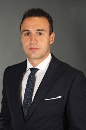 Aristotelis Kalyvas合作论文数Evaggelismos Hospital, University of Athens;Microneurosurgery Laboratory, Evaggelismos Hospital;Evaggelismos Hospital, University of Athens16Mark C. Gebhardt合作论文数16
 Jeremy Whelan合作论文数Cancer Institute, Faculty of Medical Sciences, University College London;Department of Oncology, University College London Hospitals NHS Foundation Trust16
 Sigbjørn Smeland合作论文数16
 Katherine A. Janeway合作论文数Dana-Farber Cancer Institute;Harvard Medical School15
 Matthew R. Sydes合作论文数MRC Clinical Trials Unit, University College London15
 Lisa A Teot合作论文数14Dirks Peter合作论文数14Stefan S. Bielack合作论文数14
 R. Lor Randall合作论文数Department of Orthopaedics, Huntsman Cancer Institute14
 Cindy L Schwartz合作论文数14
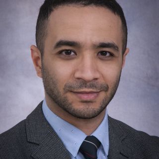 George M. Ibrahim合作论文数Department of Surgery, University of Toronto14
 Caroline Chung合作论文数Department of Radiation Oncology, Division of Radiation Oncology, The University of Texas MD Anderson Cancer Center;Institute for Data Science in Oncology, The University of Texas MD Anderson Cancer Center13
 Gabriele Calaminus合作论文数Universitätsklinikum Münster;Universitätsklinikum Bonn13Richard B. Womer合作论文数13Jennifer Glen合作论文数13Kathleen Joy Khu合作论文数13Allen Goorin合作论文数12
 Catherine Coolens合作论文数Department of Medical Biophysics, Temerty Faculty of Medicine, University of Toronto12
 Meenakshi Devidas合作论文数St. Jude Children's Research Hospital12
 Nir Lipsman合作论文数Sunnybrook Health Sciences Centre;Department of Surgery, Temerty Faculty of Medicine, University of Toronto12
 Cynthia Ménard合作论文数Department of Radiation Oncology, University of Montreal Hospital Center;University of Montreal;University of Toronto12Jessica Weiss合作论文数Princess Margaret Cancer Centre, University Health Network11
 Jeff Winter合作论文数Department of Radiation Oncology, Temerty Faculty of Medicine, University of Toronto11
 James Rutka合作论文数Department of Surgery, University of Toronto;Division of Neurosurgery, University of Toronto;The Labatt Brain Tumour Research Centre, The Hospital for Sick Children11Gordana Jovic合作论文数10
 Pirjo Manninen合作论文数Department of Anesthesia, University of Toronto10
 John H. Healey合作论文数Memorial Sloan Kettering Cancer Center;Weill Cornell Medical College, Cornell University10
 Enrique Gutierrez合作论文数Dept Radiat Oncol, Univ Toronto9
 Suneil Kalia合作论文数Department of Laboratory Medicine and Pathobiology, Temerty Faculty of Medicine, University of Toronto9
 Mojgan Hodaie合作论文数Department of Surgery, Temerty Faculty of Medicine, University of Toronto;Toronto Western Hospital9




