Lycorine (LYC), the most abundant monomeric compound from Lycoris radiata bulbs, possesses a range of biological activities, including antipulmonary fibrosis and cardioprotective effects. However, its role in regulating pathological cardiac remodeling, particularly in cardiac fibrosis, remains unclear. In this study, we used transforming growth factor-β1 (TGF-β1) to stimulate NIH/3T3 cells and primary rat cardiac fibroblasts, which were in vitro models of pathological fibroblast activation, and established a mouse model of cardiac fibrosis via intraperitoneal injection of doxorubicin (DOX). The effects of LYC on TGF-β1-induced fibroblast activation and migration were assessed using Western blotting, quantitative PCR, scratch assays, and EdU staining. Cardiac damage and fibrosis were evaluated in vivo by echocardiography, enzyme-linked immunosorbent assay (ELISA), and Masson’s trichrome staining. Through transcriptomic sequencing and bioinformatic analyses, proline-rich tyrosine kinase 2 (PYK2) was identified as a potential target of LYC. This finding was further validated using siPYK2 and the PYK2 inhibitor PF4618433 in TGF-β1-induced NIH/3T3 cells. Our results demonstrated that LYC effectively suppressed fibroblast activation, proliferation, and migration. In addition, LYC significantly ameliorated cardiac dysfunction and reduced collagen deposition in myocardial tissues, thereby attenuating the progression of cardiac fibrosis. Mechanistically, these effects were attributed to the regulation of PYK2 expression and activity by LYC. This study provides a scientific basis for understanding the mechanism of LYC in improving cardiac fibrosis and highlights PYK2 as a potential therapeutic target for the treatment of cardiac fibrosis.
BACKGROUND:Chronic kidney disease (CKD) is a significant risk factor for myocardial injury. Despite the proven clinical safety and efficacy of Shen Shuai II Recipe (SSR) in CKD management, and evidence of its renoprotective effects involving anti-inflammation and mitochondrial protection in experimental models, its potential to ameliorate CKD-related myocardial injury has not been investigated. PURPOSE:We sought to determine whether SSR confers protection against CKD-induced myocardial injury by suppressing the IL-18/IL-18R1/MyD88 pathway. METHODS:A CKD model was established in rats by 5/6 (A/I) surgery. The rats were randomly assigned to receive daily gavage of normal saline, SSR, or Losartan potassium for 8 weeks. In vitro, IL-18-stimulated H9C2 cells were treated with different concentrations of SSR, or H9C2 cells were directly treated with different concentrations of 5/6 (A/I)+SSR rat serum. The effects of SSR and rat serum on the IL-18R1/MyD88-mediated inflammatory pathway and myocardial injury were investigated via immunoblotting, luminex chip assay, histopathology and fluorescence staining. Additionally, to further elucidate the mechanisms of SSR against myocardial injury, H9C2 cells were treated with 5/6 (A/I) or 5/6 (A/I)+SSR rat serum in the presence or absence of IL-18 neutralizing antibody. Next, we delivered IL-18R1 overexpression plasmid or MyD88 inhibitor into the IL-18-treated H9C2 cells with concomitant SSR administration. Finally, using a co-culture approach, we explored whether hypoxic renal tubular cells induced myocardial injury via the IL-18R1/MyD88 pathway. RESULTS:SSR inhibited IL-18R1/MyD88-mediated inflammatory response, decreased the expression of β-MHC and ANP hypertrophy marker proteins, and attenuated myocardial injury in myocardial tissues of 5/6 (A/I) rats or IL-18-treated H9C2 cells. The same effect was also observed in H9C2 cells treated with 5/6 (A/I)+SSR rat serum. Further investigation confirmed that SSR ameliorated myocardial injury through suppression of the IL-18R1/MyD88 inflammatory pathway. More crucially, co-culture experiments demonstrated that SSR alleviated crosstalk between hypoxic tubular cells and cardiomyocytes via IL-18/IL-18R1/MyD88 pathway, thereby mitigating myocardial injury. CONCLUSION:SSR ameliorates CKD-induced myocardial injury through suppression of the IL-18/IL-18R1/MyD88 pathway.
Introduction Idiopathic pulmonary fibrosis (IPF) is a progressive and fatal lung disease characterized by chronic inflammation and fibroblast activation, with limited treatment options. The bioactive diterpenoid andrographolide (AP) exhibits anti-inflammatory and antifibrotic properties, but its direct molecular targets and precise mechanism remain unclear. Objectives This study aimed to identify the primary functional target of AP and elucidate the molecular mechanism underlying its therapeutic effects against pulmonary fibrosis. Methods We employed activity-based protein profiling (ABPP) to identify covalent targets of AP in macrophages. Functional validation was performed using siRNA knockdown, enzymatic assays, molecular docking, and biophysical analyses. The therapeutic efficacy and target dependency of AP were evaluated in bleomycin-induced pulmonary fibrosis and LPS-induced acute lung injury mouse models, utilizing lung-specific Ptges3 knockdown. Results AP covalently bound to Cys58 of prostaglandin E synthase 3 (Ptges3), an allosteric site distinct from its catalytic and Hsp90-binding regions. This binding inhibited Ptges3 enzymatic activity, reduced prostaglandin E2 (PGE2) production, and disrupted the Ptges3-Hsp90 chaperone complex, leading to suppressed NF-κB signaling. Genetic knockdown of Ptges3 significantly attenuated the anti-inflammatory and antifibrotic effects of AP both in vitro and in vivo. Conclusions Our findings establish Ptges3 as a critical functional target of AP. AP attenuates pulmonary fibrosis through a dual mechanism involving covalent inhibition of Ptges3 and disruption of the Ptges3-Hsp90-NF-κB axis, highlighting AP as a promising therapeutic agent and Ptges3 as a novel druggable target for IPF.
Background:Lignans from Schisandra chinensis, such as schisandrin B, possess antioxidant, anti-inflammatory, and protein-regulating properties, suggesting potential against muscle atrophy. However, existing animal evidence is fragmented and lacks quantitative synthesis. Objective:This meta-analysis evaluates the effects of S. chinensis on various animal models of muscle atrophy, focusing on muscle structure, function, and oxidative stress. Methods:We systematically searched PubMed, Web of Science, Embase, Cochrane Library, and Ovid from inception to April 2025 for relevant animal RCTs, selected via the PICOS framework. Study quality was assessed using the SYRCLE tool. A random-effects meta-analysis was performed on outcomes including muscle weight, fiber cross-sectional area (CSA), catalase (CAT) activity, and grip strength. Results:Eleven studies (149 experimental, 149 control animals) were included. S. chinensis significantly increased muscle weight (standardized mean difference = 1.18, 95% CI: 0.19-2.16, P = 0.020) and CAT activity (SMD = 1.77, 95% CI: 0.31-3.23, P = 0.020). cross-sectional area improvement was borderline (SMD = 2.10, 95% CI: -0.02-4.22, P = 0.050). No significant changes occurred in body weight or grip strength. High heterogeneity (I2 = 80%-95%) was observed, attributable to variations in models and interventions. Conclusion:S. chinensis improves muscle structure potentially via enhanced antioxidant defense and reduced protein degradation, though functional benefits remain unconfirmed. Future high-quality, standardized studies are needed to clarify dose-response relationships and translational potential. Systematic Review Registration:identifier INPLASY202590030.
Osteoarthritis (OA) is a prevalent age-related joint disorder with limited treatment options. Chronic activation of the innate immune response in chondrocytes plays a key role in OA progression. However, the underlying mechanisms remain incompletely understood. Here, we report that mitochondrial antiviral signaling protein (MAVS) exacerbates cartilage extracellular matrix (ECM) degradation in OA. MAVS activation is observed in chondrocytes from both OA patients and the destabilization of the medial meniscus (DMM) mouse model. Both constitutive and chondrocyte-specific MAVS knockout alleviate cartilage degradation, osteophyte formation, subchondral bone remodeling, and synovitis in DMM mice. Conversely, MAVS overexpression aggravates these OA phenotypes. Mechanistically, cytosolic accumulation of mitochondrial double-stranded RNA in chondrocytes triggers MAVS activation, leading to MAVS-nuclear factor κB-dependent ECM degradation by inducing matrix metalloproteinase 3 (MMP3) and MMP13. Pharmacologically blocking MAVS using L-lactate significantly attenuates ECM degradation and OA progression. These findings suggest that MAVS signaling is critical in OA pathogenesis and may be a potential therapeutic target for OA treatment.
Heterogeneous nuclear ribonucleoprotein K (hnRNPK) is a multifunctional RNA-binding protein implicated in various cellular processes, yet its role in fish antiviral immunity and viral replication remains poorly understood. In this study, we found that common carp (Cyprinus carpio) hnRNPK expression was markedly upregulated following SVCV infection or poly(I:C) stimulation, accompanied by its translocation from the nucleus to the cytoplasm. Overexpression of hnRNPK enhanced viral replication, as reflected by increased viral gene expression and viral titers. Mechanistically, hnRNPK interacted with the SVCV phosphoprotein (P) and promoted its stability. Additionally, hnRNPK suppressed type I interferon (IFN) production by interacting with key components of the RLR signaling pathway, including MITA, TBK1, IRF3, and IRF7. Notably, hnRNPK facilitated IRF3 degradation and impeded its nuclear translocation, thereby negatively regulating the type I IFN response. Collectively, these findings indicate that hnRNPK promotes SVCV replication through a dual mechanism: stabilizing the viral P protein and attenuating the host type I interferon response by targeting IRF3. This study provides new insights into the immune evasion strategies employed by SVCV and highlights hnRNPK as a potential target for antiviral intervention in lower vertebrates.
The endoplasmic reticulum (ER)-resident STING activation induces innate immune responses, cell death, autophagy and lysosomal biogenesis for host defense 1–3 . However, the potential function of STING in resting state is poorly understood. Here, we report that the loss of STING increases SREBP2 activation and the consequent cholesterol-biosynthetic genes expression both in vitro and in vivo . Notably, STING deficiency favors the distribution of cholesterol into the plasma membrane (PM) without affecting its overall cellular levels. Unexpectedly, STING utilizes its tandem hydrophobic pockets to recruit Aster-escorted cholesterol 4 from the PM and thus functions as a newly defined cholesterol channel (transmembrane pore) to load cholesterol into the ER membrane. Consistently, Compound 53 (C53) 5 inhibits cholesterol transport both in vivo and in vitro by blocking this channel. Either constitutive or intestinal epithelial-specific Sting1 knockout suppresses dietary cholesterol absorption, tissues accumulation and decreases systemic corticosterone levels. Our study highlights a primordial function of STING in ER for cholesterol homeostasis.
Renal fibrosis is the common pathological feature of chronic kidney diseases, which is in parallel with increasing energy demand in proximal tubular epithelial cells during the metabolic shift from fatty acid oxidation to glycolysis. Shen-Shuai-II-Recipe (SSR) is a traditional Chinese medicine formula with known renal benefits in patients with chronic kidney disease (CKD) and has been shown to intervene in renal energy metabolism in a rodent model of CKD. We aimed to explore the mechanism underlying the protective effect of SSR against CKD. A 5/6 ablation/infarction (A/I) renal failure model was established in rats, followed by 8 weeks of gavage feeding with SSR or Losartan, a positive control. For in vitro experiments, normal rat kidney-52E (NRK-52E) cells were cultured under hypoxic condition (1% O2) or normoxic condition. The expression of fibrotic markers and aerobic glycolysis-related enzymes were determined by Western blotting analysis. The concentration of metabolites was also measured. The concentration of lactic acid was increased, and the concentration of pyruvic acid was decreased in vitro and in vivo models, which were correlated with increased expression of glycolysis-related enzymes Hexokinase2 (HK2) and 6-phosphofructo-2-kinase/fructose-2,6-bisphosphatase 3 (PFKFB3), SSR treatment reversed the changes of renal glycolysis in cells and animals, which was associated with reduced expression of fibrotic markers such as fibronectin and α-SMA. Mechanistically, SSR treatment enhanced the expression of Sirtuin 1 (SIRT1) and reduced the expression of Hypoxia-Inducible Factor 1-alpha (HIF-1α) in vitro and in vivo models. Downregulation of SIRT1 by small interfering ribonucleic acid (siRNA) reversed the anti-fibrotic effect and anti-glycolysis effect of SSR in renal cells. SSR exerts an anti-fibrotic effect and inhibits renal aerobic glycolysis in CKD via the regulation of SIRT1/HIF-1α.
Japanese encephalitis (JE), caused by the Japanese encephalitis virus (JEV), remains a major global health concern. A deeper understanding of how JEV manipulates host antiviral responses is critical for developing more effective therapeutic strategies. In this study, we identified a novel immune evasion mechanism through which JEV suppresses host innate immunity to facilitate viral replication. Specifically, JEV infection induces the expression of the host translation elongation factor TUFM by activating the transcription factor JUN. We demonstrated that TUFM promotes JEV replication, while TUFM silencing significantly reduces viral load. Mechanistically, TUFM acts as a scaffold to recruit the E3 ubiquitin ligase Parkin and the autophagy receptor p62, thereby mediating selective autophagy-dependent degradation of the key innate immune adaptor TRAF3. Intriguingly, the viral NS5 protein was also found to promote TRAF3 degradation via a similar autophagy-dependent pathway, suggesting a coordinated strategy between viral and host factors. Since TRAF3 is essential for the activation of IRF3 and downstream type I interferon signaling, its degradation effectively blunts antiviral innate immune responses. Collectively, our findings reveal a previously unrecognized mechanism by which JEV subverts host innate immunity through the TUFM-NS5-Parkin-p62-TRAF3 axis to promote immune evasion and viral propagation.
Pulmonary large-cell neuroendocrine carcinoma (LCNEC) is a rare yet highly aggressive subtype of non-small cell lung carcinoma (NSCLC) with limited therapeutic options. We conduct a comprehensive proteogenomic analysis of LCNEC using tumors and paired normal adjacent tissues from 107 patients (81 pure LCNEC and 26 combined LCNEC). APOBEC mutational signatures strongly correlate processes of tumor initiation and immune suppression, and KEAP1 mutations correlate with metabolic reprogramming in LCNEC combined with NSCLC. A conflicting relationship is observed between neuroendocrine and immune phenotypes. We identify three LCNEC subtypes with unique prognosis features, microenvironment dysregulation, genetic alterations, and potential therapeutic targets. Interleukin-33 (IL-33) emerges as a critical therapeutic biomarker associated with enhanced T cell infiltration and antitumor activity. We further optimize recombinant IL-33 (rIL33) with site-directed mutagenesis and develop PEGylated rIL33, demonstrating its prolonged circulation time in cynomolgus monkeys and superior immune agonist activity in mouse models. Overall, this study offers insights into LCNEC biology and provides promising innovative immunoagonist therapy strategies for lung cancer.
The cGAS-STING pathway is emerging as an essential driver in systemic lupus erythematosus (SLE). Here, we characterize the key signature of cGAS-STING pathway and its roles in SLE by leveraging large-scale transcriptomics, cell-based assays, and two lupus-like mouse models. We identify a STING-dependent gene signature termed M7core, enabling quantitative assessment of cGAS-STING pathway activity in SLE. M7core reveals widespread cGAS-STING pathway activation in 70.4% of 3,180 SLE samples and predicts therapeutic response to STING antagonists in 74.1% of patients, with higher activity indicating greater sensitivity. Across ten independent cohorts, M7core outperforms interferon-stimulated gene signatures (mean AUROC = 0.876) and correlates with disease activity, anti-dsDNA antibodies, lymphopenia, and lupus nephritis. Hydroxychloroquine treatment reduces M7core expression and its clinical associations. Importantly, in cGAS-STING pathway-driven lupus-like mice, STING antagonist administration ameliorates multiorgan pathology and suppresses M7core genes participating in promoting inflammation, type I interferon, and cell death, including ZBP1-an established cGAS-STING pathway facilitator. Notably, ZBP1 deficiency phenocopies blocking cGAS-STING pathway-mediated autoimmune pathology exacerbation in pristane-induced lupus-like mice, underscoring its context-dependent roles in lupus pathogenesis. Together, these findings define M7core as a robust diagnostic and mechanistic biomarker and highlight the necessity of assessing pathway activity before initiating STING-targeted therapy in SLE.
THSWD has the effect of reducing inflammation, improving microcirculation, and regulating immune status in patients with hepatocellular carcinoma. Regardless of its clear therapeutic effect, the underlying mechanism of action against hepatocellular carcinoma is not clear. To identify critical gut microbiota and its associated metabolites related to THSWD inhibition against hepatocellular carcinoma progression, we assessed the microbedependent anti-hepatocellular carcinoma effects of THSWD through 16 s rRNA gene sequencing, fecal microbial transplantation and antibiotic treatment. Metabolic analyses, transcriptomic analyses, and molecular experiments were performed to explore how THSWD modulates the gut microbiota against hepatocellular carcinoma progression. As confirmed by in vivo and in vitro assays, THSWD reduced tumour growth rate and promoted apoptosis in hepatocellular carcinoma cells in hepatocellular carcinoma model mice, and liver and kidney indexes were detected and confirmed the safety of THSWD. Transcriptomic analysis revealed that the targets of THSWD were significantly enriched in multiple lysosomal autophagy signalling pathways, suggesting that lysosomal autophagy is probably associated with THSWD's therapeutic effect. Based on the integrated data analysis, THSWD delays hepatocellular carcinoma progression by increasing the intestinal microbiota Duncaniella and augmenting the metabolite glabrol, and the joint analysis of metabolic and genomic data suggests that this metabolite is associated with lysosomal autophagy, and cellular experiments confirmed that the The differential metabolite glabrol induces apoptosis in hepatocellular carcinoma cells by triggering the lysosomal autophagy-mediated apoptosis signalling pathway. Supplementation with glabrol metabolites up regulates the LTF/AMPK/mTOR/Beclin1 axis and promotes hepatocellular carcinoma cells with lysosomal autophagy and induced apoptosis in hepatocellular carcinoma cells.
Nucleotide availability is crucial for DNA replication and repair; however, the coordinating mechanisms in vivo remain unclear. Here, we show that the circadian clock in the liver controls the activity of the pentose phosphate pathway (PPP) to support de novo nucleotide biosynthesis for DNA synthesis demands. We demonstrate that disrupting the hepatic clock by genetic manipulation or mistimed feeding impairs PPP activity in male mice, leading to nucleotide imbalance. Such defects not only elicit DNA replication stress to limit liver regeneration after resection but also allow genotoxin-induced hepatocyte senescence and STING signalling-dependent inflammation. Mechanistically, the molecular clock activator BMAL1 synergizes with hypoxia-inducible factor-1α (HIF-1α) to regulate the transcription of the PPP rate-limiting enzyme glucose-6-phosphate dehydrogenase (G6PD), which is enhanced during liver regeneration. Overexpressing G6PD restores the compromised regenerative capacity of the BMAL1- or HIF-1α-deficient liver. Moreover, boosting G6PD expression genetically or through preoperative intermittent fasting potently facilitates liver repair in normal mice. Hence, our findings highlight the physiological importance of the hepatic clock and suggest a promising pro-regenerative strategy. During liver damage repair, the circadian clock in the liver is shown to act together with HIF-1α signalling to regulate pentose phosphate pathway activity and nucleotide availability.
Background: Stem cell-like properties are known to promote the recurrence and metastasis of hepatocellular carcinoma (HCC), contributing to a poor prognosis for HCC patients. Betaine, an important phytochemical and a methyl-donor related substance, has shown protective effects against liver diseases. However, its effect on HCC stem cell-like properties and the underlying mechanisms remains uninvestigated. Methods: We measured the effects of betaine on the stem cell-like properties and malignant progression of HCC using patient-derived xenografts, cell-derived xenografts, tail vein-lung metastasis models, in vitro limiting dilution, tumor sphere formation, colony formation, and transwell assays. Mechanistic exploration was conducted using western blots, dot blots, methylated RNA immunoprecipitation-qPCR, RNA stability assays, RNA immunoprecipitation-qPCR, RNA pull-down, and gene mutation assays. Results: A cohort study of HCC found that a higher serum concentration of betaine was associated with decreased levels of stemness-related markers. Furthermore, in HCC cells and xenograft mice, betaine suppressed the stem cell-like properties of HCC by activating autophagy. Mechanistically, betaine increased the m6A modification in HCC by producing S-adenosylmethionine (SAM) via betaine-homocysteine S-methyltransferase (BHMT). This increase in SAM subsequently triggered autophagy by enhancing the stability of autophagy-related protein 3 (ATG3) via YTHDF1 in an m6A-dependent manner, thereby inhibiting the stem cell-like properties of HCC cells. Conclusions: These findings indicate that betaine inhibits the stem cell-like properties of HCC via the SAM/m6A/YTHDF1/ATG3 pathway. This study underscores the potential anti-tumor effects of betaine on HCC and offers novel therapeutic prospects for HCC patients.
Interleukin (IL)-2-inducible T cell kinase (ITK) is essential for T cell receptor (TCR) signalling and plays a pivotal role in asthma pathogenesis. Thus, ITK inhibitors have therapeutic potential in T cell-derived allergic airway inflammation. Nevertheless, no ITK inhibitors are currently approved for asthma treatment, warranting the need to excavate potent small-molecule ITK inhibitors. Here, a novel small-molecule ITK inhibitor C-161 was discovered by compound screening. In silico docking and surface plasmon resonance (SPR) confirmed that C-161 directly binds to the ITK kinase domain. In vitro cellular assays demonstrated that C-161 prevents TCR-induced proinflammatory cytokine release as well as activation and differentiation of Th2 and Th17 cells in a dose-dependent manner. In vivo assays demonstrated that C-161 administration ameliorates the progression of asthma by mitigating infiltration of inflammatory cells and decreasing mucus and IgE production. Additionally, C-161 markedly suppressed airway inflammation by inhibiting Th2/Th17-related immune responses with declined IL4, IL5, IL13 and IL17A expression. Collectively, our study uncovers a novel ITK-specific small molecule inhibitor, C-161, as an attractive lead compound for developing drugs to treat asthma.
Cerebral malaria (CM) is a severe encephalopathy caused by Plasmodium parasite infection, resulting in thousands of annual deaths and neuro-cognitive sequelae even after anti-malarial drugs treatment. Despite efforts to dissect the mechanism, the cellular transcriptomic reprogramming within the spatial context remains elusive. Here, we constructed single-cell and spatial transcriptome atlases of experimental CM (ECM) male murine brain tissues with or without artesunate (ART) treatment. We identified activated inflammatory endothelial cells during ECM, characterized by a disrupted blood-brain barrier, increased antigen presentation, and leukocyte adhesion. We also observed that inflammatory microglia enhance antigen presentation pathway such as MHC-I to CD8+ cytotoxic T cells. The latter underwent an inflammatory state transition with up-regulated cytokine expression and cytotoxic activity. Multi-omics analysis revealed that the activated interferon-gamma response of injured neurons during ECM and persisted after ART treatment. Overall, our research provides valuable resources for understanding malaria parasite-host interaction mechanisms and adjuvant therapy development.
Organoids were successfully established from primary tumor and its metastatic lymph nodes of a patient. These organoids faithfully replicated tumor pathology and genetic characteristics. Organoid-based drug screening was conducted, which revealed significant difference in sensitivity to drugs between organoids dervived from primary tumor versus metastatic lymph nodes. The results guided clinical decisions for personalized treatment for the patient. This approach provides an insightful strategy for advancing treatment for gastric cancer.
The oral administrated thiazolidinediones (TZDs) have been widely reported to alleviate experimental pulmonary hypertension (PH). However, previous studies mainly focused on their beneficial effects on the cardiopulmonary vascular system but failed to determine their potential roles on gut microenvironment. This study aims to investigate the effects of pioglitazone, an oral TZD drug, on gut microbiome in classic PH rat models induced by hypoxia (HPH) or SU5416/hypoxia (SuHx-PH) and evaluate the therapeutic potential of supplementation of selective probiotics for experimental PH. Pioglitazone remarkably inhibited the PH pathogenesis in both models and reshaped the gut microbiome and plasma metabolome. Correlation analyses represented strong and unique association between the protective metabolites and bacteria genera (Roseburia, Lactobacillus, and Streptococcus) that were positively stimulated by pioglitazone. Supplementation of selective probiotics Roseburia intestinalis (R. intestinalis) partially attenuated SuHx-PH and rebuilt a novel gut microbiome and host metabolome. This study reports for the first time that oral administration of pioglitazone protects PH by regulating the gut microbiome and host metabolome, providing novel insights for the TZD drugs. The data also supports that modulation of gut microbiota by supplementation of selective probiotics could be a novel effective therapeutic strategy for the treatment of PH.
Rapid and accurate detection of protein biomarkers from tiny body fluid samples (approximately microliters) is required for both point-of-care testing (POCT) and at-home testing, but most protein biomarker assays struggle to combine small sample size, rapidity, simplicity of operation, and high sensitivity at the same time. In this paper, we proposed a proximity cleavage assay (PCA) for the highly sensitive and one-pot detection of protein biomarkers with 1-μL biological samples. Employing dual ligands labeled with DNA tags as probes, this approach triggers a cascaded signal amplification reaction upon target binding, completing the detection process within 20 min and requiring only one manual operation step of sample spiking and mixing. By real-time measurement of the fluorescence intensities in the signal amplification reaction, this method enables the detection of anti-SARS-CoV-2 spike protein antibodies and SARS-CoV-2 spike protein, with a detection limit of 7 pM for both analytes. Alternatively, a portable light-emitting diode (LED) bulb can be used to irradiate the reaction products, and then the test results can be observed with the naked eye. This allows for the detection of biomarkers without the need for expensive equipment, making PCA more suitable for POCT and at-home testing of protein biomarkers.
 Haiyang Hu (胡海洋)合作论文数School of Medicine, Tongji University;Shanghai Pulmonary Hospital, Tongji University19
 Quanyi Wang合作论文数16Ze Hong合作论文数China Pharmaceut Univ, Sch Life Sci & Technol, State Key Lab Nat Med, 639 Longmian Ave, Nanjing, Jiangsu, Peoples R China15
 Juanjuan Zhu合作论文数13
 Cheng Luo (罗成)合作论文数Shanghai Institute of Materia Medica, Chinese Academy of Sciences11Lele Zhang合作论文数11
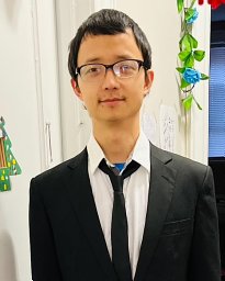 Heng Liu合作论文数Shanghai Institute for Biological Sciences Chinese Academy of Sciences, Chinese Academy of Sciences10
 Zuyi Yuan (袁祖贻)合作论文数First Affiliated Hospital of Xi 'an Jiaotong University10
 Rugang Zhang合作论文数The Wistar Institute Cancer Center;Department of Genetics, Perelman School of Medicine, University of Pennsylvania10Li Chenhui合作论文数9Shufang Cui合作论文数8
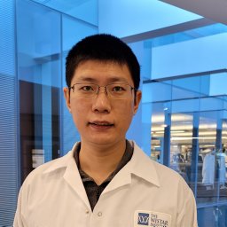 Wei Zhou合作论文数Sch Med, Nankai Univ8Hao-Yu Wu合作论文数8Chaoyang Ye合作论文数8
 Xing Liu (刘星)合作论文数Shanghai Institute of Materia Medica, Chinese Academy of Sciences8Jianqing She合作论文数7jiameng hu (胡佳萌)合作论文数6
 Wei Meng合作论文数School of Materials Science and Engineering, Anhui University of Technology6
 Bowen Lou合作论文数6
 Ronny Drapkin合作论文数Perelman School of Medicine, University of Pennsylvania;Penn Ovarian Cancer Research Center, University of Pennsylvania;United States Department of Defense6Yu Huansha合作论文数6
 Yong Yang (杨勇)合作论文数School of Basic Medicine and Clinical Pharmacy, China Pharmaceutical University;Center for New Drug Safety Evaluation and Research, China Pharmaceutical University6
 Wenbing Yao (姚文兵)合作论文数State Key Laboratory of Natural Medicines, China Pharmaceutical University6
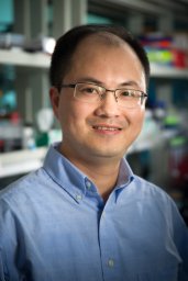 Kai Yang合作论文数Dept Immunol, St Jude Childrens Res Hosp6
 Mei Jiahao (梅家豪)合作论文数5
 Hao Jiang (姜皞)合作论文数Guangzhou Institutes of Biomedicine and Health, Chinese Academy of Sciences5Munire Maimaiti合作论文数5Jing Sun合作论文数5Xiang Zhang合作论文数5
 Iris Muller合作论文数Memorial Sloan-Kettering Cancer Center5
 Mingyu Pan (潘明玉)合作论文数School of Pharmacy, Nanjing Medical University5
 Kristian Helin合作论文数The Institute of Cancer Research5孟卫合作论文数School of Science, China Pharmaceutical University5
 Jigang Wang (王继刚)合作论文数Institute of Chinese Materia Medica, China Academy of Chinese Medical Sciences5Sun Shaogang合作论文数Department of Cancer Biology, University of Cincinnati College of Medicine5
 Bianhong Zhang (张变红)合作论文数Institute of Biomedical Sciences, School of Life Sciences, East China Normal University5
 Xiangdong Gao (高向东)合作论文数School of Life Science and Technology, China Pharmaceutical University5
 Chenyu Zhang (张辰宇)合作论文数State Key Laboratory of Pharmaceutical Biotechnology, School of Life Sciences, Nanjing University5Lirong Diao合作论文数5Gulinigaer Tuerhongjiang合作论文数4Manyun Guo合作论文数4
 Chong Qiu合作论文数4Mingxing Cheng合作论文数4
 Guang Han (韩光)合作论文数School of Pharmacy, Henan University4
 Hongwei Liang (梁宏伟)合作论文数中国药科大学生命科学与技术学院4
 Wei Guo (郭薇)合作论文数中国药科大学研究生院4Kossenkov Andrew V合作论文数4Bitler Benjamin G合作论文数4Li Senlin合作论文数4




