Objective. To explore the preoperative and intraoperative factors associated with the progression of cervical kyphosis following posterior decompressive surgery in patients with degenerative cervical kyphosis and stenosis. Methods. This retrospective study reviewed patients diagnosed with multilevel cervical spinal stenosis and degenerative cervical kyphosis who underwent posterior approach surgery. Clinical outcomes were assessed using the modified Japanese Orthopedic Association (mJOA) score, Visual Analog Scale (VAS) for neck pain, Neck Disability Index (NDI), and Short Form 36 (SF36) health survey. Radiographic parameters, including C2-C7 lordosis, cervical range of motion (flexion and extension), T1 slope, and T1S-CL, were measured preoperatively and at follow-up using cervical standing X-rays. based on whether kyphosis progressed after surgery, patients were divided into 2 groups. Results. A total of 102 patients (mean age 56.8 years; 75 men, 27 women) met the inclusion criteria. Forty-two patients (41.2
BACKGROUND CONTEXT:Current severity assessment methods for cervical spondylotic myelopathy (CSM) overlook asymptomatic gait dysfunction and potential heterogeneous gait impairment. Also, gait alterations for disease progression remain inadequately characterized. PURPOSE:This study aimed to detect asymptomatic gait dysfunction, achieve accurate detection of CSM, and discover heterogeneous gait impairment in CSM using wearable sensor-based gait evaluation. STUDY DESIGN:Cross-sectional study. PATIENT SAMPLE:A total of 45 CSM patients and 39 age-matched healthy controls (HC) were recruited for this study. OUTCOME MEASURES:Waist and lower extremity kinematics and spatiotemporal parameters. METHODS:This study utilized a 7-sensor wearable inertial measurement unit (IMU) system to obtain spatiotemporal and kinematic (waist and lower extremity joint angle time series in triaxial planes) during 2-minute walk tests (2MWT). Group comparisons were performed using the Kruskal-Wallis test to assess differences in gait parameters between lower extremity asymptomatic CSM patients CSM-ALE, cervical spondylotic myelopathy asymptomatic lower extremity (CSM-ALE) and healthy controls (HC), and between the overall CSM cohort and HC. Machine learning was used for asymptomatic gait dysfunction and CSM detection. Graph convolutional network (GCN)-based unsupervised clustering was used for heterogeneous gait impairment recognition. The Pearson correlation coefficient was used to examine the relationships between gait parameters and Japanese Orthopedic Association (JOA) scores in order to evaluate the applicability of gait metrics for monitoring CSM severity. RESULTS:For gait parameters, CSM patients showed significantly longer turn time (3.06 vs 2.60 seconds), longer gait cycle time (1.28 vs 1.21 seconds), greater waist flexion angle (4.00° vs 2.95°), shorter step length (0.66 vs 0.83 m), slower single-step speed (1.03 vs 1.38 m/s), and reduced hip (76.31 vs 105.19 °/s) and knee angular velocities (146.98 vs 180.77 °/s) compared with healthy controls (HC). Using fused kinematic time-series data, the SVM classifier achieved excellent discrimination between CSM-ALE patients and HC (AUC=0.996) as well as between all CSM patients and HC (AUC=0.997), outperforming analyses based on individual gait parameters. Unsupervised GCN clustering revealed 2 distinct gait impairment patterns. Pattern I was characterized by prolonged turning time (4.03 vs 2.71 seconds) and increased waist flexion angle (9.27° vs 2.99°) compared with Pattern II. In contrast, Pattern II was marked by reduced ankle velocity (81.05 vs 100.82 °/s) but preserved turning time (2.71 vs 2.60 seconds vs HC), suggesting distal motor impairment with relatively maintained balance control. JOA scores was correlated with gait speed (r=0.431, p=.003), swing phase duration (r=0.444, p=.002), and knee angular velocity (r=0.373, p=.012). CONCLUSION:Wearable sensor-based gait evaluation can effectively distinguish CSM patients, including CSM-ALE patients, from HC, and reveal distinct patterns of gait impairment. These findings highlight its clinical value not only for early diagnosis but also for potentially enabling intervention within a therapeutic window before significant functional decline occurs.
AIM:Disorders of consciousness (DOC) remain a major clinical challenge, and the efficacy of currently available neuromodulation strategies remains limited. The brainstem reticular formation is central to arousal regulation, but approaches directly targeting it remain technically demanding. This feasibility study aimed to explore whether epidural electrical stimulation applied via a transnasal clival approach could induce arousal and the electrocorticographic (ECoG) changes. METHODS:Three anesthetized sheep were implanted with epidural stimulating electrodes placed over the ventral brainstem via a transnasal clival approach and cortical recording electrodes on the fronto-parietal lobes. During the stimulation period, continuous behavioral monitoring was performed, and ECoG was recorded and later processed with spectral analysis. RESULTS:All three sheep successfully underwent electrode implantation without intraoperative complications, including cerebrospinal fluid leakage or neurovascular injury. Short-term stimulation reliably elicited behavioral signs of arousal. Concurrently, spectral analysis of ECoG revealed a reduction in low-frequency power (δ, θ) and an increase in high-frequency power (β, γ). CONCLUSIONS:Transnasal clival epidural stimulation is technically feasible and capable of modulating arousal-related neural activity. Further investigations with optimized electrode design and larger cohorts are required to validate its safety and efficacy.
ABSTRACT Objective To investigate the incidence and treatment of cerebrospinal fluid leakage due to late presentation of dural tears (LPDT) after lumbar surgery and provide new ideas and treatment options for clinical management. This study addresses a significant knowledge gap in the specific context of delayed CSF leaks, where diagnosis and management remain particularly challenging despite existing literature on CSF leaks in general. Methods Patients who underwent posterior lumbar surgery from January 2021 to December 2024 were selected. General patient information, cerebrospinal fluid leakage (CSF) characteristics, follow‐up results, and re‐examination data were analyzed using chi‐squared tests, Fisher's exact tests, t‐tests, and ANOVAto identify cases of LPDT, their treatment methods, complications, and clinical outcomes. Results A total of 2359 patients who underwent lumbar surgery were included in the study. CSF leakage occurred in 43 patients (1.82%). Dural tears (DT) leading to CSF leakage intraoperatively or within 5 days postoperatively were observed in 36 patients (1.53%), with 23 (0.98%) identified intraoperatively. Seven patients (0.30%) experienced CSF leakage due to LPDT: two of them underwent dural repair surgery 3 weeks and 4 months postoperatively, respectively; five patients received conservative treatment involving wound pressure bandage and bed rest. Clinical outcomes were favorable (good or excellent) in five patients (71.4%) and poor in two patients (28.6%) with LPDT, whereas in contrast, no poor outcomes were observed in non‐LPDT patients. Conclusion This study found a 0.30% incidence of CSF leakage due to LPDT following lumbar surgery, with these patients demonstrating worse outcomes compared to those with nondelayed CSF leaks. CSF leakage secondary to unrecognized LPDT is an uncommon but clinically significant complication of spine surgery, necessitating heightened awareness and appropriate management.
This study introduces eccentric cranial traction (ECT) as a traction method for congenital cervical scoliosis (CCS) at the craniovertebral junction (CVJ) and evaluates its benefits when combined with concave-side distraction surgery. A retrospective analysis was conducted on 18 patients with CCS at the CVJ treated between 2019 and 2023 using ECT followed by concave-side distraction. Radiographic parameters were assessed at five time points: pre-operation, pre-traction, post-traction, post-operation, and final follow-up. Baseline demographic and surgical data were recorded. Patients were stratified into high- and low-responder groups based on the median value of the traction correction rate, and outcomes were compared between groups. The correlation between traction correction rate and surgical correction rate was also analyzed. The median age at ECT initiation was 8 years, and the median duration of traction was 8 days. All patients presented with deformities involving the CVJ. ECT significantly reduced head–neck tilt from 16.5 ± 9.3° to 9.5 ± 5.9° (P < 0.001), yielding a mean traction correction rate of 43.1
BACKGROUND CONTEXT:For patients with high occupation rate cervical ossification of the posterior longitudinal ligament (HOR-cOPLL) that are K-line negative, laminoplasty often results in poor outcomes due to postoperative loss of cervical curvature. Preserving the posterior muscle-ligament complex (PMLC) on the hinge side during laminoplasty has been shown to reduce cervical curve loss and improve postoperative cervical alignment. We hypothesized that a modified laminoplasty (mLP) with preserving the PMLC could maintain cervical curvature and achieve similar neurological recovery to that of laminectomy with fusion (LF), regardless of K-line status. PURPOSE:To compare the clinical and radiological results between mLP and LF among patients with HOR-cOPLL during postoperative 2-year follow-up. STUDY DESIGN:Prospective observational study. PATIENTS SAMPLE:Patients with HOR-cOPLL were enrolled between December 2018 and February 2022. Among them, 30 patients underwent LF, while 34 patients received mLP. OUTCOME MEASURES:The modified Japanese Orthopedic Association (mJOA) score and the mJOA recovery rate (mJOA RR) were used for neurological improvement. The Neck Disability Index (NDI) score, Visual Analog Scale (VAS) scores were for neck and/or shoulder pain. Perioperative values included operative time, estimated blood loss, and perioperative complications. Radiological characteristics consisted of the cervical Cobb angle and cervical range of motion (ROM), etc. METHODS: Independent samples t-test and Mann-Whitney U-test were used to compare normally and nonnormally distributed continuous variables between 2 groups, respectively. Paired sample t-tests and Wilcoxon signed-rank tests were used to evaluate changes in variables before and 2 years after surgery. Subgroup based on K-line was performed to access the differences between 2 groups. RESULTS:The mLP group experienced shorter surgery times (101 min vs 122.5 min, p=.009), less blood loss (175 mL vs 200 mL, p=.020), and had complication rates comparable to those of the LF group. The mLP (12.5 vs 16.5, p<.001) and LF groups (11.5 vs 15.25, p<.001) showed significant and comparable improvements in mJOA (p=.648) and mJOA RR (65.16% vs 68.33%, p=.598) at postoperative 2-year follow-up, which were consistent in both K-line positive and negative subgroups. Postoperative NDI, VAS, and axial symptoms were also comparable between the 2 groups. While maintaining a cervical curve comparable to the LF group (3.17° vs 2.95°, p=.935), the mLP group exhibited a lesser reduction in ROM at the 2-year mark (17.77° vs 12.02°, p=.033), although this was not significant in either K-line positive or negative subgroups. CONCLUSIONS:This was the first prospective cohort to compare mLP and LF for HOR-cOPLL. Both the mLP and LF groups demonstrated significant neurological recovery and similar preservation of cervical curvature over a 2-year period, regardless of K-line status. Notably, mLP offers advantages in surgical efficiency and reduced blood loss, suggesting its potential as a less invasive alternative to LF.
Study Design Retrospective study. Objective To investigate the impact of preoperative C2 slope (C2S) on change in sagittal vertical axis (SVA) and postoperative cervical sagittal imbalance (CSI) after laminoplasty (LP). Methods Consecutive patients who underwent LP between January 2015 and January 2022 with a minimum of 12-month follow-up were reviewed. The clinical variables and radiographic measurements were assessed. Patients were divided into improvement (ΔcSVA ≤ −10 mm), stable (−10 mm ≤ ΔcSVA <10 mm), and CSI groups (ΔcSVA≥10 mm). Correlation analysis was performed to explore the associations between ΔcSVA and preoperative factors, postoperative change and outcome variables. Multivariable logistics regression was conducted to determine the factors predicting CSI. Results Over the 186 enrolled patients, 23 developed CSI. The CSI group presented with significantly lower preoperative cSVA, greater cervical lordosis (CL) and C2S. At the final follow-up, the CSI group presented significantly higher increase in neck pain, C2S and C7S, and greater loss in CL and range of motion at flexion position. Correlation analysis revealed that ΔcSVA is correlated positively to preoperative CL and surgical segments and negatively to preoperative C2S and cSVA. Multivariable regression revealed that lower preoperative C2S ( P = 0.026) was risk factor for postoperative CSI, with 8.9 as a potential cut-off value for C2S. Conclusions Lower preoperative C2S slope was risk factor for post-laminoplasty CSI. The cut-off value of C2S was 8.9. Laminoplasty should be carefully considered for patients with low C2S due to risk of postoperative CSI.
3D printed artificial vertebral bodies (AVBs) have emerged as a viable alternative to traditional titanium mesh cages (TMCs) for spinal reconstruction following ACCF. However, existing comparative studies evaluating mid-term clinical and radiographic outcomes - particularly those incorporating quantitative assessments of fusion efficacy - remain inconclusive. This study aims to compare clinical and radiological outcomes between AVB and TMC in patients undergoing single-level ACCF with matched baseline characteristics. Patients who underwent single-level ACCF for degenerative cervical myelopathy (DCM) between January 2012 and December 2022, with a minimum of 2-year clinical and radiological follow-up, were included. Patients were grouped by implant type (AVB or TMC), and propensity score matching (PSM) was used to balance baseline characteristics. Outcomes were assessed at 3-month and final follow-up (more than 2 years). Radiological assessments comprised: (1) fusion status evaluated via extra graft bridging bone (ExGBB) on computed tomography (CT) scans and interspinous motion (ISM) parameters; (2) implant subsidence; and (3) cervical alignment changes. Clinical parameters included neck pain (using Visual Analogue Scale), modified Japanese Association (mJOA) score and Neck Disability Index (NDI). A total of 93 patients were included (49 in AVB and 44 in TMC group). After 1:1 PSM, 36 patients were matched for each group, and intergroup comparisons revealed comparable baseline conditions. At 3 months, the AVB group exhibited significantly smaller ISM distance (p = 0.019) and less subsidence (p = 0.028) compared to the TMC group. The final follow-up duration was 3.57 ± 1.58 years. At final follow-up, no significant differences in subsidence and cervical alignment maintenance were observed. Furthermore, in fusion status, no significant intergroup differences were found, either in ExGBB or ISM criteria. Patients in both groups exhibited comparable neurological recovery and pain relief, while the AVB group demonstrated significantly greater 3-month (p = 0.002) and final follow-up (p = 0.011) NDI improvement. However, no significant differences were observed in the proportion of patients achieving the minimal clinically important difference (MCID) for NDI. AVB and TMC resulted in similar fusion rates, subsidence, pain alleviation and neurological recovery at mid-term. However, AVB might have superior short-term implant stability with less 3-month subsidence and better fusion.
This study aimed to investigate the knowledge, attitude and practice (KAP) of the healthy general population toward cervical spondylosis. A cross-sectional study was conducted among healthy individuals at Peking University Third Hospital between May and December 2023, utilizing a self-designed questionnaire. A total of 506 valid questionnaires were collected, of which 276 (64.94
Cervical kyphosis is a debilitating disease, and its surgical treatment involves correction to restore sagittal alignment. Few studies have explored the appropriate degree of correction, and the biomechanical impact of correction on the cervical spine is still unclear. This study aimed to compare the biomechanical changes in the cervical spine after different degrees of correction by two-level anterior cervical discectomy and fusion (ACDF). Three-dimensional finite element (FE) models of the intact cervical spine (C2–C7) with normal physiological lordosis and kyphosis were constructed. Based on the kyphotic model, three two-level ACDF in C4–6 surgical models were developed: (1) non-correction: only the intervertebral heights were restored; (2) partial correction: the cervical curvature was adjusted to straighten; (3) complete correction: the cervical curvature was adjusted to physiological lordosis. A pure moment of 1.0 Nm combined with a follower load of 73.6 N was applied to the C2 vertebra to simulate flexion, extension, lateral bending, and axial rotation. The stress of vertical bodies and facet joints, intradiscal pressure (IDP), and the overall ROMs of all models were computed. The peak von Mises stress on the upper (C4) and lower (C6) instrumented vertebral bodies in the kyphotic model was greater than that of the physiological lordosis model, with the exception of C6 under lateral bending. The maximum stress was observed in C4 during lateral bending after complete correction, which increased by 145% compared to preoperative von Mises stress. For the middle (C5) instrumented vertebral body, the peak von Mises stress increased after surgery. The maximum stress was observed in partial correction during flexion. Compared to physiological lordosis, the peak von Mises stress on the facet joints in kyphotic segments was lower; however, it was higher in the adjacent segments, except C4/5 in extension. The stress on the facet joints in kyphotic segments decreased, with the most significant decrease observed in partial correction. The IDPs in adjacent segments, except for C6/7 in flexion, showed no significant difference before and after surgery. Additionally, correction seemed to have little impact on IDPs in adjacent segments. In conclusion, for the treatment of cervical kyphosis with two-level ACDF, complete correction resulted in the highest peak von Mises stress on the upper instrumented vertebral body. Partial correction mitigated von Mises stress within the facet joints in kyphotic segments, albeit at the expense of high von Mises stress on the middle instrumented vertebral body.
The 3D-printed artificial vertebral body (AVB) was designed with theoretically better biomechanical properties than traditional implants to decrease the incidence of implant subsidence. However, implant subsidence still occurs, with the potential risk factors for AVB subsidence remaining unknown. This study aimed to identify the risk factors for early subsidence of AVB after single-level anterior cervical corpectomy and fusion (ACCF). Consecutive patients who underwent single-level ACCF using 3D-printed AVB for cervical spondylotic myelopathy between 2018 and 2022 were included. Preoperative, immediate postoperative, and 3-month postoperative X-ray scans were evaluated. Radiological assessment included segmental height (SH), intraoperative retraction height, cervical alignment parameters, bone quality and implant sagittal size ratio. Early subsidence was defined as an SH loss ≥ 2 mm based on 3-month radiographs. Of the included 98 patients (52 males, 46 females, average age: 51.8 ± 10.1), 35(35.7
Achieving bony fusion after anterior cervical corpectomy and fusion is crucial for restoring spinal stability; however, the risk factors associated with fusion failure, or pseudarthrosis, remain unclear. This study aims to identify risk factors for pseudarthrosis following anterior cervical corpectomy and fusion and evaluate the impact of C2 slope and uncovertebral joint degeneration on this condition. Patients who underwent single-level anterior cervical corpectomy and fusion between May 2015 and April 2022 and had a minimum of 2 years of computed tomography follow-up were retrospectively enrolled. Preoperative demographic, surgical, clinical, and radiographic data were collected. Patients were divided into fusion and pseudarthrosis groups based on fusion status evaluated at the final follow-up computed tomography. After identifying statistically significant variables through intergroup comparisons, multivariate logistic regression analysis was conducted to determine the risk factors for pseudarthrosis. A total of 102 patients were included in the study, with an average follow-up duration of 3.78 ± 1.70 years. At final follow-up, 37 patients (36.3
Study Design.A retrospective diagnostic test study.Objective.To establish quantitative fusion criteria for ACCF.Summary of Background Data.Currently, the criteria of fusion after ACCF remains controversial. Evaluation techniques such as observation of continuous bony trabeculae across the fusion site were qualitative and conducted by naked eyes, limiting its accuracy. Dynamic fusion indicators such as interspinous motion (ISM) have been successfully utilized in anterior cervical discectomy and fusion (ACDF) for evaluating fusion and detecting pseudarthrosis. However, the accuracy of ISM in ACCF is unclear.Methods.Patients who underwent ACCF in our hospital were reviewed. Imaging data including dynamic radiographs and computed tomography (CT) scans were collected. The distance and angle form of ISM (ISM-dis and ISM-ang) were measured on dynamic radiograph, with CT as the gold standard for fusion. The correlation between ISM methods was analyzed using Spearmann correlation. Receiver operating characteristic (ROC) curves were used to evaluate the accuracy and generate optimal cutoff values. Three observers independently measured the data twice to determine interobserver and intraobserver reliability.Results.Our study included 178 imaging datasets. ISM-dis showed positive correlation with ISM-ang (r=0.914). The area under ROC (AUC) for ISM-dis and ISM-ang was 0.926 and 0.893. Applying a cutoff value of 1.50 mm to ISM-dis yielded a sensitivity of 87.8% and specificity of 92.3% for detecting pseudarthrosis. For ISM-ang, a cutoff value of 1.75 degrees resulted in a sensitivity of 79.1% and specificity of 92.3%. Intraobserver reliabilities for ISM-dis (0.986, 0.984, and 0.972) were higher than for ISM-ang (0.935, 0.963, and 0.935), as was interobserver reliability (0.985 for ISM-dis and 0.956 for ISM-ang). Fusion rates calculated using ISM-dis and ISM-ang were 52.4% and 61.9%.Conclusions.ISM can serve as an alternative to CT for assessing fusion, with ISM-dis at a 1.50-mm cutoff and ISM-ang at 1.75 degrees, demonstrating high diagnostic accuracy.
ObjectiveThis study aimed to evaluate the clinical characteristics, surgical outcomes, and prognostic factors of cervical spondylosis in patients with dysphagia caused by anterior osteophytes.MethodsThis retrospective study included 28 patients who underwent anterior cervical surgery for dysphagia associated with anterior osteophytes between January 2006 and January 2023. Clinical data, including preoperative evaluations, surgical reports, and follow-up visits, were collected from electronic medical records. Cervical spine degeneration was confirmed by radiographic examinations. Dysphagia severity was assessed by the Functional Outcome Swallowing Scale (FOSS) preoperatively and at follow-up. Statistical analyses were conducted to compare pre- and postoperative outcomes.ResultsThe average age was 62.4 years, and 78.6% were men. All patients experienced dysphagia, with severity ranging from grades 1-4. After surgery, 89.3% of the patients showed symptomatic improvement, defined as a reduction of at least one grade on the FOSS. Complications were infrequent, primarily consisting of axial pain (10.7%), along with less common occurrences of esophageal fistula, stenosis, nutritional insufficiency, and myelitis (each at 3.6%). Both the modified Japanese Orthopaedic Association (mJOA) score and Neck Disability Index (NDI) showed significant postoperative improvement. The most frequently involved segments were C5/6 and C4/5.ConclusionAnterior cervical osteophyte resection, with or without fusion, appears effective in relieving dysphagia symptoms and improving neurological function in well-selected patients. Further prospective studies are needed to validate these findings and refine surgical strategies.
ObjectiveTo compare the biomechanical characteristics of axial traction and suspensory traction in the process of preoperative correction of cervical kyphosis.MethodsAn intact three-dimensional finite element digital model of C2-T2 with cervical kyphosis was established. The head gravity and moment were applied to the finite element model to simulate the force of skull traction and the force of suspensory traction. The changes of cervical kyphotic angle, the length of cervical spinal canal and the stress distribution of each vertebral body were analyzed under two traction modes.ResultsThe kyphotic angles of the kyphotic segments were reduced by both tractions. The C2-C5 kyphotic angle was 41° before traction, and decreased to 32° and 26° after axial traction and suspensory traction, respectively. The length of C3-C7 cervical spinal canal was 61.3 mm before traction. After axial traction, the length of C3-C7 cervical spinal canal increased to 61.8 mm; after suspensory traction, it decreased to 59. 6 mm. The high stress area of each vertebral body was located in the anterior longitudinal ligament attachment area of the vertebral body during both two kinds of traction. The maximum Mises stress of C2-C7 vertebral body in suspensory traction is generally small relative to axial traction.ConclusionCompared with axial traction, suspensory traction has better kyphotic corrective effect, while reduces the length of the cervical spinal canal and the stress on the cervical vertebral body, which decreases the possibility of nerve damage and iatrogenic fracture during traction from a biomechanical point of view.
BACKGROUND:Congenital cervical scoliosis at the craniovertebral junction is an exceedingly rare condition, with limited existing research. In this study, we aimed to elucidate the clinical characteristics, surgical strategies, and postoperative outcomes associated with this unique pathology, providing a comprehensive analysis to enhance clinical understanding and management. METHODS:We conducted a retrospective analysis involving 27 patients with congenital cervical scoliosis at the craniovertebral junction who underwent surgery at a mean age of 7.81 ± 1.52 years. The median follow-up duration was 36.00 months. Patients were categorized into Group A and Group B on the basis of the absence or presence of concomitant subaxial cervical scoliosis, respectively. Three distinct surgical strategies were employed accordingly. Radiographic parameters were measured preoperatively, within 5 days postoperatively, and at the final follow-up. Other clinical and surgical characteristics were also collected. RESULTS:Patients exhibited preoperative coronal imbalance, with a mean structural Cobb angle of 30.75° ± 13.09° and a mean head shift of 20.34 ± 13.23 mm. At the final follow-up, these parameters had significantly improved to a median of 3.00° and 8.59 mm (both p < 0.05). The mean operative time was 473.74 ± 134.29 minutes, and the mean intraoperative blood loss was 336.11 ± 166.52 mL. Among the 27 patients, 10 were in Group A and the other 17 were in Group B. Twenty-three patients/families reported being "satisfied" or "very satisfied" with the surgical outcome at the final follow-up. Postoperative complications occurred in 11 patients; no nerve root or vertebral artery injuries were observed. CONCLUSIONS:Congenital cervical scoliosis at the craniovertebral junction is a complex deformity that severely influences coronal alignment. Nevertheless, tailored surgical strategies have shown promising effectiveness in achieving satisfactory clinical and psychological results. LEVEL OF EVIDENCE:Therapeutic Level III . See Instructions for Authors for a complete description of levels of evidence.
STUDY DESIGN:A single-center retrospective study. OBJECTIVE:This study investigates the correlation between preoperative SF-36 scores and postoperative axial neck pain (ANP) in cervical spondylotic myelopathy (CSM) patients undergoing anterior cervical surgery (ACS). SUMMARY OF BACKGROUND DATA:The effects of the anterior surgical approach on ANP are not well understood. METHODS:This retrospective study included patients with CSM at Peking University Third Hospital between April 2010 and March 2016. The SF-36, visual analog scale (VAS), and Neck Disability Index (NDI) were collected preoperatively and postoperatively at 3 months, 1 year, and 2 years. RESULTS:In 107 CSM patients, ACS significantly improved pain, physical function, and quality of life over 2 years. VAS scores decreased from 6.4±2.3 to 3.8±1.9, indicating less ANP (P=0.013). SF-36 scores improved notably in social functioning and bodily pain domains. NDI scores also decreased, showing reduced neck disability (P=0.012). Patients with ANP post-ACS had consistently lower preoperative bodily pain and social functioning scores at follow-up of 3 timepoints. Correlation analysis revealed a significant negative relationship between VAS and preoperative bodily pain and social functioning scores (P<0.05), suggesting that lower preoperative scores may predict severe postoperative ANP. CONCLUSIONS:Anterior cervical surgery does not increase the incidence of postoperative ANP in patients with CSM. The BP and SF domains of the preoperative SF-36 are correlated with the occurrence of postoperative ANP.
Background: Congenital cervical scoliosis at the craniovertebral junction is an exceedingly rare condition, with limited existing research. In this study, we aimed to elucidate the clinical characteristics, surgical strategies, and postoperative outcomes associated with this unique pathology, providing a comprehensive analysis to enhance clinical understanding and management. Methods: We conducted a retrospective analysis involving 27 patients with congenital cervical scoliosis at the craniovertebral junction who underwent surgery at a mean age of 7.81 ± 1.52 years. The median follow-up duration was 36.00 months. Patients were categorized into Group A and Group B on the basis of the absence or presence of concomitant subaxial cervical scoliosis, respectively. Three distinct surgical strategies were employed accordingly. Radiographic parameters were measured preoperatively, within 5 days postoperatively, and at the final follow-up. Other clinical and surgical characteristics were also collected. Results: Patients exhibited preoperative coronal imbalance, with a mean structural Cobb angle of 30.75° ± 13.09° and a mean head shift of 20.34 ± 13.23 mm. At the final follow-up, these parameters had significantly improved to a median of 3.00° and 8.59 mm (both p < 0.05). The mean operative time was 473.74 ± 134.29 minutes, and the mean intraoperative blood loss was 336.11 ± 166.52 mL. Among the 27 patients, 10 were in Group A and the other 17 were in Group B. Twenty-three patients/families reported being “satisfied” or “very satisfied” with the surgical outcome at the final follow-up. Postoperative complications occurred in 11 patients; no nerve root or vertebral artery injuries were observed. Conclusions: Congenital cervical scoliosis at the craniovertebral junction is a complex deformity that severely influences coronal alignment. Nevertheless, tailored surgical strategies have shown promising effectiveness in achieving satisfactory clinical and psychological results. Level of Evidence: Therapeutic Level III . See Instructions for Authors for a complete description of levels of evidence.
Objectives To evaluate whether the fixation procedure is essential in posterior surgery for cervical stenosis with non-severe kyphosis. Methods A total of 109 patients diagnosed with cervical stenosis and non-severe kyphosis were included in this study. Patients were categorized based on the surgical approach into those who underwent laminoplasty (LP) alone and those who received fixation with either lateral mass screws (LMS) or pedicle screws (PS). Preoperative and postoperative assessments included the Visual Analog Scale (VAS) for neck pain, the modified Japanese Orthopaedic Association (mJOA) score, the Neck Disability Index (NDI), and the Short Form 36 (SF36) health survey. Sagittal parameters were measured using lateral cervical radiographs. Additionally, hospitalization data, surgical details, and complications were collected. Results No significant differences were observed in baseline demographic characteristics among the three groups. However, the PS group had significantly longer operative duration and greater intraoperative blood loss compared to the others groups. Postoperative mJOA scores improved significantly in all groups. However, the PS group had significantly lower follow-up mJOA scores (12.8 ± 2.9) compared to the LP (15.4 ± 1.7, P < 0.001) and LMS groups (15.6 ± 1.5, P = 0.002). VAS scores demonstrated significant pain reduction in the PS group (preoperative 3.8 ± 2.2 vs. follow-up 2.6 ± 1.8, P = 0.046), whereas changes in the LP (-0.2 ± 2.8, P = 0.877) and LMS groups (-0.3 ± 2.7, P = 0.877) were minimal. The PS group achieved the most pronounced correction in C2-7 Cobb angle (8.6 ± 11.7°) during surgeries. The LMS group did not achieve significant kyphosis correction, with a postoperative improvement in C2-7 Cobb angle of only 3.4 ± 7.7°, comparable to the LP group (3.8 ± 7.8°, P = 0.840). The overall complication rate was highest in the PS group (27.8%), followed by the LMS group (16.0%) and the LP group (9.1%). Conclusion This study suggests that fixation may not be essential in posterior surgery for cervical stenosis with non-severe kyphosis. Posterior surgeries without fixation were associated with shorter operative times, reduced blood loss, comparable neurological recovery and quality of life outcomes to those with fixation, as well as a lower incidence of postoperative complications.
 Sheng-fa Pan (潘胜发)合作论文数55
 Feifei Zhou (周非非)合作论文数Department of Orthopedics, Peking University Third Hospital51
 Yu Sun (孙宇)合作论文数Beijing Cancer Hospital50
 Yanbin Zhao (赵衍斌)合作论文数Department of Orthopedics, Peking University Third Hospital49
 Fengshan Zhang (张凤山)合作论文数,et al Department of Orthopedic Surgery,Peking University Third Hospital,Beijing,,China37
 Yinze Diao (刁垠泽)合作论文数Department of Orthopaedics, Peking University Third Hospital32
 Li Zhang (张立)合作论文数北京大学第三医院骨科 100083 北京市24
 Tian Xia合作论文数15
 Shaobo Wang (王少波)合作论文数Department of Orthopaedic, Peking University Third Hospital11
 Zhongjun Liu (刘忠军)合作论文数Peking University Third Hospital9Shuo Cao合作论文数7ruomu qu (屈若木)合作论文数6
 Weishi Li (李危石)合作论文数Peking University Third Hospital6Yilong Zhang合作论文数5
 Ben Wang合作论文数4
 Liang Jiang (姜亮)合作论文数Peking University Third Hospital4Zhen CHEN (陈振)合作论文数3QingShan Chen (陈庆山)合作论文数3
 Changman Zhou (周长满)合作论文数School of Basic Medical Sciences, Health Science Center, Peking University3Haoge Huang合作论文数2Zexiang Liu合作论文数2Jie-Sheng Liu合作论文数2JiYan Jin (金姬延)合作论文数2EnHua Yu (于恩华)合作论文数2XianZhong Shi (石献忠)合作论文数2RuiFeng Xu (许蕊凤)合作论文数2
 Lei Yang (杨磊)合作论文数School of Mathematical Sciences, Peking University2
 Feng Wei (韦峰)合作论文数Department of Orthopaedic, Peking University Third Hospital2
 Hua Zhou (周华)合作论文数Department of Orthopedics, Peking University Third Hospital2
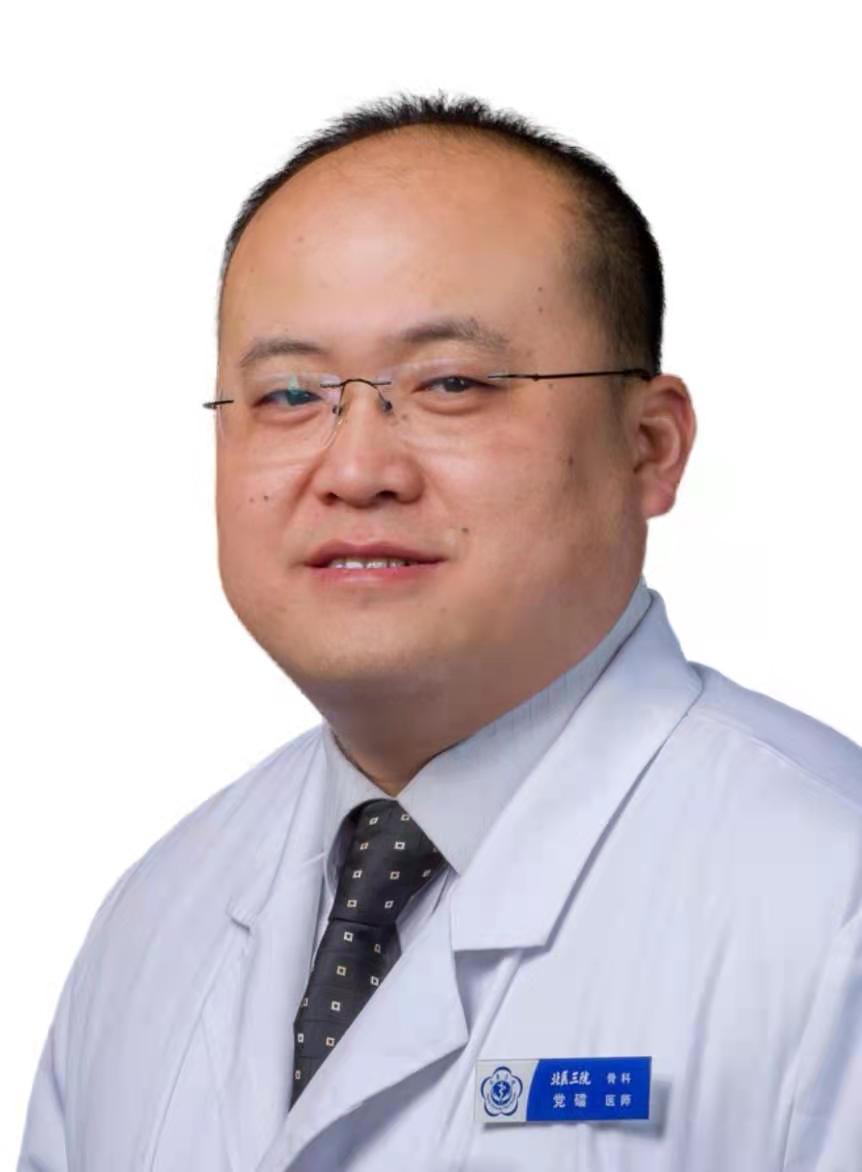 Lei Dang (党礌)合作论文数Department of Orthopaedics, Peking University Third Hospital2
 Miao Yu (于淼)合作论文数Department of Orthopedics, Peking University Third Hospital2
 Xiaoguang Liu (刘晓光)合作论文数Peking University Health Science Center;Peking University Third Hospital;Peking University International Hospital2
 Dongsheng Fan (樊东升)合作论文数Peking University Third Hospital2
 Weijia William LU (吕维加)合作论文数Department of Orthopaedics & Traumatology, School of Clinical Medicine, The University of Hong Kong;Orthopaedic Research Center, The University of Hong Kong;Chinese Academy of Sciences2Yi-latu SU (苏义拉图)合作论文数1Gangqiang Zhang合作论文数1Xiao Fan合作论文数1Wenyong Shi合作论文数1Hongyu Chen合作论文数1Xinhang Li合作论文数1Jun Yang合作论文数1Shilin Xue合作论文数1Yiyuan Yang (杨易远)合作论文数1
 Chenlong Yang合作论文数1




