Tibiotalar arthrodesis (TTA) is a common surgical method for post-traumatic arthrosis and primary osteoarthrosis of ankle. Internal screw fixation exhibits such advantages of higher fusion rate and fewer complications that it has become the mainstream surgical procedure in clinics. However, there are still disputes over the choice of screw type, number and nailing method. In this study, a 3D model of tibiotalar joint was established based on CT data. Two representative types of screws were modeled according to their real objects. Preprocessing was done in Hypermesh 2017, and the finite element simulations of different surgical schemes and postoperative ankle joint movements were performed via Abaqus 2016. The stress distribution in tibiotalar joints and screws, interarticular displacement, and screw deformation were extracted for comparative analyses. The results show that: (1) the three-screw fixation scheme with smaller displacements and higher contact pressure is superior to the two-screw one; (2) the relative displacements of the tibia and talus of full-thread screw under all loads were reduced by approximately 50% compared with those of lag screw, showing better stability; (3) the 60° scheme is more favorable for joint fusion and more stable under vertical load, while the 75° has the best bending resistance; (4) the stress and strain distributions in screws indicate that the screw break is most likely to happen near its contact region with the bone inlet, the maximum stress reaches 212.2 MPa. This study is of great significance for the selection and optimization of the surgical plan.
Background: The purpose of this article was to report the feasibility and effectiveness of 3-stage Masquelet technique and 1-stage operation for different stages of foot and ankle tuberculosis (TB). Methods: Ten consecutive patients with foot and ankle TB were retrospectively analyzed between January 2014 and December 2018. Five patients were treated with the 3-stage Masquelet technique, including thorough debridement with vacuum sealing drainage, implantation of antibiotic cement spacer, and subsequent reconstruction. Five patients were treated with a 1-stage reconstruction. The American Orthopaedic Foot & Ankle Society (AOFAS) and visual analog scale (VAS) pain scores were recorded at the last follow-up. The follow-up was 30.3 ± 17.8 months. Results: No reactivation of TB was observed in any patients. For the 3-stage operation group, 1 patient developed a distal tibia fracture. The duration of anti-TB therapy was 12.0 ± 0.8 months. The AOFAS score increased from 39.5 ± 9.9 preoperatively to 75.3 ± 7.0 postoperatively ( P < .05). The VAS pain score decreased from 6.3 ± 1.9 to 1.5 ± 1.3 ( P < .05). For the 1-stage operation, 1 patient had wound necrosis. The duration of anti-TB therapy was 13.8 ± 1.1 months. The AOFAS score increased from 51.8 ± 15.0 to 81.8 ± 6.3 ( P < .05). The VAS pain score decreased from 5.4 ± 1.1 to 1.0 ± 0.7 ( P < .05). Conclusion: Three-stage operation was effective for foot and ankle TB with stage IV, sinus tracts or other infections, and 1-stage reconstruction was effective for early-stage TBs. Level of Evidence: Level IV, case series.
Objective To investigate the surgical outcomes of one-stage repair of the ankle fracture combined with grade Ⅲ injury to the lateral ligament.Methods From April 2015 to December 2017,23 patients with ankle fracture and lateral ligament injury were treated at Department of Traumatic Reconstruction Surgery/Foot and Ankle Surgery,Zhongnan Hospital.There were 15 males and 8 females,aged from 18 to 65 years (average,37.8 years).After intraoperative treatment of their fractures,22 ruptures of the ligamentous insertion were repaired with suture anchor and one rupture of the middle ligament with Internal Brace.Routine postoperative X-rays,physical examination and Sefton grading system were used to evaluate the stability of the ankle joint and the subtalar joint.The Karlsson-Peterson and American Orthopedic Foot and Ankle Society (AOFAS) scores were used to evaluate the function and pain of the ankle.Results The follow-up for this cohort ranged from 12 to 24 months (average,13.9 months).The bone union time ranged from 6 to 24 weeks,averaging 10.1 weeks.There was no chronic instability of lateral ankle or subtalar joint in all the cases.Two cases developed osteochondral lesion of the medial talus which was treated with micro-fracture surgery one year after injury;one case suffered subtalar arthritis which was treated by subtalar arthrodesis 14 months after injury.Their Karlsson-Peterson scores averaged 84.6 points and AOFAS ankle-hindfoot scores 93.7 points with 19 excellent and 4 good cases.The Sefton grading system gave 9 cases of grade Ⅰ and 14 ones of grade Ⅱ.Conclusion One-stage repair of the ankle fracture combined with grade Ⅲ injury to the lateral ligament can lead to the stability of the ankle joint and the subtalar joint and improved function of the ankle.
Chondrosarcoma is characterized by the presence of histologically aggressive behavior, and commonly involves the scapula. Currently, limb salvage surgery is the recommended surgical treatment. Owing to the irregularity of the tumor, the suitability of an implant after tumor resection is a challenge for surgeons. Three-dimensional (3D) printing technology has the potential to make personalized limb salvage surgery a reality. We report the case of a 53-year-old man who was diagnosed with chondrosarcoma of the scapula. Considering the low-grade malignancy and lack of invasion of the glenoid, we agreed upon segmental scapula replacement as the treatment protocol. Nevertheless, reconstruction of the irregular bony defect remaining after tumor resection can be complicated. Therefore, a personalized prosthesis and navigation template corresponding to tumor was designed with 3D printing technique, and tumor resection, prosthesis implantation, and rotator cuff reconstruction were completed. The affected shoulder achieved satisfactory function during a 32-month follow-up with no tumor recurrence. 3D printing technique can help implement the individualized design of the implant and accurate reconstruction after tumor resection, simplify complicated operations, improve operational efficiency, and allow early functional recovery.
[目的]介绍跟骨结节切除带线铆钉止点重建术治疗跟腱断裂合并跟腱末端病变的手术技术.[方法]7例跟腱断裂合并跟腱末端病变患者采用跟骨结节及变性跟腱切除、带线铆钉止点重建术.术中切除足够大的跟骨骨块,切除变性坏死跟腱,对于缺损较小者(<2 cm)直接行带线铆钉缝合固定于跟骨,缺损较大者(>2 cm)采取腓肠肌腱V-Y延长技术及自体肌腱移植来恢复跟腱长度和维持后期踝关节功能.延长跟腱,带线铆钉跟腱止点重建,术后石膏固定4周.[结果]术后1例切口出现红肿,经伤口换药、升级抗生素后愈合.7例获6个月~2年随访,采用Arner-Lindholm评分标准,临床结果评定为优6足,良1足.所有患者踝关节背伸跖屈功能正常.[结论]跟骨结节及变性跟腱切除、带线铆钉止点重建术治疗跟腱断裂合并跟腱末端病具有较好的临床疗效,术中细致操作、足够大的骨块切除、变性坏死跟腱的切除、带线铆钉止点重建等是手术成功的关键.
Objective To investigate the clinical efficacy of three-stage Masquelet technique in the treatment of infective bone defects of foot and ankle.Methods A retrospective case series study was conducted on the clinical data of 19 patients with infective bone defects of foot and ankle admitted to Zhongnan Hospital of Wuhan University from December 2014 to October 2017.There were 15 males and four females,aged 18-68 years [(39.6 ± 12.3)years].Among the patients,16 patients were infected with bacteria and three patients were infected with Mycobacterium tuberculosis.The infection involved humeral end,talus and surrounding joints in 14 patients,internal hemorrhoids in two patients,midfoot and interphalangeal joints in one patient,and humerus and metatarsophalangeal joints in two.The operation included three stages:the first stage was thorough debridement,supplemented with negative pressure closed drainage (VSD) and continuous washing to clarify the pathogenic bacteria;the second stage was to fill the bone defect with targeted antibiotic bone cement to prevent or treat infection;in the third stage,after filling the antibiotic bone cement for 3 months with no sign of local wound infection,the bone cement was taken out,and the bone reconstruction operation was performed by means of internal fixation and bone grafting.The flap survival and wound healing were observed,and the time of fracture healing was recorded.The American Orthopaedic Foot and Ankle Society (AOFAS) score and the Visual Analogue Score (VAS) were used to evaluate the improvement of the function of the foot and ankle before operation and at the last follow-up,and the bone healing was evaluated according to the radiographic union scale in tibial (RUST) fractures.Results The patients were followed up for 9-12 months [(11.1 ±1.0) months].Two patients with soft tissue defects caused by preoperative infection and necrosis received posterior tibial artery perforator flap and anterolateral thigh flap repair in the second stage,and the flaps all survived.The postoperative bone healing time was 3 7 months [(3.5 ± 1.4)months].Nineteen patients underwent three-stage surgery,and the ankle and foot wound or sinus tract were all healed,with no infection recurrence during follow-up.At the last follow-up,the AOFAS score was improved significantly from preoperative (36.3 s-12.1) points to (71.4 ± 5.7) points (P < 0.05).The VAS was decreased significantly from preoperative (5.3 ± 1.2) points to (1.4 ± 0.9) points (P < 0.05).The RUST bone healing score at the last follow-up was 8-12 points [(10.2 ± 1.1) points].Conclusion In treating the infective bone defects of foot and ankle,the three-stage Masquelet technique can effectively control infection,facilitate wound healing,promote bone union,and improve foot and ankle function.
Objective To explore the application of three-dimensional (3D) printing technology in precise and individualized surgical treatment of severe distal humeral bone defect. Methods Five patients with severe distal humeral bone defects were treated with customized 3D printing prostheses between December 2010 and December 2015. There were 4 males and 1 female, with an age of 23-57 years (mean, 35 years); and the length of the bone defect was 5-12 cm (mean, 8 cm). The cause of injury was mechanical injury in 2 cases and strangulation in 3 cases. All of them were the open fracture of Gustilo type Ⅲ. There were 2 cases of radial fracture, 1 case of cubital nerve injury, and 3 cases of radial nerve injury. The time from injury to one-stage operation was 6-18 hours (mean, 10 hours). The operation time, intraoperative blood loss, and intraoperative fluoroscopy were recorded. During follow-up, the anteroposterior and lateral X-ray films of the elbow joints were performed to identify whether there was prosthesis loosening; Mayo Elbow Performance Score (MEPS) and upper extremity Enneking score were used to evaluate limb function. Results The operation time was 140-190 minutes (mean, 165 minutes). The intraoperative blood loss was 310-490 mL (mean, 415 mL). The intraoperative fluoroscopy was 1-3 times (mean, 1.6 times). Five patients were followed up 14-38 months (mean, 21 months). The wound exudate occurred in 1 case and cured after anti-inflammatory local dressing change; the subcutaneous hematoma occurred in 1 case, and improved after color Doppler ultrasound guided puncture and drainage. The MEPS scores and the Enneking scores were all significantly improved when compared with preoperative ones ( P<0.05). Except MEPS score between 6 and 12 months after operation had no significant difference ( P>0.05), there were significant differences in MEPS scores and Enneking scores between the other time points ( P<0.05). During the follow-up, no prosthetic loosening or joint dislocation occurred. Conclusion 3D printing technology can achieve personalized treatment of severe distal humeral bone defects, obtain relatively good elbow joint function, and has less postoperative complications and satisfactory effectiveness.
Objective To investigate the clinical effectivity of the muscular flap transposition and induced membrane technique in the emergency treatment for the limb salvage of Gustilo type Ⅲ B/C open fracture of lower leg. Methods From July, 2015 to December, 2017, 10 cases of Gustilo type Ⅲ B/C fracture of lower leg with bone defects were performed limb salvage surgery. Induced membrane technique was used to fill the bone defects in the emergency room.The gastrocnemius and/or soleus muscular flaps were transposed to cover the bone cement or ex-posed bone simultaneously in emergence treatment. After the wound healed completely, traditional bone grafting was used to repair the bone defects. There were 4 cases of Gustilo type Ⅲ B and 6 cases of Gustilo type Ⅲ C. The aver-age length of bone defect was (5.25±1.70) cm ranging from 3.0 cm to 11.0 cm. The gastrocnemius medial head flaps were performed in 5 cases, the combined application with the gastrocnemius medial head flaps and the medial hemimuscular flaps of soleus were performed in 2 cases, and medial hemimuscular flaps of soleus were transposed in 3 cases. Results The wounds in 6 cases were healed at one stage, but 2 cases healed by dressing because the exudate after skin grafting.In 1 case, the cross-leg flap was used to cover the exposed bone cement due to the necro-sis of soleus flap. The other 1 was performed the transposition of the lateral gastrocnemius flaps because the exposure of bone cement after the necrosis of the upper and lateral muscles in lower leg. In the second-stage, the bone defects were reconstructed by traditional bone grafting. The average healed time of bone was 7.2 months ranging from 5 months to 9 months. At the last followed-up time, all patients recovered their function of weight-bearing. The Paley's score of the adjacent joints: excellent in 8 cases and good in 2 cases. Conclusion The combination with induced membrane technique and local muscular flap transposition in emergency surgery is an effective method to limb salvage for the Gustilo type Ⅲ B/C open fracture of lower leg.
Objective To investigate the mechanism of L-Mimosine induced cell apoptosis in osteosarcoma cell lines.Methods Osteosarcoma cell lines MG-63 and U2-OS were treated with 0 μM,100 μM,200 μM,400 μM L-Mimosine,and 400 μM L-Mimosine is added with 100mg/mL FAC.CCK-8 was used to test cell proliferation,FITC-Annexin VPI was carried out to investigate cell apoptosis,Hoechst staining was used to investigate nuclear pyknosis,and western blot was executed to detect the DNA damage and repair related protein expression level.Results CCK-8 results showed that the 400 μM L-Mimosine was used to treat MG-63 and U2-OS cell lines,the cell inhibition rates were 81.1% ± 1.6 and 76.7%±2.1%,respectively.Flow cytometry test showed that,after 48 hours treatment,MG-63 and U2-OS cells apoptosis rates were 54.1%±12.6% and 46.2%±14.7%,nuclear pyknosis rates were 53.8%±10.6% and 49.2%±8.3%.In addition,FAC can attenuate the effect of L-Mimosine that increasing cell inhibition and apoptosis rate.Western blot showed that the protein expression levels of key regulators involved in DNA damage and repair process were changed significantly after treated with L-Mimosine and FAC.Conclusion L-Mimosine can effectively inhibit the proliferation of osteosarcoma cells via inducing the cell apoptosis process,and the molecular mechanism is related to iron chelation.
OBJECTIVE:To explore clinical efficacy of osteotomy and fusion in treating severe rigid equinus deformity.METHODS:From April 2010 to October 2015, 13 patients(16 feet) with severe rigid equinus deformity were treated with osteotomy and fusion by hollow screw, including 6 males and 7 females aged from 39 to 62 years old with an average of(49.6±5.3) years old;the courses of diseases ranged from 5 to 27 years with an average of (9.0±4.8) years. Six patients (9 feet) were treated with osteotomy and fusion for three joints, 4 patients(4 feet) were treated with osteotomy and fusion for four joints, and 3 patients (3 feet) were treated with osteotomy and fusion for tibiotalar and calcaneal-talar joints. All patients manifested as foot pain, heel could not touch floor and walking before operation. Postoperative complications were observed, AOFAS score were applied to evaluate clinical effect.RESULTS:Thirteen patients were followed up from 18 to 24 months with an average of 20 months. Only one patient occurred local skin necrosis after operation and healed by dressing change and anti-infective therapy. All feet obtained fracture healing, the time ranged from 12 to 16 weeks with an average of 13.2 weeks. AOFAS score were improved from 11.85±10.66 before operation to 81.38±3.69 after operation, and had significant difference(t=-25.67, P<0.05);15 feet good and 1 foot moderate.CONCLUSIONS:Tibiotalar and calcaneal-talar joint fusion, osteotomy and fusion for three and four joints could treat severe rigid equinus deformity according to patients' individual and could obtain satisfied clinical effects.
[目的]介绍成年人足舟骨坏死合并平足的手术治疗经验.[方法]回顾性研究本院在过去5年中手术治疗的足舟骨坏死患者11例.其中,合并有跟骨外翻畸形10例,另外1例合并跟骨内翻畸形.对11例足舟骨坏死患者均采取距-舟-楔关节融合及跟骨截骨矫形术.术中暴露足舟骨、距舟关节、舟楔关节,清除舟骨周围关节软骨和舟骨坏死骨质,将自体髂骨块填入舟骨缺损处,以空心钉及钢板螺钉固定.于足跟侧面垂直于跟骨长轴做一长约4cm斜形切口,楔形跟骨截骨,然后将跟骨远端向对侧翻转、下移,使胫骨力线和跟骨轴线重合,用加压螺钉固定截断的跟骨. [结果]11例患者治疗后足部疼痛消失,随访3~ 18个月,所有患者均未再次出现疼痛,日常工作及生活正常.AOFAS评分由术前(40.62±2.12)分,显著增加至末次随访时(83.00±2.03)分.[结论]距、舟、楔关节融合加跟骨截骨矫形术治疗成年人足舟骨坏死合并平足畸形临床疗效满意,可改善症状,恢复足的正常排列,但此手术开展较少,长期疗效有待进一步观察.
目的 系统评价采用钢板固定与螺钉固定治疗后踝骨折的临床疗效.方法 通过计算机检索中国知网(CNKI)、万方医学网、维普网(VIP)、中文生物医学文献数据库(CBM)、PubMed、EMbase和Central等中、英文文献数据库,搜集比较钢板固定与螺钉固定治疗后踝骨折疗效的临床对照试验.文献发表时间均为数据库建库时间至2017年4月.同时手工检索纳入文献的参考文献.评价指标包括手术时间、术中出血量、骨折愈合时间、骨折解剖复位情况、术后并发症和末次随访时的美国足踝外科协会(AOFAS)踝-后足评分等.采用RevMan5.3软件对数据进行Meta分析.结果 共纳入12项研究(808例患者).Meta分析结果显示:2种固定方式相比较,采用螺钉固定的手术时间更短[SMD=0.81,95%CI(0.40,1.23),P<0.05]、术中出血量更少[SMD=0.84,95%CI(0.17,1.50),P<0.05],钢板固定的骨折愈合时间更短[SMD=-0.35,95%CI(-0.56,-0.14),P<0.05],2种固定方式的骨折解剖复位情况差异无统计学意义[RR=1.06,95%CI(0.99,1.13),P>0.05],术后并发症的发生差异无统计学意义[RR=1.08,95%CI(0.74,1.56),P>0.05],末次随访时钢板固定的AOFAS踝-后足评分更高[RR=2.71,95%CI(0.60,4.81),P<0.05].结论 在手术治疗后踝骨折的2种固定方式中,钢板固定比螺钉固定更利于患者更早达到骨性愈合,且使踝关节在术后获得更好的功能.
Objective:To explore the application of intramedullary nail fixation combined with auxiliary plate and bone cement in the palliative treatment of pathologic fracture of extremities caused by metastatic tumors.Methods:Clinical data of 11 cases with pathologic fracture of extremities caused by metastatic tumors between April 2015 and October 2016 were retrospectively analyzed. All the patients were treated by intramedullary nail fixation combined with auxiliary plate and bone cement. There were 6 males and 5 females with an age of 54-72 years (mean, 62.9 years). The disease duration was 1.0-1.5 months. Of the 11 patients, 4 metastatic tumors were diagnosed at humerus, 6 at femur, and 1 at tibia, respectively. And the tumor infiltration length ranged from 3.3 to 5.6 cm (mean, 4.6 cm), the depth could reach the bilayer of limb bones. All the patients had suffered the limbs pain and incapability of physical movement. The preoperative visual analogue scale (VAS) score was 6.36±1.03, and the Karnofsky Performance Status (KPS) score was 42.73±10.09. The operation time, intraoperative blood loss, and postoperative complications were recorded. The VAS score, KPS score, and Musculoskeletal Tumor Society (MSTS) score were used to evaluate the effectiveness at 3 months after operation.Results:The operation time was 1.1-1.8 hours (mean, 1.5 hours), the intraoperative blood loss was 102.5-211.3 mL (mean, 135.6 mL). Postoperative limb incisions healed well without infection, necrosis, and delayed healing or other complications. All the patients were followed up 7-10 months (mean, 8.2 months). At 3 months after operation, the functions of limbs recovered. The VAS score decreased to 0.82±0.75 and the KPS score increased to 85.45±5.22, both showing significant difference when compared with preoperative ones ( t=35.218, P=0.000; t=-18.470, P=0.000); and the MSTS score was 23.91±2.47. At last follow-up, the anteroposterior and lateral X-ray films showed that all the limbs healing well and no breakage of intramedullary nail and steel plate, or loosening in bone cement, limb shortening, malalignment, or other complications occurred.Conclusion:In treating metastatic tumors of extremities, the combination of intramedullary nail fixation with auxiliary plate and bone cement will contribute to an invariable length and fixed location for limbs, resulting in biomechanical stability for skeleton. Under this premise, the tumor lesions can be eliminated and pathological pains be relieved, so as to improve patients' life quality.
目的 比较Krackow缝合法与Kessler缝合法治疗跟腱断裂的疗效及安全性.方法 检索CNKI、PubMed、Embase、CENTRAL、CBM、维普和万方数据库,搜集关于Krackow缝合法与Kessler缝合法治疗跟腱断裂的随机对照试验(RCT),检索时间均为建库至2017年3月.由2位评价员独立筛选文献、提取资料并评价纳入研究的偏倚风险后,采用RevMan 5.3软件进行Meta分析.结果 共纳入7个研究,326例患者.Meta分析结果显示:与Kessler缝合法相比,Krackow缝合法术后满意度更高[RR=8.38,95%CI(3.39,17.61),P<0.00001]、术后总的并发症差异无统计学意义[RR=1.04,95%CI(0.71,1.52),P=0.85];患者术后不同时间段内踝关节功能AOFAS评分差异无统计学意义[RR=0.92,95%CI(0.84,1.01),P=0.09];患者术后不同时间段内Arner-Lindholm评分差异无统计学意义[RR=0.54,95%CI(0.28,1.05),P=0.07].结论 与Kessler缝合法相比,Krackow缝合法术后满意度更高,但两种缝合法在术后总的并发症、不同时间段内踝关节功能AOFAS评分及不同时间段内Arner-Lindholm评分方面,临床效果相当.
跗骨窦综合征(sinus tarsi syndrome,STS)是基于关节不稳定、韧带撕裂、关节纤维化、腱鞘囊肿、关节去神经等病理改变,引起距下关节跗骨窦区疼痛的一系列疾病.1958年O'Connor[1]和1960年Brown等[2]首次提出跗骨窦综合征的概念.但多数临床医生对该疾病认识不足,往往耽误诊治的最佳时机.本文针对该病的解剖、病因、发病机制、临床表现、诊治予以综述.
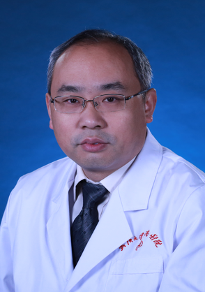 Shaobo Zhu (祝少博)合作论文数Zhongnan Hospital of Wuhan University15
 Linglong Deng合作论文数13
 Li Yu (余黎)合作论文数Zhongnan Hospital, Wuhan University9Bing Wang合作论文数6Xing Zhao (赵星)合作论文数5Wenqiang Qu合作论文数4
 Kai Deng (邓凯)合作论文数The Second Clinical College of Wuhan University/Zhongnan Hospital of Wuhan University3
 Guorong Yu (余国荣)合作论文数Zhongnan Hospital of Wuhan University3Bing WANG (汪冰)合作论文数2
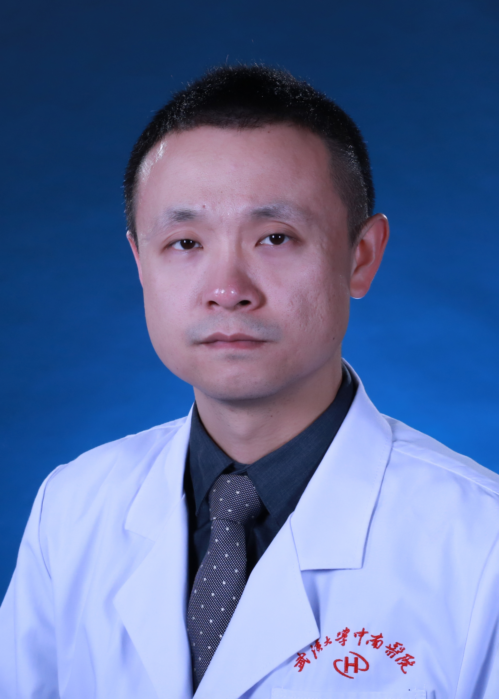 Yu Deng (邓宇)合作论文数Zhongnan Hospital of Wuhan University2Zhe Kang (亢哲)合作论文数1Panfeng Fu (付攀峰)合作论文数1Yong Zhao (赵勇)合作论文数1Zhenzi Li合作论文数1Chong Zhang合作论文数1Zhe Xie (折葉)合作论文数1Lin Jin (金林)合作论文数1ZheZhen Jiang (江哲珍)合作论文数1
 Zuoqi Zhang (张作启)合作论文数School of Civil Engineering, Wuhan University1
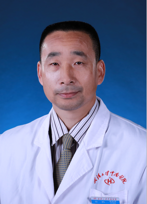 Shengxiang Tao (陶圣祥)合作论文数Zhongnan Hospital of Wuhan University1
 Pan Liu (刘攀)合作论文数Institute for Water-Carbon Cycles & Carbon Neutrality, School of Water Resources and Hydropower Engineering, Wuhan University1




