Aims:This prospective study investigated an innovative approach to the management of extruded or devitalized fragments of bone in patients with a Gustilo type III open fracture of the tibia, which enabled the efficient reconstruction of the bony defect while minimizing the requirement for iliac bone harvesting. Methods:A total of 12 patients with a Gustilo type III open fracture were enrolled into the study between June 2019 and June 2022. During initial emergency management, all patients were treated with radical debridement, the introduction of an antibiotic-loaded spacer, and stabilization of the fracture. Extruded or devitalized fragments, including detached and potentially contaminated segments, were collected and processed into autologous demineralized bone matrix (ADBM). In the second stage, after removal of the spacer, ADBM combined with iliac bone graft was implanted into the induced membrane cavity for reconstruction of the bony defect. The sterility of the ADBM specimens was assessed using microbiological culture. Volumetric analysis of iliac graft requirements was performed. Primary outcome measures including time to healing of the fracture and complications were recorded. The recovery of function was assessed using the Lower Extremity Functional Scale (LEFS) at final follow-up. Results:The mean follow-up was 32.5 months (24 to 48). No bacteria were detected in any ADBM implants. Complete reconstruction of the defect was achieved in all patients. Quantitative volume analysis revealed an autogenous bone-to-ADBM ratio of 1.36:1, reducing the autologous bone requirements. Radiological union occurred at a mean of seven months (5 to 10) after the second surgical stage. There were no complications such as rejection, infection, nonunion, or refracture. At the last follow-up, all patients had regained satisfactory function of the lower limb as recorded by a mean LEFS score of 72.2 (68 to 76). Conclusion:The processing of comminuted extruded or devitalized fragments of bone into ADBM represents a safe and effective strategy for managing severe open fractures of the tibia. When synergistically integrated with an induced membrane technique, this innovative approach considerably reduced the incidence of infection, diminished the need for iliac bone graft, and substantially enhanced the efficiency of reconstruction of the bony defect.
Traumatic soft tissue defects pose significant challenges for reconstruction due to tissue loss, impaired vascularity, and difficulties achieving durable coverage. Adipose tissue offers a practical autologous tissue source, and mechanically processed stromal vascular fraction (SVF)-enriched microfat can be prepared intraoperatively without enzymatic digestion. This study presents a standardized clinical protocol for harvesting autologous adipose tissue and processing it into SVF-enriched microfat for injection into cavity-type traumatic soft tissue defects. Fat is harvested manually from the thigh or abdomen under low negative pressure, mechanically fragmented by cutting and syringe-to-syringe emulsification, filtered to achieve uniform microfat consistency, and centrifuged to isolate the SVF-containing fraction. The processed microfat is injected throughout the wound cavity in a multilayered pattern. Postoperative assessment includes serial clinical evaluation, photographic documentation, and measurement of wound-area reduction until epithelialization. In a small cohort, the method was associated with progressive wound contraction and complete epithelialization within approximately 4-8 weeks, with no major complications. Although cellular composition and viability were not quantified, the technique provided a feasible intraoperative approach suitable for settings without access to enzymatic processing or laboratory facilities. This protocol offers a practical, minimally manipulated method for delivering SVF-enriched microfat in managing traumatic cavity-type defects and may serve as a foundation for further controlled studies.
The optimal surgical management of acute Rockwood type III acromioclavicular (AC) joint dislocations remains controversial. This study aimed to compare clinical and radiological outcomes among three techniques: hook plate (HP) fixation, isolated coracoclavicular (CC) suspension with a single endobutton, and combined AC and CC ligament reconstruction using two separate endobuttons. This retrospective cohort study included 69 patients who underwent surgical treatment for acute Rockwood type III AC joint dislocations between 2018 and 2024 at the mean follow-up of approximately 24 months. Patients were divided into three groups: hook plate fixation (Group A, n = 29), single endobutton CC fixation (Group B, n = 18), and combined AC and CC reconstruction with two separate endobuttons (Group C, n = 22). Clinical outcomes were assessed using the Constant–Murley Score (CMS), American Shoulder and Elbow Surgeons (ASES) score, Visual Analog Scale (VAS), and time to return to daily activities and sports. Radiological evaluation included acromioclavicular distance (ACD) and coracoclavicular distance (CCD). All groups demonstrated significant improvement (p < 0.05) in CMS, ASES, and VAS scores from baseline to final follow-up, which refers to the last clinical and radiographic assessment at approximately 24 months postoperatively. At the final assessment, Group C demonstrated more favorable outcomes, with significantly higher CMS and ASES scores and a lower VAS score compared to Groups A and B (p < 0.05). Importantly, the observed improvements in all three scores exceeded the reported minimal clinically important difference thresholds across all groups. Radiologically, while CCD did not differ significantly among groups, ACD showed significant intergroup differences (p < 0.05), with Group C demonstrating better maintenance of AC joint reduction. Group C also demonstrated a lower complication rate descriptively and the shortest time to return to daily activities and sports (p < 0.05). All three surgical techniques were effective in treating acute Rockwood type III AC joint dislocations. Combined AC–CC reconstruction with two endobuttons offered superior maintenance of AC joint reduction versus isolated CC suspension, a lower complication rate trend than hook plate fixation, and faster return to activities. Radiographic advantages over hook plate fixation were not statistically significant, and CCD differences were non-significant across groups.
BACKGROUND:While the induced membrane technique effectively addresses bone defect reconstruction, its reliance on autografts remains a significant limitation. Building upon the well-established osteogenic properties of platelet-rich plasma (PRP) and demineralized bone matrix (DBM), this study systematically investigated their combined potential as a functional alternative to autografts within the induced membrane microenvironment. METHODS:PRP was extracted and DBM was prepared from New Zealand White rabbits. PRP gel and a composite gel incorporating DBM into PRP were subsequently formulated. The extract solutions derived from PRP, DBM, and PRP/DBM composites were, respectively, collected to induce osteogenic differentiation of bone marrow mesenchymal stem cells (BMSCs). Alkaline phosphatase (ALP) staining was performed on day 5, while mineralization was assessed via Alizarin red staining and osteogenic gene (OCN, OPN, Runx-2, and Col-1) mRNA expression was quantified by quantitative real-time polymerase chain reaction on Days 14 and 21. The rabbits (n = 48) were randomly assigned into four experimental groups (n = 12 per group): empty defect control (no graft material), PRP gel alone, DBM alone, and PRP/DBM composite gel graft. A 20-mm large segmental bone defect was surgically created in the bilateral radius of each animal, followed by initially filling with polymethyl methacrylate spacers to maintain the defect space. After an 8-week period, the spacers were removed, and the resulting membranous cavities were implanted with the respective graft materials according to group assignment. Bone regeneration was evaluated at 6 and 12 weeks post-grafting through radiography, micro-CT imaging, fluorescence-labeled dynamic histomorphometry, histochemical staining, and immunofluorescence analysis. RESULTS:BMSCs treated with PRP/DBM composites demonstrated the most pronounced ALP activity and mineralization capacity, alongside the strongest upregulation of osteogenic genes. Minimal new bone formation, restricted primarily to the defect peripheries, was observed in both control and PRP groups at 6- and 12-week timepoints. In contrast, the DBM and PRP/DBM groups exhibited progressive, time-dependent osteogenic activity. Notably, the PRP/DBM group demonstrated superior bone regeneration, with compact trabeculae and well-organized collagen fully bridging the defect area, accompanied by robust vascularization. In comparison, the DBM group showed less mature bone formation, characterized by loose trabecular structures, immature collagen deposition, and sparse blood vessels. Furthermore, the PRP/DBM group displayed the highest mineral apposition rates and the most pronounced expression of the angiogenic marker CD31. Additionally, the PRP/DBM composite, mediated by the induced membrane, elicited stronger immunomodulatory effects within the local microenvironment and a more robust activation of the hypoxia-inducible factor/vascular endothelial growth factor pathway during bone repair compared to the other groups. CONCLUSION:Within the induced membrane microenvironment, PRP synergistically enhances the osteogenic capacity of DBM. The resulting PRP/DBM composite represents a highly promising autologous candidate for bone graft substitution.
BACKGROUND The purpose of this study was to analyze the outcomes of revascularization exceeding 12 h after arterial injury at different sites of the lower extremity. MATERIAL AND METHODS From January 2009 to April 2017, 58 patients with 58 lower-limb arterial injuries who underwent revascularization over 12 h after trauma were included in our study. Outcomes measured, including mortality, amputation, complications, and other parameters (gait, length discrepancy, the range of movement of the knee and ankle joint, and muscle wasting) were analyzed. RESULTS External iliac artery injury (EIAI) or femoral artery injury (FAI) was affected in 4 patients, superficial femoral artery injury (SFAI) in 18, and popliteal artery injury (PAI) (including proximal gastrocnemius muscle vascular (PGMV) and proximal gastrocnemius muscle vascular [PGMV]) in 36. The median time of arterial injury was 72 h (interquartile range, 59.5). No mortality was found. Amputations were performed in 16 patients due to non-viable limbs, progressing infection, or muscle necrosis. All patients were followed up (median, 52 months; interquartile range, 5.5). Of the 42 limb-salvage patients, most had a limp, muscle wasting, or ankle and knee dysfunctions, and 26 patients with knee or ankle dysfunction underwent secondary surgery. CONCLUSIONS Although limited recanalization of blood vessels may lead to limb complications or amputations over time, the high success rate of limb salvage still merits the surgeon's best efforts.
The management of diabetic wounds presents significant challenges due to persistent inflammation, microenvironmental disruptions, and impaired angiogenesis. To address these issues, this study developed a multifunctional chondroitin sulfate sponge (CSP@Cu-Mg) with anti-inflammatory properties, hemostatic effects, effusion absorption, and enhanced healing promotion. Through ion crosslinking, MgO and CuO were incorporated into the interpenetrating network structure of chondroitin sulfate and acellular dermal matrix, resulting in a sponge with impressive liquid absorption capacity (3450 %) and porosity (83 %). This sponge enabled sustained release of Mg2+/Cu2+ ions, with approximately 40 % cumulative release over 7 days. This release helped reduce inflammation, promote the proliferation and migration of skin repair-related cells, and stimulate angiogenesis. In vivo studies demonstrated that the CSP@Cu-Mg sponge significantly improved diabetic wound healing by modulating inflammation and accelerating collagen deposition, angiogenesis, and re-epithelialization. This extracellular matrix sponge, which synergistically releases Mg2+/Cu2+, presents a promising strategy for comprehensive diabetic wound management with substantial clinical implications.
The prosperity of chemodynamic therapy provides a new strategy for tumor treatment. However, the lack of reactive oxygen species and the specific reductive tumor microenvironment have limited the further development of chemodynamic therapy. Herein, we reported a Fe-based cyclically catalyzing double free radical system for tumor therapy by catalyzing exogenous potassium persulfate (K2S2O8) and endogenous hydrogen peroxide (H2O2). Sufficient amounts of Fe3+ and S2O82- were delivered to tumor sites via tumor-targeted hyaluronic acid (HA) encapsulated mesoporous silica nanoparticles (MSNs) and released under the dual stimulation of acid and hyaluronidase (HAase) in the tumor microenvironment. Fe3+ was reduced to Fe2+ by the reducing agents of loaded tannic acid (TA) and intracellular glutathione (GSH), and Fe2+ was subsequently reacted with S2O82- and endogenous H2O2 to produce two types of ROS (˙OH and SO4-˙), showing an excellent anti-tumor effect. This process not only supplied Fe2+ for the catalysis of active substances, but also reduced the concentration of reduced substances in cells, which was conducive to the existence of free radicals for the efficient killing of tumor cells. Therefore, this iron-based catalysis of exogenous and exogenous active substances to realize a dual-radical oncotherapy nanosystem would provide a new perspective for chemodynamic therapy.
Diabetic wounds are serious clinical complications which manifest wet condition due to the mass exudate, along with disturbed regulation of inflammation, severe oxidative stress and repetitive bacterial infection. Existing treatments for diabetic wounds remain unsatisfactory due to the lack of ideal dressings that encompass mechanical performance, adherence to moist tissue surfaces, quick repair, and diverse therapeutic benefits. Herein, we fabricated a wet adhesive, self-healing, glucose-responsive drug releasing hydrogel with efficient antimicrobial and pro-healing properties for diabetic wound treatment. PAE hydrogel was constructed with poly(acrylic acid-co-acrylamide) (AA-Am) integrated with a dynamic E-F crosslinker, which consisted of epigallocatechin gallate (EGCG) and 4-(2-acrylamidoethylcarbamoyl)-3-fluorophenylboronic acid (AFPBA). Due to the dynamic crosslinking nature of boronate esters, abundant catechol groups and hydrogen bonding, PAE hydrogel demonstrated excellent mechanical properties with about 1000 % elongation, robust adhesion to moist tissues, fast self-healing, and absorption of biofluids of 10 times of its own weight. Importantly, PAE hydrogel exhibited sustained and glucose-responsive release of EGCG. Together, the bioactive PAE hydrogel had effective antibacterial, antioxidative, and anti-inflammatory properties in vitro, and accelerated diabetic wound healing in rats via reducing tissue-inflammatory response, enhancing angiogenesis, and reprogramming of macrophages. Overall, this versatile hydrogel provides a straightforward solution for the treatment of diabetic wound, and shows potential for other wound-related application scenarios.
Recurrent infection of chronic wounds remains a major clinical challenge. Recently, the hydrogel antibacterial materials have attracted extensive attention for preventing infection in wound healing. In this study, a hybrid hydrogel made of polyvinyl alcohol -iodine (PAI), sodium carboxymethyl cellulose (CMC), and carbamino quantum dot (CQDs) was prepared by the cross-linking of hydrogen bonds, named as polyvinyl alcohol-iodine/ sodium carboxymethyl cellulose/carbon quantum dots (PAI/CMC/CQDs). The composite hydrogels exhibited the outstanding photothermal conversion efficiency with near infrared (NIR) light irradiation, and the high anti-bacterial activity against Staphylococcus aureus (S. aureus) and Escherichia coli (E. coli). Meanwhile, the elevated temperature of the composite hydrogels up to similar to 45 degrees C was able to stimulate the migration of epidermal cell to accelerate skin repair. Given that PAI and CQDs could respond to different pH values (5-8), the real-time would pH information was provided by the visible light and fluorescent light dual monitoring system by naked eye. Moreover, the visible-fluorescent images could be collected and transformed into RGB signals to quantify the would pH levels, avoiding secondary injuries caused by frequent dressing changes. PAI/CMC/CQDs was demonstrated the significant therapeutic effect on chronic wounds by eliminating bacterial infections and pro-moting skin repair under the smart RGB monitoring system.
Background Internal fixation for severe open articular bone defects is sometimes ineffective or dangerous. In the emergency stage, radical debridement and infection prevention are demanded to provide a good tissue base and the space‐occupying effect to provide enough necessary space to avoid soft‐tissue contraction for the reconstruction. In addition, the 3D printing technology makes individual limb reconstruction a reality. Case Presentation Here, we present a 31‐year‐old patient with an open fracture and severe bone defect of his left elbow caused by traffic accident, classified as Gustilo–Anderson IIIB. We adopted aggressive debridement and insertion of polymethyl methacrylate (PMMA) to prevent the infection and temporarily construct the bone defect in the emergency stage. Secondly, the total elbow arthroplasty was performed using a unique three‐dimensional (3D) printed prosthesis to reconstruct the elbow joint. During the follow‐up, the elbow movement function was satisfactory. Conclusions The modified Masquelet technique assisting 3D printing of personalized elbow joint makes the satisfactory functional reconstruction for open high‐energy injuries come true. It could be promoted for the similar surgery of other open joints fractures with severe bone defects.
Objective:To establish a model of epiphyseal plate injury in juvenile rabbits and explore the effect of periosteum flap with saphenous artery on preventing the formation of bone bridge after epiphyseal plate injury.Methods:From July 2017 to January 2018, 30 young New Zealand immature rabbit were randomly grouped into 3 groups (group A, B and C, with 10 rabbits per group). The blood vessels of knee joint were dissected and a periosteum flap with saphenous artery were designed. A model of distal femoral epiphyseal plate injury was established in immature rabbits with a 3.0 mm Kirschner wire on one side of the distal femoral epiphyseal plate as experimental side, and the other side of the distal femoral epiphyseal plate was assigned as control side. The injured epiphysis were prepared and described as follow: in experimental side of group A, a piece of periosteum with the same size as that in group C was resected and discarded. In experimental side of group B, a periosteal flap without vascular pedicle was filled in the injured area. And in experimental side of group C, a periosteum flap with saphenous artery was filled in the injured area. The length and varus angle of femur specimens of length and varus angle of femur specimens wihthin groups were measured at 24 weeks after surgery to evaluate the effect on bone growth in epiphyseal plate injury. The effect of the periosteum flap with saphenous artery on preventing a formation of bone bridge was assessed by section view of decalcified specimens, HE staining and toluidine blue staining. The measured data were expressed as Mean and standard deviation (Mean±SD), and data were compared within and between the groups using one-way analysis of variance (ANOVA). P<0.05 was considered as statistically significant. Results:The femur of experimental side of group A and B showed obvious short with valgus deformity compared with that of control side, and the difference of length and varus angle of femur specimens wihthin groups was statistically significant ( P<0.05). There were no significant differences in the length and valgus angle of the femur between experimental side and its control side in group C ( P>0.05). In experimental side of group A, there were bone bridges in the epiphyseal lesion area, with a clear boundary to the surrounding epiphyseal plate. In experimental side of group B, the periosteum flap in the injured area was absorbed, accompanied by the formation of a few pale white bone tissue, and with a clear boundary to the surrounding epiphyseal plate. In experimental side of group C, the lesion area was filled with milky white cartilage tissue with the same colour as the epiphyseal plate, and with an unclear boundary with the epiphyseal plate. HE stain and toluidine blue staining showed that the injured area of group A was filled with a large number of bone tissue, but no obvious cartilage tissue was observed. In group B, the area of lesion was filled mainly with fibrous tissue, with a small amount of bone tissue. In group C, a large number of hyaline cartilage tissues were formed along the tunnel of the injury area, and closely connected with the normal epiphyseal plate. Conclusion:Filling a periosteum flap with saphenous artery after epiphyseal plate injury can prevent the formation of bone bridge and prevent the affected limb form shortening and angulation.
Objective:To screen osteosarcoma related microRNA (miRNA, miR) and study the effect and mechanism on osteosarcoma cells.Methods:Bioinformatics screened the difference of osteosarcoma-related miRNA expression in GSE39058 and identified miR-329 as the research goal. The effect of miR-329 on the proliferation of osteosarcoma 143B cells was analyzed by cell counting kit-8 (CCK-8), and the effect of miR-329 on the invasion of osteosarcoma 143B cells was compared by Transwel. Double luciferase reporter gene assay was used to observe the regulatory effect of miR-329 on G3BP1. The expression levels of G3BP1, E-cadherin, N-cadherin and vimentin before and after miR-329 transfection were analyzed by Western blotting. T-test was used for comparison between groups. Results:Totally, 90 differential miRNAs were obtained, among which the functional enrichment of miR-329 was higher. Polymerase chain reaction (PCR) showed that the expression of miR-329 in osteosarcoma cells was lower than that in osteoblasts hFOB1.19 ( P<0.05). CCK-8 showed that the proliferation rate of 143B cells in the miR-329 mimics group was significantly lower than that in the control group (0.571±0.019 vs. 0.975±0.022). Transwell analysis showed that the number of invasive cells in the miR-329 mimics transfection group was significantly less than that in the control group (28.83±4.55 vs. 58.17±6.25, t=6.220, P<0.05). Double luciferase reporter gene analysis showed that the relative activity of luciferase in the cells cotransfected with G3BP1 mRNA 3′untranslated regions (3′UTR) wt and miR-329 plasmids was significantly lower than that in the control group (0.45±0.06 vs. 1.01±0.09). Western blotting showed that the expression level of E-cadherin in miR-329 mimic transfected group was higher than that in control group, and that of Vimentin and N-cadherin was lower than that in control group (0.13±0.03 vs. 0.25±0.06). Conclusion:MiR-329 inhibits the proliferation and invasion of osteosarcoma 143B cells in vitro by targeting inhibition of G3BP1 expression and epithelial mesenchymal transformation of tumor cells.
The synthesis of drug delivery systems based on hollow mesoporous silica nanoparticles (MSNs) is still a major challenge. In this work, the hollow hybrid MSNs were successfully prepared by cetyltrimethylammonium bromide-directed sol-gel process and one-step hydrothermal treatment process. The hollow hybrid MSNs had hybrid ethane-bridged frameworks with the uniform particle size (250 nm) and mesoporous pore diameter (3.7 nm). The polyacrylic acid (PAA) encapsulated drug delivery system based on hollow hybrid MSNs was prepared by using silanization, surface modification, doxorubicin hydrochloride (DOX) loading, and PAA coating. Drug encapsulation and release behavior at different temperatures and pH were studied by using DOX as a model guest molecule. The results displayed that the modified hollow ethane-bridged MSNs possessed good biocompatibility and excellent thermal/pH-dual-sensitive drug release property. This novel thermal/pH-sensitive drug delivery system based on hollow ethane-bridged MSNs has the advantages of feasible synthesis, no cytotoxicity, and good drug loading capacity, which may have potential applications in the anticancer therapy.
Su, Xing MM1; Luo, Binyang MM1; Li, Yu BN1; Wu, Junchao MD1; Yang, Jinlin MD1; Deng, Kai MD1 Author Information
Background: Ankle fracture is often accompanied by injuries of lateral collateral ligaments, which results in ankle instability and traumatic arthritis in the later period, causing pain and discomfort in patients. However, the repair of the lateral collateral ligament is still inconclusive. The aim of this study was to investigate the operative outcome of one-stage simultaneous repair of the foot and ankle fracture combined with the III grade injury of lateral ligament. Methods: 15 males and 8 females (mean age 37.8 [10.5] years) underwent X-ray and CT scan to determine the type of fracture. MRI, Ultrasound and intraoperative fluoroscopic stress testing were applied to confirm the ligament injury. After the operative treatment of fracture, the ruptured ligament was repaired with suture anchor in 21 cases, meanwhile 1 case combined with chronic ankle instability was repaired with modified Broström-Gould method and another case with middle ligament rupture was applied with the InternalBrace approach. Routine postoperative X-rays and physical examination were carried out, and the American Orthopaedic Food & Ankle Society score was recorded to assess the function and the pain of the wounded ankle. Results: After the operation, the range of ankle motion in all patients was improved, and the bone healing time of the patients was 6-24 (10.09±3.61) weeks. There was no chronic instability of lateral ankle or subtalar joint in all the cases. Moreover, 2 cases presented as cartilage lesions of the medial talus, and the micro-fracture surgery were performed one year later; 1 case suffered from subtalar arthritis, and underwent subtalar arthrodesis 14 months after operation. Furthermore, the patients’ last follow-up AOFAS score was (83.70±4.69) points. There was no occurrence of adverse events and complications such as neurovascular injury, infection or delayed healing of operative incision.Conclusions: One-stage simultaneous repair of the foot and ankle fracture combined with the III grade injury of lateral ligament contributes to the stability of ankle and subtalar joint, and improves the joint function of ankle. It proved to be safe and effective.
Background: The purpose of this article was to report the feasibility and effectiveness of 3-stage Masquelet technique and 1-stage operation for different stages of foot and ankle tuberculosis (TB). Methods: Ten consecutive patients with foot and ankle TB were retrospectively analyzed between January 2014 and December 2018. Five patients were treated with the 3-stage Masquelet technique, including thorough debridement with vacuum sealing drainage, implantation of antibiotic cement spacer, and subsequent reconstruction. Five patients were treated with a 1-stage reconstruction. The American Orthopaedic Foot & Ankle Society (AOFAS) and visual analog scale (VAS) pain scores were recorded at the last follow-up. The follow-up was 30.3 ± 17.8 months. Results: No reactivation of TB was observed in any patients. For the 3-stage operation group, 1 patient developed a distal tibia fracture. The duration of anti-TB therapy was 12.0 ± 0.8 months. The AOFAS score increased from 39.5 ± 9.9 preoperatively to 75.3 ± 7.0 postoperatively ( P < .05). The VAS pain score decreased from 6.3 ± 1.9 to 1.5 ± 1.3 ( P < .05). For the 1-stage operation, 1 patient had wound necrosis. The duration of anti-TB therapy was 13.8 ± 1.1 months. The AOFAS score increased from 51.8 ± 15.0 to 81.8 ± 6.3 ( P < .05). The VAS pain score decreased from 5.4 ± 1.1 to 1.0 ± 0.7 ( P < .05). Conclusion: Three-stage operation was effective for foot and ankle TB with stage IV, sinus tracts or other infections, and 1-stage reconstruction was effective for early-stage TBs. Level of Evidence: Level IV, case series.
目的 探讨应用硬膜外穿刺导针引导,微创小切口缝合急性闭合性跟腱断裂的手术疗效.方法 急性跟腱断裂病人29例,根据治疗方式不同分为导针引导微创小切口缝合组16例,传统开放缝合组13例.病人于伤后5小时~5天内行跟腱手术修补,术后行个性化长腿及短腿石膏固定,功能锻炼1~2个月;所有病人随访至少1年;比较病人手术时间,术后并发症(切口感染,腓肠神经损伤,跟腱再断裂)和术前、术后1年美国矫形足踝协会评分(AOFAS)及跟腱完全断裂评分(ATe).结果 传统开放缝合组手术时间为(48.91±12.56)分钟,微创组(41.78±9.56)分钟,两者比较差异无统计学意义(P>0.05);两组均未出现术后腓肠神经损伤及跟腱再断裂情况,传统组在随访期间出现3例皮肤感染坏死/延迟愈合;截至随访结束,两组病人术前术后AOFAS评分比较,差异有统计学意义(P<0.05),但两组间术后AOFAS及ATRS评分比较差异无统计学意义(P>0.05).结论 经导针引导下微创治疗跟腱断裂手术时间略长,但与常规手术比较,能有效避免术后并发症.
Background: Gustilo-Anderson type III traumas have been described as high-energy injuries with severe bone defects and extensive soft tissue damage, which remain a challenging entity, due to an inherent risk of infection, nonunion and even amputation. The emergency management of such severe trauma presents additional difficulties. Our study attempts to retrospectively evaluate the Masquelet technique combined with the muscle flap for the management of Gustilo type III trauma of the lower limb with segmental bone loss in emergencies and assess key points of success in this technique. Material and methods: From June 2014 to December 2017, 17 patients of Gustilo type IIIA/B/C trauma of lower limb with segmental bone loss, were recruited for our studies. All the cases experienced thorough debridement, stabilization of fracture and antibiotic-impregnated cement spacer insertion. When necessary, muscle flap surgeries were performed immediately. After wound healing, cement spacers were removed, and cancellous bone was filled to repair bone defects. Procedures were performed by two experienced orthopedic surgeons. Results: Among the patients studied, retrograde translocations of the medial head of the gastrocnemius were performed in 5 cases, medial hemimuscular flaps of soleus in 3 cases, and medial head of the gastrocnemius combined with medial hemimuscular flaps of soleus in 4 cases. One patient developed a necrotic soleus flap and was treated with the cross-leg flap. Using a mean 28.2 months of follow-up, results were analyzed radiologically and clinically. Failures (include infection and nonunion) were not noted. And all the patients returned to full weight bearing without pain. According to the Paley fracture healing score, 15 patients showed excellent results and 2 patients displayed good results regarding bone outcomes. When considering functional outcomes, 14 patients exhibited excellent results and 3 patients displayed good results. Conclusions: The muscle flap is synergistic with the Masquelet technique in the emergency management of severe complex fractures. The combination of both techniques in emergency surgery demonstrates an alternative option for the treatment of acute Gustilo type III trauma of the lower limb with segmental bone loss, which can effectively prevent bone infection and amputation. We also demonstrate that firm fixation is key to the Masquelet technique.
Retraction requested by the author due to an admission of research fraud. Reference: Chong Zhang, Chunquan Zhu, Guorong Yu, Kai Deng, Li Yu. Management of Infected Bone Defects of the Lower Extremities by Three-Stage Induced Membrane Technique. Med Sci Monit 2020; 26: e919925. 10.12659/MSM.919925.
Objective To explore the mental experience during the surgical treatment of elderly patients with hallux valgus, so as to provide a basis for their health education and psychological nursing. Methods Twelve elderly patients with hallux valgus undergoing surgeries between November 2016 and March 2017 were selected using the purposive sampling method and given semi-structured interviews. The data were collected using live recordings and analyzed using the Colaizzi seven-step analysis method. Results The psychological experience during the surgical treatment for elderly patients with hallux valgus can be grouped into 6 themes: the cognitive defect in their own illnesses, physiological and psychological discomfort resulting from the illness, disagreement with the surgery method, hesitation and anxiety when choosing the surgical treatment, worries during surgery, as well as confusion and expectation after surgery. Conclusions The knowledge about foot diseases should be publicized so that people can develop healthy living habits and take good care of their feet, and patients with hallux valgus will seek medical treatment without any delay. Moreover, medical staff should attach importance to the physical and mental health of such patients, communicating with them and providing them with information, medical and mental support, so as to promote their physical and mental health.
 Guorong Yu (余国荣)合作论文数Zhongnan Hospital of Wuhan University30
 Linglong Deng合作论文数18
 Shengxiang Tao (陶圣祥)合作论文数Zhongnan Hospital of Wuhan University18
 Shaobo Zhu (祝少博)合作论文数Zhongnan Hospital of Wuhan University17
 Kai Deng (邓凯)合作论文数The Second Clinical College of Wuhan University/Zhongnan Hospital of Wuhan University15Chi Wei合作论文数9
 Lin Cai (蔡林)合作论文数Zhongnan Hospital of Wuhan University9Chunquan Zhu合作论文数8
 Aixi Yu (喻爱喜)合作论文数Zhongnan Hospital of Wuhan University7Xing Zhao (赵星)合作论文数6
 Xuan Xiao合作论文数4ZhongHua Ceng (曾中华)合作论文数4GuangHui Lu (卢光辉)合作论文数4ZhenGuang Chen (陈振光)合作论文数4
 John Fisher合作论文数Fischell Department of Bioengineering, A. James Clark School of Engineering, University of Maryland;Center for Engineering Complex Tissue, University of Maryland3Wenqiang Qu合作论文数3Yaowei CAO (曹耀威)合作论文数3Chong Zhang合作论文数3LiangBo Jiang (江良波)合作论文数3BaiWen Qi (漆白文)合作论文数3QiongHua Feng (冯琼华)合作论文数3Shan Zhao (赵剡)合作论文数3ZheZhen Jiang (江哲珍)合作论文数3
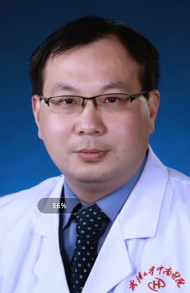 Xiaobing Song (宋小兵)合作论文数Zhongnan Hospital of Wuhan University3
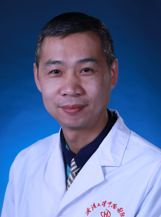 Zhenyu Pan (潘振宇)合作论文数Zhongnan Hospital of Wuhan University3
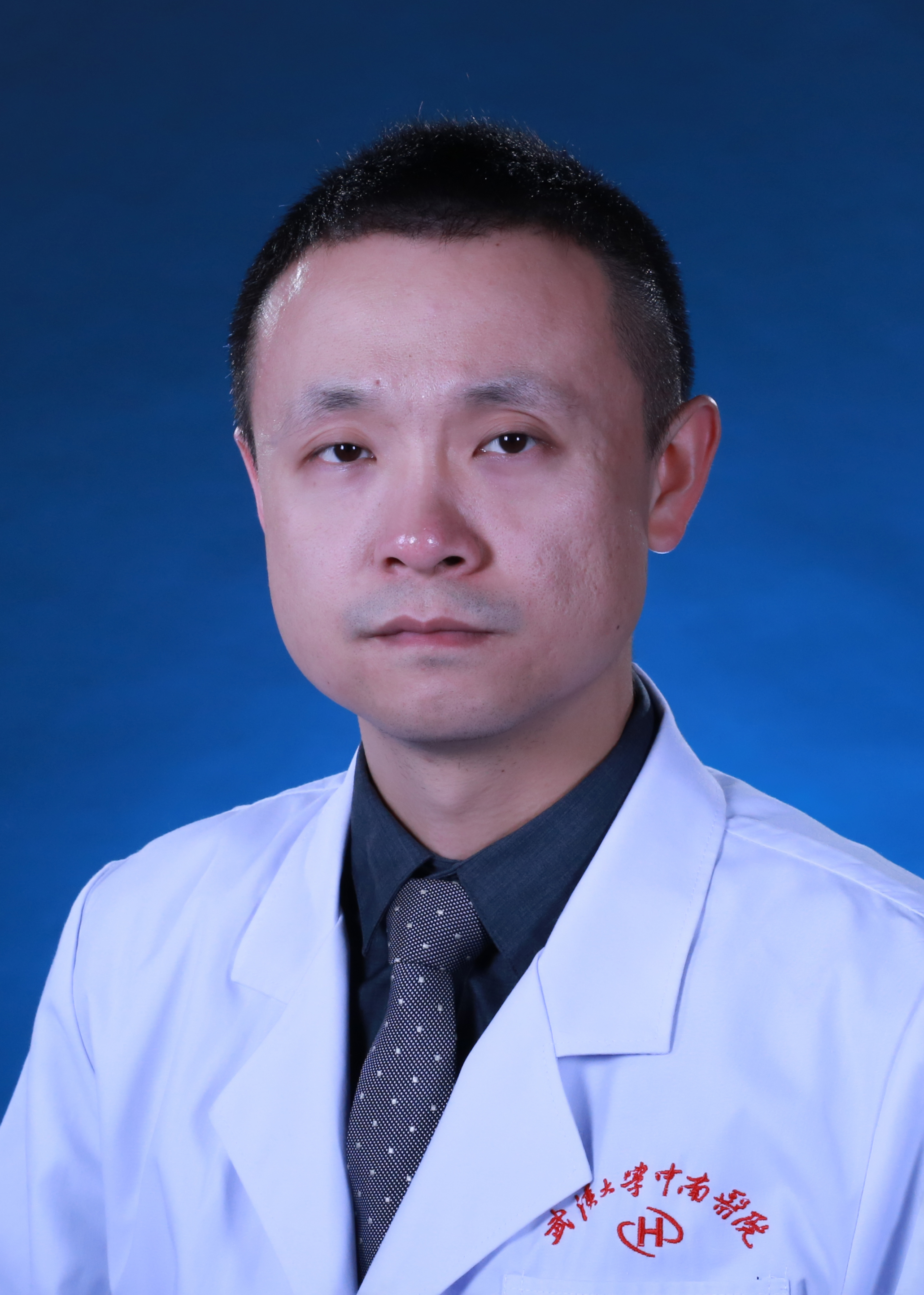 Yu Deng (邓宇)合作论文数Zhongnan Hospital of Wuhan University3Yuanyuan Yang合作论文数2yingmei huang (黄迎梅)合作论文数2Bing Wang合作论文数2Bing WANG (汪冰)合作论文数2XiaoFei Jian (简小飞)合作论文数2XianXun Wang (王显勋)合作论文数2Yun Xie (谢昀)合作论文数2XiaoHui Zheng (郑晓晖)合作论文数2QiSheng Yang (杨奇盛)合作论文数2Hao Hu (胡昊)合作论文数2XiuQing Chen (陈秀清)合作论文数2Hong Qin (秦红)合作论文数2Jun Ceng (曾俊)合作论文数2Ferlin Kimberly M合作论文数2
 Nguyen Bao-Ngoc B合作论文数2Zhang Yi合作论文数2Jianjiao Liu (刘建交)合作论文数2
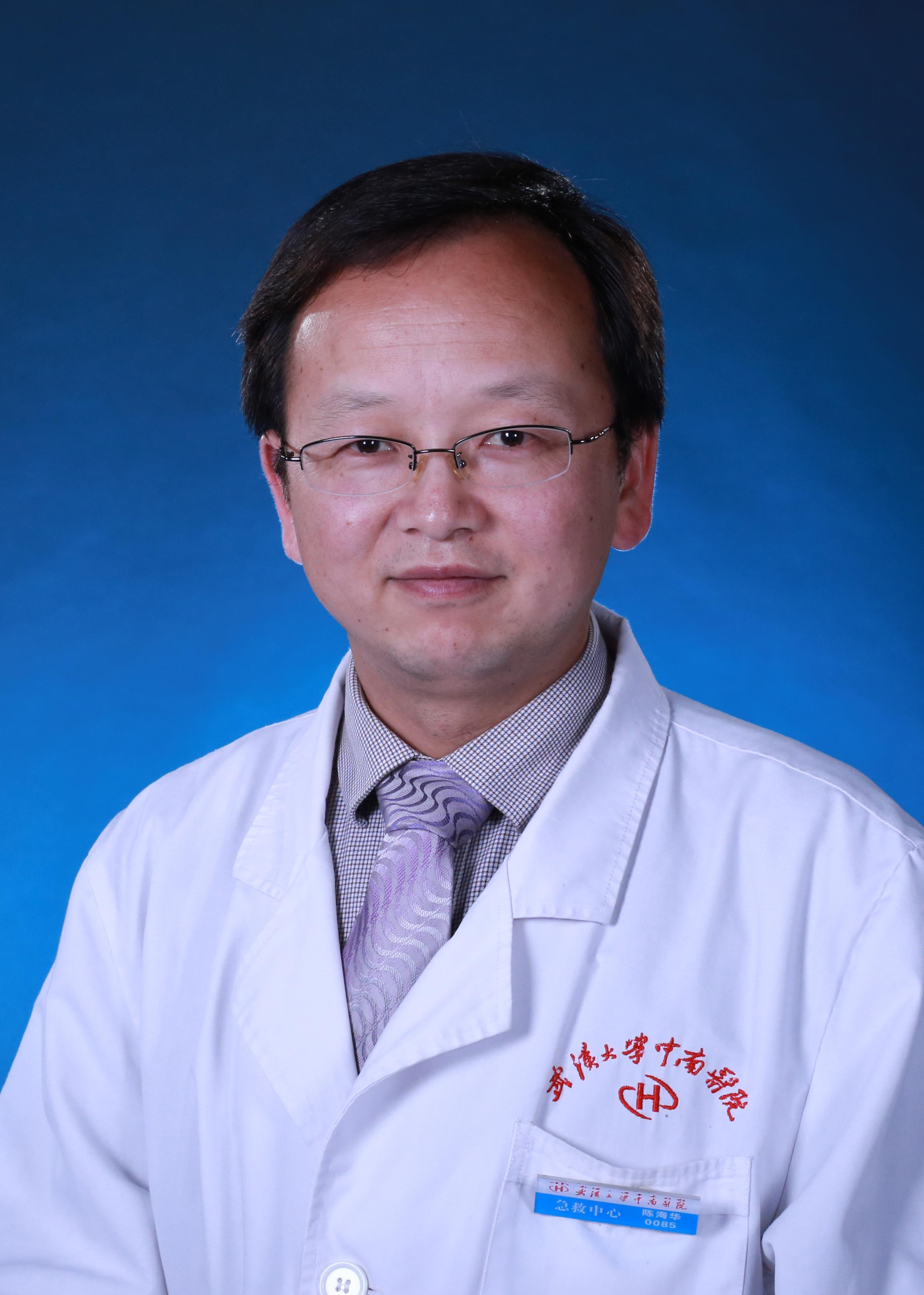 Haihua Chen (陈海华)合作论文数Zhongnan Hospital of Wuhan University2
 Jun Lei (雷军)合作论文数Zhongnan Hospital of Wuhan University2Zhengqi Pan (潘正启)合作论文数2Dianxiong Xu (许典雄)合作论文数2
 Zhaoxia Chen (陈朝霞)合作论文数College of Chemistry and Chemical Engineering, Hubei University2Hang Chen合作论文数1




