The temporal signal-to-noise ratio (tSNR) of functional magnetic resonance imaging (fMRI) is particularly poor in ventral anterior temporal and orbitofrontal regions because of B 0 and B 1 + magnetic field inhomogeneity, a problem that is exacerbated at higher field strengths. In this 7T-fMRI study we compared three methods of improving sensitivity in these areas: parallel transmit, which uses multiple transmit elements, controlled independently, to homogenise the flip angle experienced by the tissue; multi-echo, which entails collection of multiple volumes at different echo times following a single radiofrequency pulse; and multiband, in which multiple slices are acquired simultaneously. We found that parallel transmit and multi-echo increased the magnitude of the BOLD signal change, but only multi-echo increased BOLD magnitude in areas prone to susceptibility artefacts. Multiband and denoising of multi-echo data with independent components analysis (ICA) both improved precision of GLM fit. Exploratory results suggested that multi-echo and ICA denoising can both benefit multivariate analyses. In conclusion, a multi-echo, multiband sequence improved fMRI quality in areas prone to susceptibility artefacts while maintaining sensitivity across the whole brain. We recommend this approach for studies investigating the functional roles of ventral temporal and orbitofrontal regions with 7T fMRI.
Background:Somatic, cognitive and mental health issues have been identified in three-quarters of people 5 months after hospitalisation for severe acute SARS-CoV-2 (COVID-19) infection. The underlying neuroanatomical basis of these symptoms remains unclear, but recent studies suggest a role for altered brainstem physiology. We aimed to test the hypothesis that brainstem neurochemical profiles differ in patients who had been hospitalised for COVID-19 compared to matched controls using 7T magnetic resonance spectroscopy (MRS). Methods:This prospective case-control study recruited 34 individuals who were hospitalised for COVID-19 and 15 healthy controls with no history of COVID-19 infection from two major UK hospitals before vaccines became available. The participants underwent 7T semi-adiabatic localization by adiabatic selective refocusing (sLASER) 1H-MRS at the ponto-medullary junction. Water-referenced metabolite concentrations were compared between the patients and controls and correlated with infection severity, as measured by maximum C-reactive protein (CRPmax) assay during inpatient admission. Linear mixed modelling was used with a 0.05 significance level. Results:Spectral quality was high/acceptable in 44/49 participants according to the MRS Consensus criteria. The magnitude of inflammation during patient admission (i.e., CRPmax) correlated positively with myo-inositol concentration (β = 0.005, p = 0.035), as did patient-reported symptoms (β = -0.564, p = 0.023). However, metabolite concentrations were not significantly different between the patients and controls. Conclusion:We show the feasibility of assessing brainstem neurochemical profiles using 7T 1H-MRS in a multi-centre study. Technical limitations at one site's 7T MRI led to variable repetition times, which limited our statistical power and should be avoided in future studies. Our findings highlight the need for further investigation into the role of neuroinflammation in post-acute COVID-19.
Introduction Progressive supranuclear palsy (PSP) is a devastating neurodegenerative disease characterised by cognitive, behavioural and motor problems. Motor symptoms are highly disabling, while cognitive and behavioural changes have a major impact on carer burden, quality of life and prognosis. Apathy and impulsivity are very common, often coexistent in PSP, and negatively predict survival. In preclinical models and other diseases, apathy and impulsivity are associated with noradrenergic deficits, which can be severe in PSP.Methods and analysis Noradrenaline for Progressive Supranuclear Palsy Syndromes trial is a randomised, double-blind, placebo-controlled, crossover design, Phase IIb clinical trial to evaluate the efficacy and safety of oral atomoxetine for the treatment of cognitive and behavioural changes in PSP. Participants receive atomoxetine 40 mg (10 mg/mL oral solution) once daily or a matched placebo solution, in random order, each for 8 weeks. An ‘informant’, who knows the patient with PSP well, is co-recruited to complete some of the trial outcome measures. Participants remain in the trial for 22 weeks after randomisation. The primary objectives are to assess (1) safety and tolerability and (2) efficacy versus placebo on challenging behaviours as reported in a subscale of the Cambridge Behavioural Inventory. Secondary and exploratory measures relate to cognition, the PSP Rating Scale, mood and potential baseline predictors of individual response to atomoxetine computed from imaging, genetic and cognitive measures at baseline.Ethics and dissemination The trial was approved by the South Central-Oxford B Research Ethics Committee (REC) and the Medicines and Healthcare products Regulatory Agency (REC reference: 20/SC/0416). Dissemination will include publication in peer-reviewed journals, presentations at academic and public conferences and engagement with patients, the public, policymakers and practitioners.Trial registration number ISRCTN99462035; DOI: https://doi.org/10.1186/ISRCTN99462035; EudraCT (European Union Drug Regulating Authorities Clinical Trials Database)/CTIS (Clinical Trial Information System) number: 2019-004472-19; IRAS (Integrated Research Application System) number: 272063; Secondary identifying numbers: CPMS (Central Portfolio Management System) 44441.
BACKGROUND AND PURPOSE: Charcot-Bouchard aneurysms (CBAs) are tiny aneurysms arising from small perforating arteries. Despite the potentially catastrophic consequences of rupture of these aneurysms, the existence and prevalence of CBAs are controversial. The literature in this area is sparse with most previous studies based on radiologic case reports of single hemorrhage or histopathologic analysis. 7T MRI provides higher spatial resolution than 3T MRI, which enables imaging of the small perforating arteries. We determined whether CBAs could be detected in vivo by using 7T MRI. MATERIALS AND METHODS: Ninety-four patients with ischemic stroke collected in the Cambridge 7T Cerebral Small Vessel Disease study prospective cohort were retrospectively included; 75 of them had lacunar infarcts due to presumed small vessel disease, and 19 had nonlacunar infarcts due to presumed cardioembolism or large artery disease. Contrast-enhanced 3D time-of-flight angiography (MRA) and structural sequences were performed by 7T MRI. Two neuroradiologists independently reviewed the MR scans to identify aneurysms on the lenticulostriate arteries (LSA) bilaterally. RESULTS: In 4 of the 94 subjects, CBAs were detected in the LSA; of these, 3 had a single CBA, and 1 had 2. The diameter of the parent vessel ranged from 0.26 mm-0.37 mm and the maximum diameter of the CBA ranged from 0.73 mm-1.39 mm. Use of 3D images allowed differentiation of looped vessels, which could mimic aneurysms on 2D images, from true CBA. CONCLUSIONS: We have demonstrated that 7T MRI can detect CBAs in vivo in humans. This technique could allow further longitudinal studies to determine the true prevalence and prognostic significance of CBAs.
The lenticulostriate arteries (LSAs) supply critical subcortical brain structures and are affected in cerebral small vessel disease (CSVD). Changes in their morphology are linked to cardiovascular risk factors and may indicate early pathology. 7T Time-of-Flight MR angiography (TOF-MRA) enables clear LSA visualisation. We aimed to develop a semi-automated pipeline for quantifying 3D LSA morphology from 7T TOF-MRA in CSVD patients. We used data from a local 7T CSVD study to create a pipeline, LUMEN, comprising two stages: vessel segmentation and LSA quantification. For segmentation, we fine-tuned a deep learning model, DS6, and compared it against nnU-Net and a Frangi-filter pipeline, MSFDF. For quantification, centrelines of LSAs within basal ganglia were extracted to compute branch counts, length, tortuosity, and maximum curvature. This pipeline was applied to 69 subjects, with results compared to traditional analysis measuring LSA morphology on 2D coronal maximum intensity projection (MIP) images. For vessel segmentation, fine-tuned DS6 achieved the highest test Dice score (0.814±0.029) and sensitivity, whereas nnU-Net achieved the best balanced average Hausdorff distance and precision. Visual inspection confirmed that DS6 was most sensitive in detecting LSAs with weak signals. Across 69 subjects, the pipeline with DS6 identified 23.5 ± 8.5 LSA branches. Branch length inside the basal ganglia was 26.4 ± 3.5 mm, and tortuosity was 1.5 ± 0.1. Extracted LSA metrics from 2D MIP analysis and our 3D analysis showed fair-to-moderate correlations. Outliers highlighted the added value of 3D analysis. This open-source deep-learning-based pipeline offers a validated tool quantifying 3D LSA morphology in CSVD patients from 7T-TOF-MRA for clinical research.
PURPOSE:We introduce a novel commercial phosphorus-31 (31P) dipole-loop array coil, describing the coil hardware and testing its performance on phantoms. We used this coil to assess cardiac metabolism per region in healthy volunteers. METHODS:B1 + field maps were simulated and compared to maps measured with a set of CSI sequences with varying voltages. Seventeen volunteers were scanned with 7 T phosphorus-31 magnetic resonance spectroscopic imaging (31P-MRSI). Reproducibility was assessed in nine of these volunteers. Strain was measured for six of these volunteers at 3 T. RESULTS:Blood- and saturation-corrected Phosphocreatine/γ-adenosine triphosphate (PCr/ATP) ratios were measured for four regions of the left ventricle: 1.86 in septum, 2.25 in anterior wall, 1.41 in inferior wall, and 1.53 in lateral wall, respectively. These are in the expected range compared to previous studies. B1 + maps show good signal uniformity around the position of the heart (0.13 ± 0.06 μT/sqrt(W)). Intrasession and intersession coefficients of reproducibility were 0.22-0.88 and 0.29-0.79, respectively. Linear modeling shows that regional PCr/γATP correlates with circumferential strain but not radial strain. This requires corroboration by a larger study including patients with impaired function and energetics. CONCLUSION:Dipole-loop array coils present a promising new approach for human cardiac 31P-MRSI at 7 T. Their favorable B1 + uniformity at depth and specific absorption rate over loop arrays and improved SNR when combined with loops for reception could be beneficial for further clinical studies measuring energetics by 31P-MRSI at 7 T. The new capability to assess PCr/γATP ratios across the whole left ventricle could enable clinical studies to investigate regional changes in cardiac energetics for the first time.
P-31 protocols, even at 7 T, are long, severely limiting the time available to collect flip angle maps needed for metabolite quantification across the heart. Here we aimed to evaluate the feasibility of accelerating P-31 B-1(+) mapping in the myocardium at 7 T. Using the Bloch-Siegert B-1 mapping technique, flip angle maps were estimated using Cartesian 2D-MRSI (FA(CSI)) and concentric ring trajectory-based 3D-MRSI (FA(CRT)) and compared in the thigh of 4 participants. FA(CRT) was also estimated in the myocardium of 3 participants. We have found a non-identity linear relationship between FA(CSI) and FA(CRT), raising the need for a correction. This correction yielded homogeneous flip angle values in the myocardium. Our 3D B-1(+) mapping approach, using half the acquisition time of a 2D Cartesian CSI-MRSI can significantly shorten the length of P-31 protocols at 7 T while allowing absolute quantification of myocardial energetics.
PURPOSE:Turbo spin echo (TSE) is important clinically. Unfortunately, 7 T TSE suffers from B1 +-induced signal dropouts. Magnitude-based parallel transmit (pTx) pulse design algorithms cannot enforce phase patterns complying with the Carr-Purcell-Meiboom-Gill conditions (90° phase shift between excitation and refocusing). We introduce scalable spokes pTx pulses for 7 T TSE imaging. THEORY:We define scalable spokes pulses as having time-symmetric RF waveforms, antisymmetric in-plane gradients, and rephased subpulse slice-selection gradients. They produce flip angles that are approximately proportional to the applied voltage with voltage-independent phase patterns. METHODS:Scalable spokes pulses were designed for a phantom. Scaling behavior was characterized via Bloch simulations. Performance in terms of TSE echo homogeneity was assessed by extended phase graph simulations using in vivo field maps. Performance was validated for TSE acquisitions in a phantom and in vivo. Hippocampal TSE imaging was performed for four subjects comparing circularly polarized (CP), RF shimming, and scalable spokes pulses. RESULTS:Scalable spokes pTx pulses show similar scaling behavior to previously proposed 3D kT-point pulses. Scalable three-spoke pulses decrease flip-angle RMS error across subjects compared to CP mode pulses (11% vs. 23% for 120° pulses). TSE images with these pulses recover signals in cerebellum and temporal lobes. CONCLUSION:Scalable spokes pTx pulses produce flip angles that vary approximately linearly with peak voltage while maintaining consistent spatial patterns of phase. Together with their spatial flip-angle homogeneity, these pulses enable high-fidelity 2D slice-by-slice TSE imaging at 7 T, albeit with reduced slice coverage with our choice of homogeneity target under the current vendor-provided specific absorption rate constraints on 7 T MRI scanners.
How individuals process and respond to uncertainty has important implications for cognition and mental health. Here we use computational phenotyping to examine individualised 'uncertainty fingerprints' in relation to neurometabolites and trait anxiety in humans. We introduce a novel categorical state-transition extension of the Hierarchical Gaussian Filter (HGF) to capture implicit learning in a four-choice probabilistic sensorimotor reversal learning task by tracking beliefs about stimulus transitions. Using 7-Tesla Magnetic Resonance Spectroscopy, we measured baseline neurotransmitter levels in the primary motor cortex (M1). Model-based results revealed dynamic belief updating in response to environmental changes. We further found region-specific relationships between M1 glutamate+ glutamine levels and prediction errors and volatility beliefs, revealing an important neural marker of probabilistic reversal learning in humans. High trait anxiety was associated with faster post-reversal responses. By integrating computational modelling with neurochemical assessments, this study provides novel insights into the neurocomputations that drive individual differences in processing uncertainty. ### Competing Interest Statement The authors have declared no competing interest.
OBJECTIVE:To implement parallel transmit (pTx) 7T magnetic resonance imaging (MRI) in the pre-surgical evaluation of 3T-negative patients with drug-resistant focal epilepsy, and to compare quality to conventional single transmit (specifically, circularly polarized [CP]) 7T MRI. METHODS:We implemented a comparative protocol comprising both pTx and CP 7T MRI in consecutive adult candidates for epilepsy surgery who had negative or equivocal 3T MRI imaging. Here we report the outcomes from the first 31 patients. We acquired pTx and CP T1, T2, fluid-attenuated inversion recovery (FLAIR) and edge-enhancing gradient echo (EDGE) images, all in the same three-dimensional (3D) 0.8 mm isotropic space. Two-dimensional (2D) high-resolution T2 and T2*-weighted sequences were acquired only in CP mode due to current technological limitations. Two neuroradiologists, a neurologist, and a neurosurgeon made independent, blinded quality and preference ratings of pTx vs CP images. Quantitative methods were used to assess signal dropout. RESULTS:7T revealed previously-unseen structural lesions in nine patients (29%), confirmed 3T-equivocal lesions in four patients (13%), and disproved 3T-equivocal lesions in four patients (13%). Lesions were better visualized on pTx than CP in 57% of cases, and never better visualized on CP. Clinical management was altered by 7T in 18 cases (58%). Nine cases were offered surgical resection and one laser interstitial thermal therapy (LITT). Three cases were removed from the surgical pathway because of bilateral or extensive lesions. Five cases were offered stereo-electroencephalography (sEEG) with better targeting (in three because the 7T lesion was deemed equivocal by the multi-disciplinary team (MDT), and in two because the lesion was extensive). Blinded comparison confirmed significantly better overall quality of pTx FLAIR images (F(2, 184) = 13.7, p = 2.88 × 10⁻⁶), whereas pTx MP2RAGE images were subjectively non-inferior and had improved temporal lobe coverage with quantitatively less signal drop-out. SIGNIFICANCE:pTx-7T is implementable in a clinical pathway, changed management in 58% of patients where 3T + FDG-PET had not enabled resection, and is superior to single transmit 7T MRI.
The lenticulostriate arteries (LSA) supply important subcortical structures in the brain and are affected in cerebral small vessel disease (CSVD), leading to changes in their morphology. 7 Tesla Time-of-Flight magnetic resonance angiography (7T-TOF-MRA) now allows their visualisation in humans, but current analysis of LSA morphology largely relies on manual tracing on 2D coronal maximum-intensity-projection (MIP) images, which discards significant information from the third spatial dimension. We aimed to develop a semi-automatic pipeline for quantifying the 3D morphology of LSAs from 7T-TOF-MRA in patients with CSVD. We used contrast-enhanced 7T-TOF-MRA data from 15 subjects enrolled in a local CSVD study. Our pipeline consists of two main stages: vessel segmentation and LSA quantification. For segmentation, we fine-tuned a state-of-the-art deep learning model, DS6, for vessel segmentation and compared its performance against a classical Frangi filter-based pipeline, Multi-Scale Frangi Diffusive Filter (MSFDF). Both methods were evaluated against manually labelled ground-truth masks in LSA regions. In the LSA quantification stage, the user defines a region-of-interest around LSAs and checks the segmentation. Based on this, the LSA centrelines are extracted, and branch counts, length, tortuosity, and curvature are computed. Additionally, we conducted the traditional LSA analysis using 2D coronal MIPs, and we evaluated the correlation between the results from the 2D and 3D analyses. For vessel segmentation, the fine-tuned DS6 model achieved a mean Dice similarity coefficient (DSC) of 0.814±0.029 during testing, outperforming MSFDF on DSC, sensitivity, and balanced average Hausdorff distance in terms of both mean value and stability. Visual inspection confirmed that DS6 was more sensitive in detecting LSA branches with weak signals. On average, the 15 subjects had 5.9±1.6 LSA stems and 28.7±9.9 branches. The mean length of an LSA branch was 42.5±5.7mm, and mean tortuosity was 1.9±0.2. Finally, the branch counts from 2D and 3D analyses correlated well (ρ=0.741, p=2.816e-06), whereas the stem count, branch length and tortuosity measurements were significantly different, showing the insufficiency of MIP analysis (stem: ρ=0.230, p=2.207e-01; length: r=0.565, p=1.153e-03; tortuosity: r=0.400, p=2.847e-02). We have developed an open-source semi-automatic pipeline using deep learning for evaluating the 3D morphology of LSAs in CSVD patients from 7T-TOF-MRA. We show that analysing LSA morphology in 3D reveals previously inaccessible aspects of morphology. Our pipeline offers a valuable tool for clinical research studies to characterise the 3D morphology of LSAs in CSVD. ### Competing Interest Statement The authors have declared no competing interest. ### Funding Statement This work was supported by a British Heart Foundation project grant [PG/19/74/34670]. HSM receives Infrastructural support from the Cambridge British Heart Foundation Centre of Research Excellence [RE/24/130011]. HSM and CTR are supported by the Cambridge University Hospitals NIHR Biomedical Research Centre [NIHR203312]. The views expressed in this publication are those of the authors and not necessarily those of the NIHR, NHS, or UK Department of Health and Social Care. CTR receives research support from Siemens Healthcare for a different project. The 7T MRI was supported by an MRC Clinical Research Infrastructure Award [MR/M008983/1]. RL was supported by a PhD studentship awarded by Trinity College, University of Cambridge, UK. These funding sources were not involved in the study design, data collection, analysis, interpretation of data, writing, or manuscript submission for this study. For the purpose of open access, the author has applied a CC BY public copyright licence to any Author Accepted Manuscript version arising from this submission. ### Author Declarations I confirm all relevant ethical guidelines have been followed, and any necessary IRB and/or ethics committee approvals have been obtained. Yes The details of the IRB/oversight body that provided approval or exemption for the research described are given below: This study was approved by the Institutional Review Board of East of England Cambridge Central Research Ethics Committee (REC Ref: 19/EE/0219). I confirm that all necessary patient/participant consent has been obtained and the appropriate institutional forms have been archived, and that any patient/participant/sample identifiers included were not known to anyone (e.g., hospital staff, patients or participants themselves) outside the research group so cannot be used to identify individuals. Yes I understand that all clinical trials and any other prospective interventional studies must be registered with an ICMJE-approved registry, such as ClinicalTrials.gov. I confirm that any such study reported in the manuscript has been registered and the trial registration ID is provided (note: if posting a prospective study registered retrospectively, please provide a statement in the trial ID field explaining why the study was not registered in advance). Yes I have followed all appropriate research reporting guidelines, such as any relevant EQUATOR Network research reporting checklist(s) and other pertinent material, if applicable. Yes The finetuned DS6 model is available at https://huggingface.co/soumickmj/SMILEUHURA\_DS6\_CamSVD\_UNetMSS3D\_wDeform (retrieved on 20/09/2024). Code for our pipeline and detailed instructions are available at https://github.com/RuiLiGitLove/LSA\_3D\_Morph (retrieved on 20/09/2024). [https://github.com/RuiLiGitLove/LSA\_3D\_Morph][1] [https://huggingface.co/soumickmj/SMILEUHURA\_DS6\_CamSVD\_UNetMSS3D\_wDeform][2] [1]: https://github.com/RuiLiGitLove/LSA_3D_Morph [2]: https://huggingface.co/soumickmj/SMILEUHURA_DS6_CamSVD_UNetMSS3D_wDeform
We present a sequence building block (SBB) that embeds magnetic resonance spectroscopy (MRS) into another sequence on the Siemens VE platform without any custom hardware. This enables dynamic studies such as functional MRS (fMRS), dynamic shimming and frequency correction, and acquisition of navigator images for motion correction. The SBB supports nonlocalised spectroscopy (free induction decay), STimulated Echo Acquisition Mode single voxel spectroscopy, and 1D, 2D and 3D phase-encoded chemical shift imaging. It can embed 1H or X-nuclear MRS into a 1H sequence; and 1H-MRS into an X-nuclear sequence. We demonstrate integration into the vendor's gradient-recalled echo sequence. We acquire test data in phantoms with three coils (31P/1H, 13C/1H and 2H/1H) and in two volunteers on a 7-T Terra MRI scanner. Fifteen lines of code are required to insert the SBB into a sequence. Spectra and images are acquired successfully in all cases in phantoms, and in human abdomen and calf muscle. Phantom comparison of signal-to-noise ratio and linewidth showed that the SBB has negligible effects on image and spectral quality, except that it sometimes produces a nuclear Overhauser effect (NOE) signal enhancement for multinuclear applications in line with conventional 1H NOE pulses. Our new SBB embeds MRS into a host imaging or spectroscopy sequence in 15 lines of code. It allows homonuclear and heteronuclear interleaving. The package is available through the standard C2P procedure. We hope this will lower the barrier for entry to studies applying dynamic fMRS and for online motion correction and B0-shim updating.
AbstractObjectiveTo implement parallel transmit (pTx) 7T MRI in the pre-surgical evaluation of patients with drug resistant focal epilepsy, and to compare quality and diagnostic yield to conventional single transmit (specifically, circularly polarised, CP) 7T MRI.MethodsWe implemented a comparative protocol comprising both pTx and CP 7T MRI in consecutive adult candidates for epilepsy surgery who had negative or equivocal 3T MRI imaging. Here we report the outcomes from the first 31 patients.We acquired pTx and CP T1, T2, FLAIR and EDGE images, all in the same 3D 0.8mm isotropic space. 2D high-resolution T2and T2*-weighted sequences were acquired only in CP mode due to current technological limitations.Two neuroradiologists, a neurologist and a neurosurgeon made independent, blinded quality and preference ratings of pTx vs CP images. Quantitative methods were used to assess signal dropout.ResultsBlinded comparison confirmed significantly better overall quality of pTx FLAIR images (F(2,184)=13.7, p=2.88×10⁻⁶), while pTx MP2RAGE images were subjectively non-inferior and had improved temporal lobe coverage with quantitatively less signal drop-out.7T-pTx revealed previously-unseen structural lesions in 9 patients (29%), confirmed 3T-equivocal lesions in 4 patients (13%), and disproved 3T-equivocal lesions in 4 patients (13%).Lesions were better visualised on pTx than CP in 57% of cases, and never better visualised on CP.Clinical management was altered by pTx-7T in 18 cases (58%). 9 cases were offered surgical resection and 1 LITT. 3 cases were removed from the surgical pathway because of bilateral or extensive lesions. 5 cases were offered sEEG with better targeting (in 3 because the 7T lesion was deemed equivocal by the MDT, and in 2 because the lesion was extensive).SignificanceParallel transmit 7T MRI is implementable in a clinical pathway, is superior to single transmit 7T MRI, and changed management in 58% of patients scanned.Key pointsWe scanned 31 patients with parallel transmit and conventional 7T MRI, finding previously-unreported structural lesions in 9 patients (29% of cases).In 13% of cases pTx 7T MRI showed that an equivocal lesion at 3T MRI was likely significant.In 13% of cases pTx 7T MRI showed that an equivocal lesion at 3T MRI could be disregarded.Both qualitative and quantitative quality assessments indicate superiority of pTx images over CP.Future clinical implementations of 7T MRI for epilepsy should utilise parallel transmit where possible.
PURPOSE:Spoke pulses improve excitation homogeneity in parallel-transmit MRI. We propose an efficient global optimization algorithm, Bayesian optimization of gradient trajectory (BOGAT), for single-slice and simultaneous multislice imaging. THEORY AND METHODS:BOGAT adds an outer loop to optimize kT-space positions. For each position, the RF coefficients are optimized (e.g., with magnitude least squares) and the cost function evaluated. Bayesian optimization progressively estimates the cost function. It automatically chooses the kT-space positions to sample, to achieve fast convergence, often coming close to the globally optimal spoke positions. We investigated the typical features of spokes cost functions by a grid search with field maps comprising 85 slabs from 14 volunteers. We tested BOGAT in this database, and prospectively in a phantom and in vivo. We compared the vendor-provided Fourier transform approach with the same magnitude least squares RF optimizer. RESULTS:The cost function is nonconvex and seen empirically to be piecewise smooth with discontinuities where the underlying RF optimum changes sharply. BOGAT converged to within 10% of the global minimum cost within 30 iterations in 93% of slices in our database. BOGAT achieved up to 56% lower flip angle RMS error (RMSE) or 55% lower pulse energy in phantoms versus the Fourier transform approach, and up to 30% lower RMSE and 29% lower energy in vivo with 7.8 s extra computation. CONCLUSION:BOGAT efficiently estimated near-global optimum spoke positions for the two-spoke tests, reducing flip-angle RMSE and/or pulse energy in a computation time (˜10 s), which is suitable for online optimization.
Ultra-high field (7T) MRI allows scans at sub-millimetre resolution with exquisite signal-to-noise ratio (SNR). As 7T MRI becomes more widely used clinically, the challenge of patient motion must be overcome. Retrospective motion correction is used successfully for some protocols, but for acquisitions such as slice-by-slice scans only prospective motion correction can deliver the full potential of 7T MRI. We report the first implementation of prospective 3D Fat Navigator (“FatNav”) motion correction for the Siemens 7T Terra MRI. We implemented a modular Sequence Building Block for FatNav and embedded it into the vendor's gradient-recalled echo (GRE) sequence. We modified the reconstruction pipeline to reconstruct FatNav images online, coregistering them and sending motion updates to the host sequence online. We tested five registration algorithms for performance and accuracy on synthetic FatNav data. We implemented the best three of these in our sequence and tested them online. We acquired T 1 and T 2 * weighted brain images of healthy volunteers correcting every other image for motion to visualise the effectiveness of online motion correction. Data were acquired with and without head immobilisation. We also tested performance while correcting every measurement for motion. Our implementation uses a 1.23 s 3D FatNav acquisition module and delivers motion updates in less than 3 s, which is sufficient for motion updates every few k -space lines in typical scans. Corrected images are crisper with fewer visible motion artefacts. This improved sharpness is reflected quantitatively by an increase in the variance of the image Laplacian which is 1.59 x better for corrected vs uncorrected images. Profiles across the cerebral falx are 33% steeper for corrected vs uncorrected images. Prospective FatNav improves GRE image quality in the brain. Our modular Sequence Building Block provides a simple method to integrate motion correction in 7T MRI pulse sequences.
Neuroferritinopathy is a disorder of neurodegeneration with brain iron accumulation that has no proven disease-modifying treatments. Clinical trials require biomarkers of iron deposition. We examined brain iron accumulation in one presymptomatic FTL mutation carrier, two individuals with neuroferritinopathy and one healthy control using ultra-high-field 7T MRI. There was increased magnetic susceptibility, suggestive of iron deposition, in superficial and deep gray matter in both presymptomatic and symptomatic neuroferritinopathy. Cavitation of the putamen and globus pallidus increased with disease stage and at follow up. The widespread brain iron deposition in presymptomatic and early disease provides an opportunity for monitoring disease-modifying intervention.
Post-mortem studies have shown that patients dying from severe acute respiratory syndrome coronavirus (SARS-CoV-2) infection frequently have pathological changes in their CNS, particularly in the brainstem. Many of these changes are proposed to result from para-infectious and/or post-infection immune responses. Clinical symptoms such as fatigue, breathlessness, and chest pain are frequently reported in post-hospitalized coronavirus disease 2019 (COVID-19) patients. We propose that these symptoms are in part due to damage to key neuromodulatory brainstem nuclei. While brainstem involvement has been demonstrated in the acute phase of the illness, the evidence of long-term brainstem change on MRI is inconclusive. We therefore used ultra-high field (7 T) quantitative susceptibility mapping (QSM) to test the hypothesis that brainstem abnormalities persist in post-COVID patients and that these are associated with persistence of key symptoms. We used 7 T QSM data from 30 patients, scanned 93-548 days after hospital admission for COVID-19 and compared them to 51 age-matched controls without prior history of COVID-19 infection. We correlated the patients' QSM signals with disease severity (duration of hospital admission and COVID-19 severity scale), inflammatory response during the acute illness (C-reactive protein, D-dimer and platelet levels), functional recovery (modified Rankin scale), depression (Patient Health Questionnaire-9) and anxiety (Generalized Anxiety Disorder-7). In COVID-19 survivors, the MR susceptibility increased in the medulla, pons and midbrain regions of the brainstem. Specifically, there was increased susceptibility in the inferior medullary reticular formation and the raphe pallidus and obscurus. In these regions, patients with higher tissue susceptibility had worse acute disease severity, higher acute inflammatory markers, and significantly worse functional recovery. This study contributes to understanding the long-term effects of COVID-19 and recovery. Using non-invasive ultra-high field 7 T MRI, we show evidence of brainstem pathophysiological changes associated with inflammatory processes in post-hospitalized COVID-19 survivors.
Diffusion MRI (dMRI) is inherently limited by SNR. Scanning at 7 T increases intrinsic SNR but 7 T MRI scans suffer from regions of signal dropout, especially in the temporal lobes and cerebellum. We applied dynamic parallel transmit (pTx) to allow whole-brain 7 T dMRI and compared with circularly polarized (CP) pulses in 6 subjects. Subject-specific 2-spoke dynamic pTx pulses were designed offline for 8 slabs covering the brain. We used vendor-provided B0 and B1+ mapping. Spokes positions were set using the Fourier difference approach, and RF coefficients optimized with a Jacobi-matrix high-flip-angle optimizer. Diffusion data were analyzed with FSL. Comparing whole-brain averages for pTx against CP scans: mean flip angle error improved by 15% for excitation (2-spoke-VERSE 15.7° vs CP 18.4°, P = 0.012) and improved by 14% for refocusing (2-spoke-VERSE 39.7° vs CP 46.2°, P = 0.008). Computed spin-echo signal standard deviation improved by 14% (2-spoke-VERSE 0.185 vs 0.214 CP, P = 0.025). Temporal SNR increased by 5.4% (2-spoke-VERSE 8.47 vs CP 8.04, P = 0.004) especially in the inferior temporal lobes. Diffusion fitting uncertainty decreased by 6.2% for first fibers (2-spoke VERSE 0.0655 vs CP 0.0703, P < 0.001) and 1.3% for second fibers (2-spoke VERSE 0.139 vs CP 0.141, P = 0.01). In conclusion, dynamic parallel transmit improves the uniformity of 7 T diffusion-weighted imaging. In future, less restrictive SAR limits for parallel transmit scans are expected to allow further improvements.
INTRODUCTION:Entorhinal cortex (EC) is the first cortical region to exhibit neurodegeneration in Alzheimer's disease (AD), associated with EC grid cell dysfunction. Given the role of grid cells in path integration (PI)-based spatial behaviors, we predicted that PI impairment would represent the first behavioral change in adults at risk of AD. METHODS:We compared immersive virtual reality (VR) PI ability to other cognitive domains in 100 asymptomatic midlife adults stratified by hereditary and physiological AD risk factors. In some participants, behavioral data were compared to 7T magnetic resonance imaging (MRI) measures of brain structure and function. RESULTS:Midlife PI impairments predicted both hereditary and physiological AD risk, with no corresponding multi-risk impairment in episodic memory or other spatial behaviors. Impairments associated with altered functional MRI signal in the posterior-medial EC. DISCUSSION:Altered PI may represent the transition point from at-risk state to disease manifestation in AD, prior to impairment in other cognitive domains.
 Stefan Neubauer合作论文数Radcliffe Department of Medicine, Medical Sciences Division, University of Oxford59
 William L. Clarke合作论文数University of Virginia Medical Center50
 Matthew Robson合作论文数Perspectum Diagnostics Ltd38
 Oliver Rider合作论文数Medical Sciences Division, Radcliffe Department of Medicine, University of Oxford30
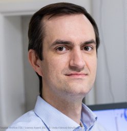 Valkovič Ladislav合作论文数Dept Biomed Imaging & Image Guided Therapy, Med Univ Vienna29
 Catarina Rua合作论文数25
 Eylem Levelt合作论文数15Jennifer J. Rayner合作论文数Univ Oxford, Radcliffe Dept Med, Oxford Ctr Clin Magnet Resonance Res, Div Cardiovasc Med, Oxford, England13
 Damian Tyler合作论文数Department of Physiology, Anatomy and Genetics, University of Oxford;Somerville College, University of Oxford13
 Masliza Mahmod合作论文数Radcliffe Department of Medicine, University of Oxford12Jabrane Karkouri合作论文数Univ Claude Bernard Lyon 1, Univ Lyon11
 Moritz Hundertmark合作论文数Oxford Centre for Clinical Magnetic Resonance Research, University of Oxford11Jane M. Francis合作论文数11Guy B. Williams合作论文数Addenbrooke's Hospital, Cambridge University11Mark A Peterzan合作论文数10
 James B. Rowe合作论文数Cambridge Centre for Frontotemporal Dementia and Related Disorders, Department of Clinical Neurosciences, University of Cambridge;Dementias Platform UK;Alzheimer's Research UK;Medical Research Council Cognition and Brain Sciences Unit, University of Cambridge10
 Rina Ariga合作论文数Radcliffe Department of Medicine, University of Oxford10
 Kieran Clarke合作论文数Department of Physiology, Anatomy and Genetics, Division of Medical Sciences, University of Oxford;TdeltaS10
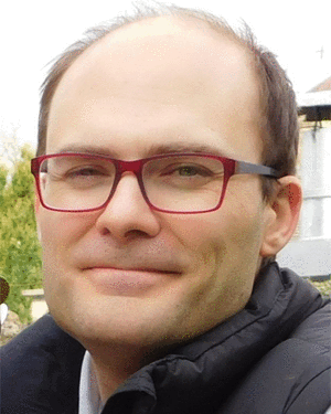 Aaron Hess合作论文数Oxford University Centre for Integrative Neuroimaging10
 Peter Hore合作论文数Department of Chemistry, University of Oxford;Corpus Christi College, University of Oxford10Belinda Ding合作论文数9
 Betty Raman合作论文数Queen Elizabeth Hospital, University of Adelaide9
 Theodoros D. Karamitsos合作论文数1st Department of Cardiology, School of Medicine, Aristotle University of Thessaloniki9Christiane R Timmel合作论文数8Jane Ellis合作论文数8Liu Alexander合作论文数8Cameron J Holloway合作论文数8
 Victoria Stoll合作论文数Institute of Cardiovascular Sciences, University of Birmingham8
 Douglas Tucker合作论文数Fermi National Accelerator Laboratory8Lucian A B Purvis合作论文数7
 Timothy Beers合作论文数Department of Physics and Astronomy, College of Science, University of Notre Dame7
 Stuart Clare合作论文数Nuffield Department of Clinical Neurosciences, Medical Sciences Division, University of Oxford;Wellcome Centre for Integrative Neuroimaging, University of Oxford7
 Saul G. Myerson合作论文数Radcliffe Department of Medicine, University of Oxford7
 John Thomas Vaughan (王唐明)合作论文数Zuckerman Institute, Columbia University in the City of New York;Department of Biomedical Engineering, Fu Foundation School of Engineering and Applied Science, Columbia University in the City of New York;MRAccess Inc.7Carl J. Snyder合作论文数Unversity of Minnesota7
 M. Dall'Ora合作论文数INAF—Osservatorio Astronomico di Capodimonte6Dragonu Iulius合作论文数Department of Radiology, University Medical Center Freiburg6
 Miller Jack J合作论文数Clarendon Laboratory, University of Oxford6
 Graeme A. Keith合作论文数6
 Lau Justin Y C合作论文数Department of Medical Biophysics, University of Toronto;Imaging Research, Sunnybrook Research Institute;Sunnybrook Research Institute, University of Toronto6
 Zoe Kourtzi合作论文数Department of Psychology, University of Cambridge6
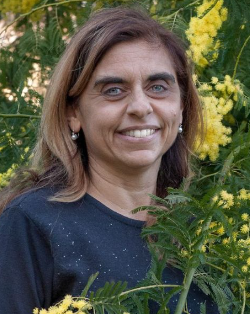 Marcella Marconi合作论文数INAF – Astronomical Observatory of Capodimonte6
 Wolfgang Bogner合作论文数Department of Radiology, Medical University6
 Gisella Clementini合作论文数Osservatorio di Astrofisica e Scienza dello Spazio di Bologna, Istituto Nazionale di Astrofisica6Kevin B. Henbest合作论文数6
 Craig A. Lygate合作论文数Radcliffe Department of Medicine, University of Oxford6Charles A. Kuehn合作论文数6V. Ripepi合作论文数6
 Sayeed Rana合作论文数5




