Currently, there is substantial ongoing discussion around the functional role of the thalamus in consciousness. What is missing in the literature, however, is a systematic investigation of the relevance of specific thalamic nuclei in pharmacologically and pathologically altered states of consciousness in humans. Using resting-state functional magnetic resonance imaging in both healthy anaesthetized volunteers (n = 16) and patients with disorders of consciousness (n = 22), we sought to identify which specific thalamic subregions in both cohorts may be differentially significant for loss of consciousness. Our findings revealed that, among all nuclei, the pulvinar was found to have the strongest functional connectivity change with loss of consciousness following anaesthesia, while demonstrating distinct functional connectivity patterns related to higher-order default mode and executive control networks. Remarkably, in loss of consciousness in disorders of consciousness patients, the ventral-latero-ventral was found to have the strongest connectivity change in comparison with healthy controls, while exhibiting discrete functional connectivity patterns related to higher-order default mode, executive control and frontoparietal networks. Furthermore, we provide evidence that this neural connectivity biomarker in patients also mirrored the changes observed at the behavioural level, which could have clinical implications for targeted deep brain stimulation in therapy for disorders of consciousness.
The precise neuromodulatory mechanisms underlying consciousness and its disorders, despite growing evidence in both animals and humans, remain poorly understood at the subcortical level. The basal ganglia, a key component of the cortico-basal ganglia-thalamo-cortical loop, are known to play a crucial role in mediating consciousness and behavioural responsiveness to the environment. Here, we aimed to present a comprehensive mapping of the distinct contributions of basal ganglia nuclei and their associated neurotransmitter systems in pharmacologically induced and pathological loss of responsiveness. We used functional MRI to provide a systematic investigation of 9 major nuclei (putamen, caudate, nucleus accumbens, globus pallidus external, globus pallidus internal, substantia nigra pars compacta, substantia nigra pars reticulata, ventral pallidus, and subthalamic nucleus) and their functional relationship with 19 major neurotransmitter systems in loss of responsiveness, in both healthy volunteers under propofol anaesthesia (n = 16) and patients with disorders of consciousness (DOC) (i.e. minimally conscious, unresponsive wakefulness syndrome) (n = 22). We found that in pharmacologically-induced loss of responsiveness, the functional connectivity changes of the putamen (Pu) correlated with the norepinephrine transporter (NET), and the globus pallidus external (GPe) with the serotonin transporter (5-HTT) and vesicular acetylcholine transporter (VAChT); additionally, altered connectivity in the ventral pallidum (VeP) was associated with the norepinephrine transporter (NET). In contrast, with pathological-induced loss of responsiveness, we found that connectivity changes of the caudate were associated with the serotonin transporter (5-HTT), dopamine transporter (DAT), and GABAA receptor, and connectivity changes of the substantia nigra pars compacta (SNc) were associated with the cannabinoid (CB1) receptor. Finally, we report that among all basal ganglia nuclei, a key dopamine-rich area of the basal ganglia-the substantia nigra-was found to have the largest DOC subgroup difference in functional connectivity, following stratification based on mental-imagery task responsiveness. Critically, we provide evidence that pharmacological and pathological loss of responsiveness involve distinct neurotransmitter system contributions across individual basal ganglia nuclei, offering a novel framework for targeted therapeutic interventions in patients with disorders of consciousness.
How is the information-processing architecture of the human brain organised, and how does its organisation support consciousness? Here, we combine network science and a rigorous information-theoretic notion of synergy to delineate a ‘synergistic global workspace’, comprising gateway regions that gather synergistic information from specialised modules across the human brain. This information is then integrated within the workspace and widely distributed via broadcaster regions. Through functional MRI analysis, we show that gateway regions of the synergistic workspace correspond to the human brain’s default mode network, whereas broadcasters coincide with the executive control network. We find that loss of consciousness due to general anaesthesia or disorders of consciousness corresponds to diminished ability of the synergistic workspace to integrate information, which is restored upon recovery. Thus, loss of consciousness coincides with a breakdown of information integration within the synergistic workspace of the human brain. This work contributes to conceptual and empirical reconciliation between two prominent scientific theories of consciousness, the Global Neuronal Workspace and Integrated Information Theory, while also advancing our understanding of how the human brain supports consciousness through the synergistic integration of information.
INTRODUCTION:Cerebral small vessel disease (SVD) is a common cause of stroke/vascular dementia with few effective treatments. Neuroinflammation and increased blood-brain barrier (BBB) permeability may influence pathogenesis. In rodent models, minocycline reduced inflammation/BBB permeability. We determined whether minocycline had a similar effect in patients with SVD. METHODS:MINERVA was a single-center, phase II, randomized, double-blind, placebo-controlled trial. Forty-four participants with moderate-to-severe SVD took minocycline or placebo for 3 months. Co-primary outcomes were microglial signal (determined using 11C-PK11195 positron emission tomography) and BBB permeability (using dynamic contrast-enhanced MRI). RESULTS:Forty-four participants were recruited between September 2019 and June 2022. Minocycline had no effect on 11C-PK11195 binding (relative risk [RR] 1.01, 95% confidence interval [CI] 0.98-1.04), or BBB permeability (RR 0.97, 95% CI 0.91-1.03). Serum inflammatory markers were not affected. DISCUSSION:11C-PK11195 binding and increased BBB permeability are present in SVD; minocycline did not reduce either process. Whether these pathophysiological mechanisms are disease-causing remains unclear. INTERNATIONAL CLINICAL TRIALS REGISTRY PORTAL IDENTIFIER:ISRCTN15483452 HIGHLIGHTS: We found focal areas of increased microglial signal and increased blood-brain barrier permeability in patients with small vessel disease. Minocycline treatment was not associated with a change in these processes measured using advanced neuroimaging. Blood-brain barrier permeability was dynamic but MRI-derived measurements correlated well with CSF/serum albumin ratio. Advanced neuroimaging is a feasible outcome measure for mechanistic clinical trials.
Background Even patients with normal computed tomography (CT) head imaging may experience persistent symptoms for months to years after mild traumatic brain injury (mTBI). There is currently no good way to predict recovery and triage patients who may benefit fi t from early follow-up and targeted intervention. We aimed to assess if existing prognostic models can be improved by serum biomarkers or diffusion tensor imaging metrics (DTI) from MRI, and if serum biomarkers can identify patients for DTI. Methods We included 1025 patients aged >18 years with a Glasgow Coma Score >12 and normal CT from the Collaborative European NeuroTrauma Effectiveness Research in Traumatic Brain Injury (CENTER-TBI) study which recruited between December 19,2014 and December 17, 2017 (NCT02210221). Biomarkers (GFAP, NFL, S100B) were obtained at a median of 8.8 h (Q1-Q3 - Q3 4.2-16.7) - 16.7) and DTI at 13 days (3-19) - 19) after injury. DTI metrics were available in 153 patients for 48 white matter tracts (ICBM-DTI-81 atlas). Incomplete recovery at three months was defined fi ned as an extended Glasgow Outcome Scale score <8. Existing prognostic models were fi tted with and without biomarkers, or with and without DTI, and internally validated using bootstrapping. Findings 385 (38%) patients had incomplete recovery. Adding biomarkers did not improve performance beyond the best existing clinical prognostic model [optimism-corrected AUC 0.69 (95% CI 0.65-0.72) - 0.72) and R2 2 17% (11-22)]. - 22)]. Adding DTI metrics significantly fi cantly enhanced all models [best optimism-corrected AUC 0.82 (0.79-0.85) - 0.85) and R2 2 75% (39-100)]. - 100)]. The top three prognostic tracts were the left posterior thalamic radiation, left superior cerebellar peduncle and right uncinate fasciculus. Serum biomarkers could have avoided 1 in 5 DTI scans, with GFAP <12 h and NFL 12-24 - 24 h from injury performing best. Interpretation DTI substantially improved existing prognostic models for functional outcome in patients with mTBI and a normal CT, and biomarkers could help select patients for MRI. If validated, DTI could allow for targeted follow- up and enrichment of clinical trials of early interventions to improve outcome.
Changes in the brain's physiology in Alzheimer's disease are thought to occur early in the disease's trajectory. In this study our aim was to investigate the brain's neurochemical profile in a midlife cohort in relation to risk factors for future dementia using single voxel proton magnetic resonance spectroscopy. Participants in the multi-site PREVENT-Dementia study (age range 40-59 year old) underwent 3T magnetic resonance spectroscopy with the spectroscopy voxel placed in the posterior cingulate/precuneus region. Using LCModel, we quantified the absolute concentrations of myo-inositol, total N-acetylaspartate, total creatine, choline, glutathione and glutamate-glutamine for 406 participants (mean age 51.1; 65.3% female). Underlying partial volume effects were accounted for by applying a correction for the presence of cerebrospinal fluid in the magnetic resonance spectroscopy voxel. We investigated how metabolite concentrations related to apolipoprotein epsilon 4 genotype, dementia family history, a risk score (Cardiovascular Risk Factors, Aging and Incidence of Dementia -CAIDE) for future dementia including non-modifiable and potentially-modifiable factors and dietary patterns (adherence to Mediterranean diet). Dementia family history was associated with decreased total N-acetylaspartate and no differences were found between apolipoprotein epsilon 4 carriers and non-carriers. A higher Cardiovascular Risk Factors, Aging, and Incidence of Dementia score related to higher myo-inositol, choline, total creatine and glutamate-glutamine, an effect which was mainly driven by older age and a higher body mass index. Greater adherence to the Mediterranean diet was associated with lower choline, myo-inositol and total creatine; these effects did not survive correction for multiple comparisons. The observed associations suggest that at midlife the brain demonstrates subtle neurochemical changes in relation to both inherited and potentially modifiable risk factors for future dementia. Dounavi and McKiernan et al. used Magnetic Resonance Spectroscopy in a large mid-life cohort with varying future dementia risk. Lower N-acetylaspartate associated with parental dementia; lifestyle risk correlated with higher myo-inositol, choline, creatine and glutamate+glutamine. Neurochemical changes were found at mid-life in relation to inherited and potentially modifiable dementia risk. Graphical abstract
Post-mortem studies have shown that patients dying from severe acute respiratory syndrome coronavirus (SARS-CoV-2) infection frequently have pathological changes in their CNS, particularly in the brainstem. Many of these changes are proposed to result from para-infectious and/or post-infection immune responses. Clinical symptoms such as fatigue, breathlessness, and chest pain are frequently reported in post-hospitalized coronavirus disease 2019 (COVID-19) patients. We propose that these symptoms are in part due to damage to key neuromodulatory brainstem nuclei. While brainstem involvement has been demonstrated in the acute phase of the illness, the evidence of long-term brainstem change on MRI is inconclusive. We therefore used ultra-high field (7 T) quantitative susceptibility mapping (QSM) to test the hypothesis that brainstem abnormalities persist in post-COVID patients and that these are associated with persistence of key symptoms. We used 7 T QSM data from 30 patients, scanned 93-548 days after hospital admission for COVID-19 and compared them to 51 age-matched controls without prior history of COVID-19 infection. We correlated the patients' QSM signals with disease severity (duration of hospital admission and COVID-19 severity scale), inflammatory response during the acute illness (C-reactive protein, D-dimer and platelet levels), functional recovery (modified Rankin scale), depression (Patient Health Questionnaire-9) and anxiety (Generalized Anxiety Disorder-7). In COVID-19 survivors, the MR susceptibility increased in the medulla, pons and midbrain regions of the brainstem. Specifically, there was increased susceptibility in the inferior medullary reticular formation and the raphe pallidus and obscurus. In these regions, patients with higher tissue susceptibility had worse acute disease severity, higher acute inflammatory markers, and significantly worse functional recovery. This study contributes to understanding the long-term effects of COVID-19 and recovery. Using non-invasive ultra-high field 7 T MRI, we show evidence of brainstem pathophysiological changes associated with inflammatory processes in post-hospitalized COVID-19 survivors.
Background Predicting dementia early has major implications for clinical management and patient outcomes. Yet, we still lack sensitive tools for stratifying patients early, resulting in patients being undiagnosed or wrongly diagnosed. Despite rapid expansion in machine learning models for dementia prediction, limited model interpretability and generalizability impede translation to the clinic. Methods We build a robust and interpretable predictive prognostic model (PPM) and validate its clinical utility using real-world, routinely-collected, non-invasive, and low-cost (cognitive tests, structural MRI) patient data. To enhance scalability and generalizability to the clinic, we: 1) train the PPM with clinically-relevant predictors (cognitive tests, grey matter atrophy) that are common across research and clinical cohorts, 2) test PPM predictions with independent multicenter real-world data from memory clinics across countries (UK, Singapore). Findings PPM robustly predicts (accuracy: 81.66%, AUC: 0.84, sensitivity: 82.38%, specificity: 80.94%) whether patients at early disease stages (MCI) will remain stable or progress to Alzheimer's Disease (AD). PPM generalizes from research to real-world patient data across memory clinics and its predictions are validated against longitudinal clinical outcomes. PPM allows us to derive an individualized AI-guided multimodal marker (i.e. predictive prognostic index) that predicts progression to AD more precisely than standard clinical markers (grey matter atrophy, cognitive scores; PPM-derived marker: hazard ratio = 3.42, p = 0.01) or clinical diagnosis (PPM-derived marker: hazard ratio = 2.84, p < 0.01), reducing misdiagnosis. Interpretation Our results provide evidence for a robust and explainable clinical AI-guided marker for early dementia prediction that is validated against longitudinal, multicenter patient data across countries, and has strong potential for adoption in clinical practice. Funding Wellcome Trust, Royal Society, Alzheimer’s Research UK, Alzheimer’s Drug Discovery Foundation Diagnostics Accelerator, Alan Turing Institute.
The spectrum, pathophysiology, and recovery trajectory of persistent post-COVID-19 cognitive deficits are unknown, limiting our ability to develop prevention and treatment strategies. We report the one-year cognitive, serum biomarker, and neuroimaging findings from a prospective, national longitudinal study of cognition in 351 COVID-19 patients who had required hospitalisation, compared to 2,927 normative matched controls. Cognitive deficits were global and associated with elevated brain injury markers and reduced anterior cingulate cortex volume one year after admission. The severity of the initial infective insult, post-acute psychiatric symptoms, and a history of encephalopathy were associated with greatest deficits. There was strong concordance between subjective and objective cognitive deficits. Treatment with corticosteroids during the acute phase appeared protective against cognitive deficits. Together, these findings support the hypothesis that brain injury in moderate to severe COVID-19 is immune-mediated, and should guide the development of therapeutic strategies.
The spectrum, pathophysiology and recovery trajectory of persistent post-COVID-19 cognitive deficits are unknown, limiting our ability to develop prevention and treatment strategies. We report the 1-year cognitive, serum biomarker and neuroimaging findings from a prospective, national study of cognition in 351 COVID-19 patients who required hospitalization, compared with 2,927 normative matched controls. Cognitive deficits were global, associated with elevated brain injury markers and reduced anterior cingulate cortex volume 1 year after COVID-19. Severity of the initial infective insult, postacute psychiatric symptoms and a history of encephalopathy were associated with the greatest deficits. There was strong concordance between subjective and objective cognitive deficits. Longitudinal follow-up in 106 patients demonstrated a trend toward recovery. Together, these findings support the hypothesis that brain injury in moderate to severe COVID-19 may be immune-mediated, and should guide the development of therapeutic strategies. A national prospective study of patients requiring hospitalization for COVID-19 demonstrates global cognitive deficits at 1 year, associated with elevated brain injury markers and reduced gray matter volume.
Image-to-image reconstruction problems with free or inexpensive metadata in the form of class labels appear often in biological and medical image domains. Existing text-guided or style-transfer image-to-image approaches do not translate to datasets where additional information is provided as discrete classes. We introduce and implement a model which combines image-to-image and class-guided denoising diffusion probabilistic models. We train our model on a real-world dataset of microscopy images used for drug discovery, with and without incorporating metadata labels. By exploring the properties of image-to-image diffusion with relevant labels, we show that class-guided image-to-image diffusion can improve the meaningful content of the reconstructed images and outperform the unguided model in useful downstream tasks.
Background: Recent studies have demonstrated increased microglial activation using 11C-PK11195 positron emission tomography imaging, indicating central nervous system inflammation, in cerebral small vessel disease. However, whether such areas of neuroinflammation progress to tissue damage is uncertain. We determined whether white matter destined to become white matter hyperintensities (WMH) at 1 year had evidence of altered inflammation at baseline. Methods: Forty subjects with small vessel disease (20 sporadic and 20 cerebral autosomal dominant arteriopathy with subcortical infarcts and leukoencephalopathy) and 20 controls were recruited to this case-control observational study from in- and out-patient clinics at Addenbrooke’s Hospital, Cambridge, UK and imaged at baseline with both 11C-PK11195 positron emission tomography and magnetic resonance imaging; and magnetic resonance imaging including diffusion tensor imaging was repeated at 1 year. WMH were segmented at baseline and 1 year, and areas of new lesion identified. Baseline 11C-PK11195 binding potential and diffusion tensor imaging parameters in these voxels, and normal appearing white matter, was measured. Results: Complete positron emission tomography-magnetic resonance imaging data was available for 17 controls, 16 sporadic small vessel disease, and 14 cerebral autosomal dominant arteriopathy with subcortical infarcts and leukoencephalopathy participants. 11C-PK11195 binding in voxels destined to become new WMH was lower than in normal appearing white matter, which did not progress to WMH (−0.133[±0.081] versus −0.045 [±0.044]; P<0.001). Mean diffusivity was higher and mean fractional anisotropy lower in new WMH voxels than in normal appearing white matter (900 [±80]×10−6 versus 1045 [±149]×10−6 mm2/s and 0.37±0.05 versus 0.29±0.06, both P<0.001) consistent with new WMH showing tissue damage on diffusion tensor imaging a year prior to developing into new WMH; similar results were seen across the 3 groups. Conclusions: White matter tissue destined to develop into new WMH over the subsequent year is associated with both lower neuroinflammation, and white matter ultrastructural damage at baseline. Our results suggest that this tissue is already damaged 1 year prior to lesion formation. This may reflect that the role of neuroinflammation in the lesion development process occurs at an early stage, although more studies over a longer period would be needed to investigate this further.
Recent advances in machine learning highlight the potential of Ai-guided tools for early dementia prediction with major implications for timely clinical management. Yet, we still lack robust tools for predicting neurocognitive decline early in clinical practice. We have developed a predictive prognostic model based on Generalised Matrix Learning Vector Quantisation (GMLVQ) that combines multimodal biological and cognitive data to predict cognitive decline ( Giorgio et al., 2020 ). Here we test the clinical validity of this model in predicting individualised cognitive trajectories on real-world memory clinic data. We trained our model (Fig.1) to classify sMCI (individuals with stable MCI diagnosis within a 3-year period; n = 290) vs. pMCI (individuals who progressed from MCI to AD within a 3-year period; n = 120). We tested the model (out-of-sample validation) on a) an asymptomatic sample (Berkley Ageing Cohort: BACS, n = 187 healthy controls), b) two clinical samples: National University of Singapore (NUS; AD: n = 32, non-AD neurodegenerative disease: n = 15, MCI moderate: n = 42, MCI mild: n = 53, Control: n = 27); Quantitative MRI in NHS Memory Clinics; AD: n = 72, MCI: n = 50, Non-AD: n = 30, Functional/attentional memory symptoms: n = 30, other cognitive disorders: n = 10). We trained and tested the model with baseline structural MRI (medial temporal grey matter density) and cognitive data (following data harmonization across training and test samples). Using a scalar projection method, we generated a prognostic index that indicates distance from the stable prototype and classifies individuals to Clinically Stable vs. Declining (slowly vs. rapidly). Our trained model on ADNI data classified sMCI vs. pMCI with 79.87% class-balanced cross-validated accuracy (sensitivity = 83.33%, specificity = 76.52%). For the clinical NUS and QMIN-MC samples, most AD patients were classified as rapidly progressive compared to MCI patients that were mostly classified as stable or slowly progressive (Fig.2). In contrast, for the BACS asymptomatic sample most individuals were classified as stable. Survival analysis on the NUS sample— for which longitudinal assessments (up to 6 years) are available— validated the model predictions, showing that patients with higher model-derived prognostic index decline faster (Fig.3). Validating our AI-guided tool for early dementia prediction in real-world patient data provides evidence for its clinical utility.
A central question in neuroscience is how consciousness arises from the dynamic interplay of brain structure and function. Here we decompose functional MRI signals from pathological and pharmacologically-induced perturbations of consciousness into distributed patterns of structure-function dependence across scales: the harmonic modes of the human structural connectome. We show that structure-function coupling is a generalisable indicator of consciousness that is under bi-directional neuromodulatory control. We find increased structure-function coupling across scales during loss of consciousness, whether due to anaesthesia or brain injury, capable of discriminating between behaviourally indistinguishable sub-categories of brain-injured patients, tracking the presence of covert consciousness. The opposite harmonic signature characterises the altered state induced by LSD or ketamine, reflecting psychedelic-induced decoupling of brain function from structure and correlating with physiological and subjective scores. Overall, connectome harmonic decomposition reveals how neuromodulation and the network architecture of the human connectome jointly shape consciousness and distributed functional activation across scales.
Cerebral hemodynamic alterations have been observed in apolipoprotein ε4 (APOE4) carriers at midlife, however the physiological underpinnings of this observation are poorly understood. Our goal was to investigate cerebral blood flow (CBF) and its spatial coefficient of variation (CoV) in relation to APOE4 and a measure of erythrocyte anisocytosis (red blood cell distribution width – RDW) in a middle-aged cohort. Data from 563 participants in the PREVENT-Dementia study scanned with 3 T MRI cross-sectionally were analysed. Voxel-wise and region-of-interest analyses within nine vascular regions were run to detect areas of altered perfusion. Within the vascular regions, interaction terms between APOE4 and RDW in predicting CBF were examined. Areas of hyperperfusion in APOE4 carriers were detected mainly in frontotemporal regions. The APOE4 allele differentially moderated the association between RDW and CBF, an association which was more prominent in the distal vascular territories (p – [0.01, 0.05]). The CoV was not different between the considered groups. We provide novel evidence that in midlife, RDW and CBF are differentially associated in APOE4 carriers and non-carriers. This association is consistent with a differential hemodynamic response to hematological alterations in APOE4 carriers.
The prediction of functional outcome after mild traumatic brain injury (mTBI) is challenging. Conventional magnetic resonance imaging (MRI) does not do a good job of explaining the variance in outcome, as many patients with incomplete recovery will have normal-appearing clinical neuroimaging. More advanced quantitative techniques such as diffusion MRI (dMRI), can detect microstructural changes not otherwise visible, and so may offer a way to improve outcome prediction. In this study, we explore the potential of linear support vector classifiers (linearSVCs) to identify dMRI biomarkers that can predict recovery after mTBI. Simultaneously, the harmonization of fractional anisotropy (FA) and mean diffusivity (MD) via ComBat was evaluated and compared for the classification performances of the linearSVCs. We included dMRI scans of 179 mTBI patients and 85 controls from the Collaborative European NeuroTrauma Effectiveness Research in Traumatic Brain Injury (CENTER-TBI), a multi-center prospective cohort study, up to 21 days post-injury. Patients were dichotomized according to their Extended Glasgow Outcome Scale (GOSE) scores at 6 months into complete (n = 92; GOSE = 8) and incomplete (n = 87; GOSE <8) recovery. FA and MD maps were registered to a common space and harmonized via the ComBat algorithm. LinearSVCs were applied to distinguish: (1) mTBI patients from controls and (2) mTBI patients with complete from those with incomplete recovery. The linearSVCs were trained on (1) age and sex only, (2) non-harmonized, (3) two-category-harmonized ComBat, and (4) three-category-harmonized ComBat FA and MD images combined with age and sex. White matter FA and MD voxels and regions of interest (ROIs) within the John Hopkins University (JHU) atlas were examined. Recursive feature elimination was used to identify the 10% most discriminative voxels or the 10 most discriminative ROIs for each implementation. mTBI patients displayed significantly higher MD and lower FA values than controls for the discriminative voxels and ROIs. For the analysis between mTBI patients and controls, the three-category-harmonized ComBat FA and MD voxel-wise linearSVC provided significantly higher classification scores (81.4% accuracy, 93.3% sensitivity, 80.3% F1-score, and 0.88 area under the curve [AUC], p < 0.05) compared with the classification based on age and sex only and the ROI approaches (accuracies: 59.8% and 64.8%, respectively). Similar to the analysis between mTBI patients and controls, the three-category-harmonized ComBat FA and MD maps voxelwise approach yields statistically significant prediction scores between mTBI patients with complete and those with incomplete recovery (71.8% specificity, 66.2% F1-score and 0.71 AUC, p < 0.05), which provided a modest increase in the classification score (accuracy: 66.4%) compared with the classification based on age and sex only and ROI-wise approaches (accuracy: 61.4% and 64.7%, respectively). This study showed that ComBat harmonized FA and MD may provide additional information for diagnosis and prognosis of mTBI in a multi-modal machine learning approach. These findings demonstrate that dMRI may assist in the early detection of patients at risk of incomplete recovery from mTBI.
In image-based profiling, software extracts thousands of morphological features of cells from multi-channel fluorescence microscopy images, yielding single-cell profiles that can be used for basic research and drug discovery. Powerful applications have been proven, including clustering chemical and genetic perturbations on the basis of their similar morphological impact, identifying disease phenotypes by observing differences in profiles between healthy and diseased cells and predicting assay outcomes by using machine learning, among many others. Here, we provide an updated protocol for the most popular assay for image-based profiling, Cell Painting. Introduced in 2013, it uses six stains imaged in five channels and labels eight diverse components of the cell: DNA, cytoplasmic RNA, nucleoli, actin, Golgi apparatus, plasma membrane, endoplasmic reticulum and mitochondria. The original protocol was updated in 2016 on the basis of several years’ experience running it at two sites, after optimizing it by visual stain quality. Here, we describe the work of the Joint Undertaking for Morphological Profiling Cell Painting Consortium, to improve upon the assay via quantitative optimization by measuring the assay’s ability to detect morphological phenotypes and group similar perturbations together. The assay gives very robust outputs despite various changes to the protocol, and two vendors’ dyes work equivalently well. We present Cell Painting version 3, in which some steps are simplified and several stain concentrations can be reduced, saving costs. Cell culture and image acquisition take 1–2 weeks for typically sized batches of ≤20 plates; feature extraction and data analysis take an additional 1–2 weeks. This protocol is an update to Nat. Protoc. 11, 1757–1774 (2016): https://doi.org/10.1038/nprot.2016.105 We provide an updated protocol for image-based profiling with Cell Painting. A detailed procedure, with standardized conditions for the assay, is presented, along with a comprehensive description of parameters to be considered when optimizing the assay.
Background:Recent studies have demonstrated increased microglial activation using C-11-PK11195 positron emission tomography imaging, indicating central nervous system inflammation, in cerebral small vessel disease. However, whether such areas of neuroinflammation progress to tissue damage is uncertain. We determined whether white matter destined to become white matter hyperintensities (WMH) at 1 year had evidence of altered inflammation at baseline. Methods:Forty subjects with small vessel disease (20 sporadic and 20 cerebral autosomal dominant arteriopathy with subcortical infarcts and leukoencephalopathy) and 20 controls were recruited to this case-control observational study from in- and out-patient clinics at Addenbrooke's Hospital, Cambridge, UK and imaged at baseline with both C-11-PK11195 positron emission tomography and magnetic resonance imaging; and magnetic resonance imaging including diffusion tensor imaging was repeated at 1 year. WMH were segmented at baseline and 1 year, and areas of new lesion identified. Baseline C-11-PK11195 binding potential and diffusion tensor imaging parameters in these voxels, and normal appearing white matter, was measured. Results:Complete positron emission tomography-magnetic resonance imaging data was available for 17 controls, 16 sporadic small vessel disease, and 14 cerebral autosomal dominant arteriopathy with subcortical infarcts and leukoencephalopathy participants. C-11-PK11195 binding in voxels destined to become new WMH was lower than in normal appearing white matter, which did not progress to WMH (-0.133[+/- 0.081] versus -0.045 [+/- 0.044]; P<0.001). Mean diffusivity was higher and mean fractional anisotropy lower in new WMH voxels than in normal appearing white matter (900 [+/- 80]x10(-6) versus 1045 [+/- 149]x10(-6) mm(2)/s and 0.37 +/- 0.05 versus 0.29 +/- 0.06, both P<0.001) consistent with new WMH showing tissue damage on diffusion tensor imaging a year prior to developing into new WMH; similar results were seen across the 3 groups. Conclusions:White matter tissue destined to develop into new WMH over the subsequent year is associated with both lower neuroinflammation, and white matter ultrastructural damage at baseline. Our results suggest that this tissue is already damaged 1 year prior to lesion formation. This may reflect that the role of neuroinflammation in the lesion development process occurs at an early stage, although more studies over a longer period would be needed to investigate this further.
High-level brain functions are widely believed to emerge from the orchestrated activity of multiple neural systems. However, lacking a formal definition and practical quantification of emergence for experimental data, neuroscientists have been unable to empirically test this long-standing conjecture. Here we investigate this fundamental question by leveraging a recently proposed framework known as "Integrated Information Decomposition," which establishes a principled information-theoretic approach to operationalise and quantify emergence in dynamical systems — including the human brain. By analysing functional MRI data, our results show that the emergent and hierarchical character of neural dynamics is significantly diminished in chronically unresponsive patients suffering from severe brain injury. At a functional level, we demonstrate that emergence capacity is positively correlated with the extent of hierarchical organisation in brain activity. Furthermore, by combining computational approaches from network control theory and whole-brain biophysical modelling, we show that the reduced capacity for emergent and hierarchical dynamics in severely brain-injured patients can be mechanistically explained by disruptions in the patients' structural connectome. Overall, our results suggest that chronic unresponsiveness resulting from severe brain injury may be related to structural impairment of the fundamental neural infrastructures required for brain dynamics to support emergence.
 David Menon合作论文数Division of Anaesthesia, Department of Medicine, Queens' College, University of Cambridge63
 John T. O’Brien合作论文数Department of Psychiatry, School of Clinical Medicine, University of Cambridge;Cambridge Centre for Parkinson-Plus, University of Cambridge;Faculty of Medical Sciences, Newcastle University46
 A. T. C. Carpenter合作论文数39
 Su Li合作论文数Department of Psychiatry, University of Cambridge36
 Mak Elijah合作论文数Department of Psychiatry, University of Cambridge35Tim Fryer合作论文数Wolfson Brain Imaging Centre, Department of Clinical Neurosciences, University of Cambridge34
 John Pickard合作论文数Department of Clinical Neurosciences, St Catharine's College, University of Cambridge32Peter J. Nestor合作论文数32
 Virginia Newcombe合作论文数Division of Anaesthesia, University of Cambridge;National Institute for Health and Care Research;Royal College of Emergency Medicine32
 Jonathan Coles合作论文数Department of Medicine, University of Cambridge30
 James B. Rowe合作论文数Cambridge Centre for Frontotemporal Dementia and Related Disorders, Department of Clinical Neurosciences, University of Cambridge;Dementias Platform UK;Alzheimer's Research UK;Medical Research Council Cognition and Brain Sciences Unit, University of Cambridge27
 Franklin Aigbirhio合作论文数Yusuf Hamied Department of Chemistry, University of Cambridge;Molecular Imaging Chemistry Laboratory, University of Cambridge27
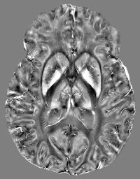 Julio Acosta-Cabronero合作论文数Department of Clinical Neuroscience, University of Cambridge27Young T. Hong合作论文数Addenbrooke's Hospital, University of Cambridge21
 Peter Jones合作论文数Wolfson College, University of Cambridge;Department of Psychiatry, School of Clinical Medicine, University of Cambridge20
 Peter J A Hutchinson合作论文数Department of Clinical Neurosciences, Robinson College, University of Cambridge18
 Ed Bullmore合作论文数Behavioural and Clinical Neuroscience Institute, University of Cambridge;Department of Psychiatry, University of Cambridge18
 Luca Passamonti合作论文数Lantheus18
 Marta Correia合作论文数MRC Cognition and Brain Sciences Unit, University of Cambridge17Joanne Outtrim合作论文数Wolfson Brain Imaging Centre, University of Cambridge17Judith Allanson合作论文数Department of Public Health and Primary Care, University of Cambridge16
 Catarina Rua合作论文数15W Richard Bevan-Jones合作论文数Dept Psychiat, Univ Cambridge14
 Craig Ritchie合作论文数Department of Psychiatry, Centre for Clinical Brain Sciences, University of Edinburgh14
 Adrian M. Owen合作论文数Centre for Brain and Mind, Western University;Institute for Neuroscience, Western University14Karen D. Ersche合作论文数Department of Psychiatry, University of Cambridge, Herchel Smith Building for Brain and Mind Sciences, Cambridge Biomedical Campus, Cambridge, CB2 0SZ UK14
 Ajenthan Surendranathan合作论文数13
 Paola Finoia合作论文数Department of Medicine, Division of Anaesthesia, University of Cambridge;Department of Clinical Neurosciences, University of Cambridge13
 Barbara Sahakian合作论文数Department of Psychiatry, University of Cambridge13
 George Pengas合作论文数13Manktelow Anne合作论文数12
 Lorina Naci合作论文数Brain & Mind Inst, Univ Western Ontario12Ritchie Karen合作论文数12
 Michael Firbank合作论文数Centre for In Vivo Imaging, Newcastle University12Doris A. Chatfield合作论文数12
 Emmanuel Stamatakis合作论文数School of Health Sciences, Faculty of Medicine and Health, The University of Sydney;Charles Perkins Centre, Faculty of Medicine and Health, The University of Sydney11
 Silvy Gabel合作论文数11Wells Katie合作论文数11
 John R Hodges合作论文数Brain and Mind Centre, The University of Sydney11Sally G. Harding合作论文数11
 Richard E Ansorge合作论文数University of Cambridge11
 Chris Rodgers合作论文数Department of Clinical Neurosciences, University of Cambridge11
 Andrea Luppi合作论文数Univ Cambridge, Addenbrookes Hosp, Sch Clin Med, Div Anaesthesia, Hills Rd, Cambridge CB2 0SP, England10
 Patricia Vazquez Rodriguez合作论文数Department of Clinical Neurosciences, University of Cambridge10
 Peter Smielewski合作论文数Division of Neurosurgery, University of Cambridge10
 Trevor W. Robbins合作论文数Department of Psychology, University of Cambridge;Behavioural and Clinical Neuroscience Institute, University of Cambridge;Institute of Science and Technology for Brain-inspired Intelligence, Fudan University10
 Zoe Kourtzi合作论文数Department of Psychology, University of Cambridge10Michael M. Craig合作论文数9
 Stefan Winzeck合作论文数Biomedical Image Analysis Group, Imperial College London9




