Polydimethylsiloxane (PDMS) is an elastomeric polymer frequently used as implant material, for flexible tubing and in microfluidic devices. The pronounced hydrophobic surface of this unique material impedes many applications where a good wetting behavior is required. Consequentially, various ways of surface modifications have been used to introduce new properties. Plasma treatment is the most popular technique in this respect, but is not generally applicable, especially if hardly accessible surfaces are to be modified. A novel wet-chemistry-based modification scheme yielding an amino-functionalized PDMS surface using a combined alcoholysis/aminolysis reaction is presented. Biological applications are exemplified by the conjugation of the RGD peptide, or polyethylene glycol (PEG) and heparin, yielding surfaces with cell-adhesive or nonthrombogenic properties, respectively. The effect of subsequent conjugation with an adhesive peptide is tested in cell culture. Additionally, two antifouling surfaces generated by coupling heparin and polyethylene glycol respectively are shown to improve the materials resistance to platelet adhesion drastically while simultaneously preventing hydrophobic recovery of the PDMS surface. The findings provide a versatile means of surface functionalization of PDMS substrates and is suitable for many biomedical applications.
Tissue engineering concepts, which are concerned with the attachment and growth of specific cell types, frequently employ immobilized ligands that interact preferentially with cell types of interest. Creating multicellular grafts such as heart valves calls for scaffolds with spatial control over the different cells involved. Cardiac heart valves are mainly constituted out of two cell types, endothelial cells and valvular interstitial cells. To have control over where which cell type can be attracted would enable targeted cell settlement and growth contributing to the first step of an engineered construct. For endothelial cells, constituting the outer lining of the valve tissue, several specific peptide ligands have been described. Valvular interstitial cells, representing the bulk of the leaflet, have not been investigated in this regard. Two receptors, the integrin α9β1 and CD44, are known to be highly expressed on valvular interstitial cells. Here, we demonstrate that by covalently grafting the corresponding peptide and polysaccharide ligand onto an erodible, polycaprolactone (PCL), and a non-degradable, polytetrafluoroethylene (PTFE), polymer, surfaces were generated that strongly support valvular interstitial cell colonization with minimal endothelial cell and reduced platelet adhesion. The technology for covalent binding of corresponding ligands is a key element towards tissue engineered cardiac valves for in vitro applications, but also towards future in vivo application, especially in combination with degradable scaffold material.
The surface modification of polyvinylidene difluoride (PVDF) for various biomedical uses is notoriously hampered by the chemical inertness of the polymer. A wet chemical approach aiming at covalently grafting biomolecules was demonstrated by means of an elimination reaction of fluorine from the polymer backbone followed by subsequent modification steps. Exemplified as a possible biological application, the coupling of the peptide REDV rendered the material adhesive for endothelial cells while adhesion of thrombocytes was dramatically reduced.
Mice have been used as accepted tools for investigating complex human diseases and new drug therapies because of their shared genetics and anatomical characteristics with humans. However, the tissues in mice are different from humans in that human cells have a natural mutation in the α1,3 galactosyltransferase (α1,3GT) gene and lack α-Gal epitopes on glycosylated proteins, whereas mice and other nonprimate mammals express this epitope. The lack of α-Gal epitopes in humans results in the loss of immune tolerance to this epitope and production of abundant natural anti-Gal Abs. These natural anti-Gal Abs can be used as an adjuvant to enhance processing of vaccine epitopes to APCs. However, wild-type mice and all existing humanized mouse models cannot be used to test the efficacy of vaccines expressing α-Gal epitopes because they express α-Gal epitopes and lack anti-Gal Abs. Therefore, in an effort to bridge the gap between the mouse models and humans, we developed a new humanized mouse model that mimics humans in that it lacks α-Gal epitopes and secretes human anti-Gal Abs. The new humanized mouse model (Hu-NSG/α-Galnull) is designed to be used for preclinical evaluations of viral and tumor vaccines based on α-Gal epitopes, human-specific immune responses, xenotransplantation studies, and in vivo biomaterials evaluation. To our knowledge, our new Hu-NSG/α-Galnull is the first available humanized mouse model with such features.
The chemical coupling of growth factors to solid substrates are discussed as an alternative to delivery systems. Utilizing entire proteins for this application is hampered by safety and stability considerations. Instead, growth factor mimicking peptides are of great interest for biomedical applications, such as tissue engineering, due to their purity and stability. The human cathelicidin derived antimicrobial peptide LL37, beside its microbicidal activity, was shown to stimulate endothelial cell growth when used in a soluble form. Here, in a novel approach, spacer mediated immobilization, but not direct conjugation of LL37, to a gold substrate was shown to result in a pronounced mitogenic effect on endothelial cells, comparable to that of soluble vascular endothelial growth factor.
The apelin peptide is described as one of the most potent inotropic agents, produced endogenously in a wide range of cells, including cardiomyocytes. Despite positive effects on cardiac contractility in multicellular preparations, as well as indications of cardio-protective actions in several diseases, its effects and mechanisms of action at the cellular level are incompletely understood. Here, we report apelin effects on dynamic mechanical characteristics of single ventricular cardiomyocytes, isolated from mouse models (control, apelin-deficient [Apelin-KO], apelin-receptor KO mouse [APJ-KO]), and rat. Dynamic changes in maximal velocity of cell shortening and relaxation were monitored. In addition, more traditional indicators of inotropic effects, such as maximum shortening (in mechanically unloaded cells) or peak force development (in auxotonic contracting cells, preloaded using the carbon fibre technique) were studied. The key finding is that, using Apelin-KO cardiomyocytes exposed to different preloads with the 2-dcarbon fibre technique, we observe a lowering of the slope of the end-diastolic stress-length relation in response to 10 nM apelin, an effect that is preload-dependent. This suggests a positive lusitropic effect of apelin, which could explain earlier counter-intuitive findings on an apelin-induced increase in contractility occurring without matching rise in oxygen consumption. (C) 2017 The Authors. Published by Elsevier Ltd.
BACKGROUND: Survival after sudden cardiac arrest is limited by postarrest myocardial dysfunction, but understanding of this phenomenon is constrained by a lack of data from a physiological model of disease. In this study, we established an in vivo model of cardiac arrest and resuscitation, characterized the biology of the associated myocardial dysfunction, and tested novel therapeutic strategies.METHODS: We developed rodent models of in vivo postarrest myocardial dysfunction using extracorporeal membrane oxygenation resuscitation followed by invasive hemodynamics measurement. In postarrest isolated cardiomyocytes, we assessed mechanical load and Ca2+-induced Ca2+ release (CICR) simultaneously using the microcarbon fiber technique and observed reduced function and myofilament calcium sensitivity. We used a novel fiberoptic catheter imaging system and a genetically encoded calcium sensor, GCaMP6f, to image CICR in vivo.RESULTS: We found potentiation of CICR in isolated cells from this extracorporeal membrane oxygenation model and in cells isolated from an ischemia/reperfusion Langendorff model perfused with oxygenated blood from an arrested animal but not when reperfused in saline. We established that CICR potentiation begins in vivo. The augmented CICR observed after arrest was mediated by the activation of Ca2+/calmodulin-dependent protein kinase II (CaMKII). Increased phosphorylation of CaMKII, phospholamban, and ryanodine receptor 2 was detected in the postarrest period. Exogenous adrenergic activation in vivo recapitulated Ca2+ potentiation but was associated with lesser CaMKII activation. Because oxidative stress and aldehydic adduct formation were high after arrest, we tested a small-molecule activator of aldehyde dehydrogenase type 2, Alda-1, which reduced oxidative stress, restored calcium and CaMKII homeostasis, and improved cardiac function and postarrest outcome in vivo.CONCLUSIONS: Cardiac arrest and reperfusion lead to CaMKII activation and calcium long-term potentiation, which support cardiomyocyte contractility in the face of impaired postarrest myofilament calcium sensitivity. Alda-1 mitigates these effects, normalizes calcium cycling, and improves outcome.
The effects of temperature on cardiac cells have been previously explored. It is well characterised that on single cell level the action potential measured at room temperature has longer duration compared with that obtained at body temperature. The duration of the AP (APD) is a parameter which is related to the ability of cells to cope with faster pacing rates. APD prolongation by drug or disease condition may lead to arrhythmia. However so far, the comparison has been done mostly on data obtained at body and room temperature. One common explanation for the prolongation of the APD at room temperature is the slower kinetics of underlying ion channels, exchangers and pumps (please check for pump). We measured AP from live cardiac ventricular slices after incubating in ice cold solution for over 60min. The AP from rabbit cardiac tissue slices, were measured shortly after transfer into body temperature solution. These AP exhibit a very short APD (e.g. 89.6ms APD, 6min after warming up). Only after incubation in body temperature of about 1h or more, the AP shape and duration are back to the values under control conditions (205+/-21ms, at 2Hz pacing). The mechanisms for the changes in AP are still not clear however an impaired ion gradient control and subsequently increased intracellular sodium concentration could be responsible for these changes. The temperature effects of ice cold solution are of different origin compared to the reduction to room temperature settings. This could be one of the main reasons why implanted hearts can adapt to the new environment without exhibiting prolonged AP, once coming from cold incubation. However, further investigations are needed to explain the effects in total.
Heart failure is a growing endemic in the aging Western population with a prevalence of over 20 million people worldwide1. Existing heart failure therapies are unable to reverse heart failure and do not address its fundamental cause, the loss of cardiomyocytes2. In order to induce myocardial regeneration for the myocardium and the heart valve, facilitate self-repair, improve tissue salvage, reduce or reverse the adverse-remodeling and ultimately achieve long-term functional stabilization and improvement in the heart function, novel strategies for therapeutic regeneration are being developed which are aiming to compensate for the insufficient and low intrinsic regenerative ability of the adult heart3. Similarly, valve replacement with mechanical or biological substitutes meets numerous hurdles. New approaches using multicellular approaches and new material are extensively studied. Most of those strategies depend on biomaterials that help to achieve functional integrated vasculogenesis and myogenesis in the heart/tissue. Especially for failed heart valve function a number of therapeutic approaches are common from corrective intervention to complete replacement4. However the complexity of the heart valve tissue and its high physical exposure has led to a variety of approaches, however therapeutic regeneration needs to be established. Beside other approaches alginate has been identified as one building block to achieve therapeutic regeneration. Alginate is a versatile and adaptable biomaterial that has found numerous biomedical applications which include wound healing, drug delivery and tissue engineering. Due to its biologically favorable properties including the ease of gelation and its biocompatibility, alginate-based hydrogels have been considered a particularly attractive material for the application in cardiac regeneration and valve replacement techniques. Here, we review current applications of alginate in cardiac regeneration as well as perspectives for the alginate-dependent, cardiac regeneration strategies.
Purpose(i) To optimize an MR‐compatible organ perfusion setup for the nondestructive investigation of isolated rat hearts by placing the radiofrequency (RF) coil inside the perfusion chamber; (ii) to characterize the benefit of this system for diffusion tensor imaging and proton (1H‐) MR spectroscopy.MethodsCoil quality assessment was conducted both on the bench, and in the magnet. The benefit of the new RF‐coil was quantified by measuring signal‐to‐noise ratio (SNR), accuracy, and precision of diffusion tensor imaging/error in metabolite amplitude estimation, and compared to an RF‐coil placed externally to the perfusion chamber.ResultsThe new design provided a 59% gain in signal‐to‐noise ratio on a fixed rat heart compared to using an external resonator, which found reflection in an improvement of living heart data quality, compared to previous external resonator studies. This resulted in 14–29% improvement in accuracy and precision of diffusion tensor imaging. The Cramer–Rao lower bounds for metabolite amplitude estimations were up to 5‐fold smaller.ConclusionOptimization of MR‐compatible perfusion equipment advances the study of rat hearts with improved signal‐to‐noise ratio performance, and thus improved accuracy/precision. Magn Reson Med 73:2398–2405, 2015. © 2014 The Authors. Magnetic Resonance in Medicine Published by Wiley Periodicals, Inc. on behalf of International Society of Medicine in Resonance.
Introduction: Cardiac tissue slices, a pseudo two-dimensional organo-typic experimental model, are an increasingly popular model to study cardiac biophysics in-vitro. This preparation benefits from moderate complexity, native cell-type presence, and locally preserved cell-cell connections. We used optical mapping of tissue slices to monitor transmembrane potential (Vm) and intracellular Ca2+ concentration to study the effects of stretch on action potential duration (APD) and calcium transient (CaT) dynamics with a relatively high spatio-temporal resolution, to explore mechanisms of stretch-induced arrhythmias. Methods: Langendorff perfused rabbit hearts were loaded with dyes for Vm (di-4-ANBDQPQ) and CaT (Rhod-2-AM). Thereafter, 350 µm tissue slices were vibratome-cut from left and right ventricular free wall in an epicardium-tangential plane. Slices were attached at their ends to a manual stretcher and optically mapped using an EMCCD camera with LED excitation light sources. Slices were field stimulated at 2Hz, and Vm and CaT were measured before, during (up to 50min) and after application of stretch. Results: Cardiac tissue slices (n=9), exposed to stretch (by 4-12%) showed an initial shortening in both APD and CaT duration (APD80 and APD50 reduced by 10.7% and 11.4%, respectively; CaT80 and CaT50 reduced by 6% and 5.3%, respectively). During maintained stretch, a gradual re-lengthening of APD and CaT duration was observed. After release of stretch, APD and CaT duration reverted to shorter values. Conclusion: Living cardiac tissue slices offer a promising experimental model for the study of cardiac mechano-electric coupling. The methodology described can be refined (e.g. using a computer-controlled motorised stage to synchronise electrical and mechanical events, and by use of fiducial markers to track local tissue deformation rather than only input strain levels) and extended (e.g. exploring effects of stretch directionality, relative to prevailing cell orientation in a slice).
Cardiac tissue slices are becoming increasingly popular as a model system for cardiac electrophysiology and pharmacology research and development. Here, we describe in detail the preparation, handling, and optical mapping of transmembrane potential and intracellular free calcium concentration transients (CaT) in ventricular tissue slices from guinea pigs and rabbits. Slices cut in the epicardium-tangential plane contained well-aligned in-slice myocardial cell strands (“fibers”) in subepicardial and midmyocardial sections. Cut with a high-precision slow-advancing microtome at a thickness of 350 to 400 μm, tissue slices preserved essential action potential (AP) properties of the precutting Langendorff-perfused heart. We identified the need for a postcutting recovery period of 36 min (guinea pig) and 63 min (rabbit) to reach 97.5% of final steady-state values for AP duration (APD) (identified by exponential fitting). There was no significant difference between the postcutting recovery dynamics in slices obtained using 2,3-butanedione 2-monoxime or blebistatin as electromechanical uncouplers during the cutting process. A rapid increase in APD, seen after cutting, was caused by exposure to ice-cold solution during the slicing procedure, not by tissue injury, differences in uncouplers, or pH-buffers (bicarbonate; HEPES). To characterize intrinsic patterns of CaT, AP, and conduction, a combination of multipoint and field stimulation should be used to avoid misinterpretation based on source-sink effects. In summary, we describe in detail the preparation, mapping, and data analysis approaches for reproducible cardiac tissue slice-based investigations into AP and CaT dynamics.
The objective of this article is to present an in vitro model of atrial cardiac tissue that could serve to study the mechanisms of remodeling related to atrial fibrillation (AF). We analyze the modification on gene expression and modifications on rotor dynamics following tissue remodeling. Atrial murine cells (HL-1 myocytes) were maintained in culture after the spontaneous initiation of AF and analyzed at two time points: 3.1 ± 1.3 and 9.7 ± 0.5 days after AF initiation. The degree of electrophysiological remodeling (i.e., relative gene expression of key ion channels) and structural inhomogeneity was compared between early and late cell culture times both in nonfibrillating and fibrillating cell cultures. In addition, the electrophysiological characteristics of in vitro fibrillation [e.g., density of phase singularities (PS/cm(2)), dominant frequency, and rotor meandering] analyzed by means of optical mapping were compared with the degree of electrophysiological remodeling. Fibrillating cell cultures showed a differential ion channel gene expression associated with atrial tissue remodeling (i.e., decreased SCN5A, CACN1C, KCND3, and GJA1 and increased KCNJ2) not present in nonfibrillating cell cultures. Also, fibrillatory complexity was increased in late- vs. early stage cultures (1.12 ± 0.14 vs. 0.43 ± 0.19 PS/cm(2), P < 0.01), which was associated with changes in the electrical reentrant patterns (i.e., decrease in rotor tip meandering and increase in wavefront curvature). HL-1 cells can reproduce AF features such as electrophysiological remodeling and an increased complexity of the electrophysiological behavior associated with the fibrillation time that resembles those occurring in patients with chronic AF.
The heart is subject to multiple sources of stress. To maintain its normal function, and successfully overcome these stresses, heart muscle is equipped with fine-tuned regulatory mechanisms. Some of these mechanisms are inherent within the myocardium itself and are known as intrinsic mechanisms. Over a century ago, Otto Frank and Ernest Starling described an intrinsic mechanism by which the heart, even ex vivo, regulates its function on a beat-to-beat basis. According to this phenomenon, the higher the ventricular filling is, the bigger the stroke volume. Thus, the Frank-Starling law establishes a direct relationship between the diastolic and systolic function of the heart. To observe this biophysical phenomenon and to investigate it, technologic development has been a pre-requisite to scientific knowledge. It allowed for example to observe, at the cellular level, a Frank-Starling like mechanism and has been termed: Length Dependent Activation (LDA). In this review, we summarize some experimental systems that have been developed and are currently still in use to investigate cardiac biophysical properties from the whole heart down to the single myofibril. As a scientific support, investigation of the Frank-Starling mechanism will be used as a case study.
Background: The purpose of this study was to report the prevalence and the significance of clinically recognized chronic obstructive pulmonary disease (COPD) or bronchial asthma among patients hospitalized with acute heart failure (AHF). Methods: Data were derived from a prospective, multicenter, multinational study of 5005 patients hospitalized with AHF from February 2013 to November 2012. Data were analyzed according to the presence or absence of COPD or bronchial asthma. Demographic, management, in-hospital and 1-year outcomes were compared. Results: The prevalence of COPD or bronchial asthma was 10%. When compared to non-COPD patients, COPD patients were older and more likely to be female, have diabetes, hypertension, dyslipidemia, chronic kidney disease and sleep apnea requiring therapy; they were less likely to receive monitoring and were at higher risk of developing systemic infections that require antibiotics. And were more likely to be prescribed ivabrdine. There were no significant differences between the two groups in regards β-blockers use. COPD patients were more likely to undergo coronary revascularization and more likely to receive non-invasive ventilator support. Approximately 14.6% of COPD patients presented with atrial fibrillation and they too had higher left ventricular ejection fraction (LVEF) when compared to non-COPD patients. Conclusion: A large proportion of AHF patients presented with concomitant COPD, had different clinical characteristics/co-morbidities than those with non-COPD, COPD patients had longer hospital stay and a higher one year mortality than non-COPD patients
Living cardiac tissue slices, a pseudo two-dimensional (2D) preparation, have received less attention than isolated single cells, cell cultures, or Langendorff-perfused hearts in cardiac biophysics research. This is, in part, due to difficulties associated with sectioning cardiac tissue to obtain live slices. With moderate complexity, native cell-types, and well-preserved cell-cell electrical and mechanical interconnections, cardiac tissue slices have several advantages for studying cardiac electrophysiology. The trans-membrane potential (Vm) has, thus far, mainly been explored using multi-electrode arrays. Here, we combine tissue slices with optical mapping to monitor Vm and intracellular Ca(2+) concentration ([Ca(2+)]i). This combination opens up the possibility of studying the effects of experimental interventions upon action potential (AP) and calcium transient (CaT) dynamics in 2D, and with relatively high spatio-temporal resolution. As an intervention, we conducted proof-of-principle application of stretch. Mechanical stimulation of cardiac preparations is well-established for membrane patches, single cells and whole heart preparations. For cardiac tissue slices, it is possible to apply stretch perpendicular or parallel to the dominant orientation of cells, while keeping the preparation in a constant focal plane for fluorescent imaging of in-slice functional dynamics. Slice-to-slice comparison furthermore allows one to assess transmural differences in ventricular tissue responses to mechanical challenges. We developed and tested application of axial stretch to cardiac tissue slices, using a manually-controlled stretching device, and recorded Vm and [Ca(2+)]i by optical mapping before, during, and after application of stretch. Living cardiac tissue slices, exposed to axial stretch, show an initial shortening in both AP and CaT duration upon stretch application, followed in most cases by a gradual prolongation of AP and CaT duration during stretch maintained for up to 50 min. After release of sustained stretch, AP duration (APD) and CaT duration reverted to shorter values. Living cardiac tissue slices are a promising experimental model for the study of cardiac mechano-electric interactions. The methodology described here can be refined to achieve more accurate control over stretch amplitude and timing (e.g. using a computer-controlled motorised stage, or by synchronising electrical and mechanical events) and through monitoring of regional tissue deformation (e.g. by adding motion tracking).
Introduction: The role of tissue remodeling in the reentrant activity during atrial fibrillation (AF) is not well understood. The aim of this study is to evaluate in an in-vitro model of AF the role of tissue remodeling in the mechanisms of perpetuation of this arrhythmia.
 Peter Kohl合作论文数43
 Rebecca A B Burton合作论文数Medical Sciences Division, Department of Pharmacology, University of Oxford16
 Jürgen E. Schneider合作论文数Dept Cardiovasc Med, Univ Oxford13
 Patrick W. Hales合作论文数Department of Cardiovascular Medicine, University of Oxford13
 Peter D Lee合作论文数Department of Mechanical Engineering, Faculty of Engineering Science, University College London12
 David Gavaghan合作论文数Department of Computer Science, University of Oxford;New College, University of Oxford11Klaus Benndorf合作论文数11
 Magdi Yacoub合作论文数National Heart & Lung Institute, Faculty of Medicine, Imperial College London;Aswan Heart Centre10
 Vicente Grau Colomer合作论文数Institute of Biomedical Engineering, Department of Engineering Science, University of Oxford;Mansfield College, University of Oxford9
 Ken Wang合作论文数8T Zimmer合作论文数8Gil Bub合作论文数7
 Leslie M. Loew合作论文数UConn Health;Department of Cell Biology, University of Connecticut7
 T Alexander Quinn合作论文数Columbia University7
 Jassim Al Suwaidi合作论文数6
 Alan Garny合作论文数Department of Physiology, Anatomy and Genetics, University of Oxford6
 Patrizia Camelliti合作论文数University Laboratory of Physiology, Oxford, OX1 3PT, UK6Haitham Amin合作论文数Mohammed Bin Khalifa Cardiac Ctr, Manama, Bahrain5Derek A Terrar合作论文数5
 Wael Al Mahmeed合作论文数Heart, Vascular & Thoracic Institute, Cleveland Clinic Abu Dhabi5
 Nidal Asaad合作论文数5
 Blanca Rodriguez合作论文数Department of Computer Science, University of Oxford5
 Alawi Alsheikh-Ali合作论文数College of Medicine, Mohammed Bin Rashid University of Medicine and Health Sciences5
 Euan A. Ashley合作论文数Department of Biomedical Data Science, School of Medicine, Stanford University;Department of Medicine, Stanford University5
 Andreu M. Climent合作论文数Universidad Politecnica de Valencia5
 Paul Ewart合作论文数Department of Physics, Oxford University/Worcester College/Faraday Institute for Science and Religion5Martin Bishop合作论文数Oxford5Christopher E Woods合作论文数4
 Christophe Michel Raynaud合作论文数4Matthias Gabriel合作论文数4
 María S. Guillem合作论文数COR Group - ITACA Institute, Universitat Politècnica de València4
 Pedro L. Sánchez合作论文数Centro de Investigación Biomédica en Red Enfermedades Cardiovasculares (CIBERCV), Spain;Servicio de Cardiología, Hospital Clínico Universitario de Salamanca;Centro de Investigación Biomédica en Red Enfermedades Cardiovasculares (CIBERCV),, Hospital Clínico Universitario de Salamanca4Ping Yan合作论文数Potentiometr Probes LLC4
 Felipe Alonso-Atienza合作论文数Client Solut Adv Analyt, BBVA4Bazargani Nooshin合作论文数4Bulbanat Bassam合作论文数4
 Fleur E Mason合作论文数4
 Abdelfatah Elasfar合作论文数Prince Salman Heart Center, Tanta University4
 Mohammad Al-Jarallah合作论文数4
 María Eugenia Fernández Santos合作论文数Instituto de Investigación Sanitaria Gregorio Marañón4Al Faleh Husam合作论文数4
 Francisco Fernández - Avilés合作论文数Centro de Investigación Biomedica en Red en Enfermedades Cardiovasculares;Cardiology Department, Hospital General Universitario Gregorio Marañon;Centro Cardiovascular Madrid4
 Christoph Biskup合作论文数4Mustafa Ridha合作论文数4
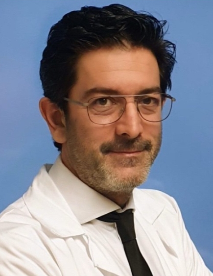 Ricardo Sanz-Ruiz合作论文数Servicio de Cardiología, Hospital General Universitario Gregorio Marañón;University of Valladolid4
 Ahmed Al-Motarreb合作论文数Faculty of Medicine, Sana'a University4Kadhim Sulaiman合作论文数4
 Prashanth Panduranga合作论文数NMC Hospital Ghubra Muscat Oman4
 Khalid F. AlHabib合作论文数Faculty, Cardiac Sciences, College of Medicine, King Saud University4




