BACKGROUND AND AIM:Randomized trials conducted in the early 2000s established the survival benefit of primary prevention implantable cardioverter-defibrillator (ICD) therapy in patients with reduced left ventricular ejection fraction (LVEF) after myocardial infarction. However, management of myocardial infarction and heart failure has substantially evolved since that time. We investigated whether the estimated association between primary prevention ICD implantation in post-myocardial infarction patients with reduced LVEF and mortality reduction has changed over time. METHODS:We analyzed individual participant data from 32,214 patients with LVEF ≤35% after myocardial infarction included in the PROFID pooled cohort, comprising 7,477 patients carrying a primary prevention ICD (ICD patients) and 24,737 patients without an ICD (non-ICD patients). The primary endpoint was all-cause mortality. Propensity scores were estimated using multivariable logistic regression including age, sex, LVEF, renal function, and diabetes, and overlap weighting was applied to balance treatment groups. Time period-specific analyses were performed across three prespecified time periods defined by inclusion year: 1995-2004, 2005-2014, and 2015-2020. Weighted cumulative mortality curves were generated for each time period. Temporal changes in the estimated association between ICD implantation and mortality reduction were assessed using a weighted Cox proportional hazards model. RESULTS:A total of 12,097 deaths occurred during a mean follow-up of 43.7 months. The estimated association between ICD implantation and mortality changed significantly across time (P for interaction <0.001). In weighted time period-specific analyses, the estimated mortality reduction associated with ICD implantation progressively decreased over more recent periods. The hazard ratio for ICD versus non-ICD patients was 0.54 (95% CI 0.47-0.62; P<0.001) in 1995-2004, 0.67 (95% CI 0.62-0.72; P<0.001) in 2005-2014, and 0.89 (95% CI 0.73-1.07; P=0.221) in 2015-2020, with negligible separation of the weighted cumulative mortality curves in the most recent time period. CONCLUSIONS:In this analysis including a large cohort of post-myocardial infarction patients with reduced LVEF, the estimated mortality reduction associated with primary prevention ICD implantation progressively decreased over time.
Background:Pulsed field ablation (PFA) using a pentaspline catheter is increasingly applied for pulmonary vein (PV) isolation, but standardized methods to assess durable lesion formation are lacking. Objective:We aimed to investigate the impact of incremental PFA applications on electrogram (EGM) characteristics and determine whether EGM signatures serve as markers for effective lesions. Methods:PV EGMs of a cohort of consecutive patients were analyzed before and after single, double, and quadruple PFA applications. Bipolar voltage amplitude (BVA) and signal power in different frequency bands (>50 Hz, >75 Hz, and >125 Hz [HF125]) were quantified. EGMs were reassessed after 60 seconds, and pacing was performed to evaluate PV pace capture. Results:This analysis included 4230 EGMs of 83 patients. Immediately after ablation, no differences in BVA and frequency power were observed between groups. After 60 seconds, significant differences were present for all features in the single, double, and quadruple groups: BVA (0.27 mV vs 0.26 mV vs 0.13 mV), >50 Hz (39 μV2 vs 35 μV2 vs 8 μV2), >75 Hz (10 μV2 vs 13 μV2 vs 3 μV2), and HF125 (3 μV2 vs 2 μV2 vs 1 μV2). EGM features recovered within 60 seconds in the single and double groups but not in the quadruple group. Pace capture at 5 V was best predicted by an HF125 threshold of 2.5 μV2 and a BVA threshold of 0.28 mV. Conclusion:Only quadruple PFA applications ensured persistent lesion effects over the observation period. A combined end point using an HF125 cutoff of 2.5 μV2 and BVA cutoff of 0.28 mV may provide accurate confirmation of PV isolation with the pentaspline catheter.
Aims:Pacing-induced cardiomyopathy (PICM) is a recognized complication of pace-and-ablate therapy. Early identification of patients at risk may allow tailored strategies, including upfront biventricular or conduction system pacing. Purpose:This study aimed to identify predictors of PICM in patients undergoing pace-and-ablate therapy, combining expert consensus with clinical validation. Methods and results:A three-round Delphi process involving 12 electrophysiologists prioritized candidate predictors of PICM. Consecutive patients undergoing atrioventricular node ablation with non-cardiac resynchronization therapy (CRT) devices at Liverpool Heart and Chest Hospital (2015-2019) were retrospectively analysed. The primary endpoint was PICM, defined as ≥10% reduction in left ventricular ejection fraction (LVEF) to <50%, adjudicated by two independent cardiologists. Cox regression was used to identify independent predictors. The Delphi process prioritized baseline LVEF, intrinsic and paced QRS duration, RV lead position, and key comorbidities. Among 658 ablation patients, 323 (median age 76 years, 67% female) had non-CRT devices. Over a median follow-up of 192 days (IQR, 63-1003 days), 33 patients (9.8%) developed PICM. In multivariable analysis, RV free wall vs. septal lead position [hazard ratio (HR) 4.896, 95% confidence interval (CI) 1.263-18.973, P = 0.022], intrinsic QRS duration (HR 1.247 per 10 ms, 95% CI 1.064-1.462, P = 0.006), independently predicted PICM. Exploratory analyses suggested that higher intrinsic QRS duration was associated with increased PICM risk; however, these findings were based on a small number of events and should be interpreted with caution. Conclusion:Intrinsic QRS duration and free wall RV lead position independently predict PICM in pace-and-ablate patients. These findings support early risk stratification and may guide personalized pacing strategies.
BACKGROUND AND AIMS:Stroke and systemic embolism are recognized but poorly characterized complications of atrial fibrillation (AF) and left atrial flutter (AFL) catheter ablation. Their incidence, outcomes, and procedural associations remain insufficiently defined. METHODS:Retrospective global registry was initiated by the European Heart Rhythm Association Scientific Committee. A secure database captured data from ablation centres worldwide on general AF and left AFL ablation activity, as well as detailed information on patients with symptomatic embolic events between 2017 and 2024. RESULTS:Overall, 204 centres reported 335 743 ablation procedures and 550 embolic events (incidence 0.16%). Centre-level rate ranged from 0% to 3.1%. Most events were cerebral (94%), followed by peripheral (5%), and combined (1%). Coronary air embolism accounted for 62% of all peripheral events. Intraprocedural events occurred in 93 patients (17%), within 72 h in 357 (78%) and beyond 72 h in 100 (22%); 128 cases (23%) were diagnosed after hospital discharge. Nonparoxysmal AF (0.23% vs 0.15%; P < .001), centre experience, and ablation energy source (radiofrequency, 0.16%; cryoballoon, 0.16%; pulsed field ablation, 0.25%; laserballoon, 0.88%; P < .001) were associated with embolic risk. Median NIHSS score after stroke was 4 (interquartile range, 2-9). Three-month follow-up was available for 518 patients with embolism (95%): 181 (35%) had sequelae and 16 (3%) died. CONCLUSIONS:Symptomatic embolic events are infrequent but associated with significant morbidity and mortality. Most events occur early, though a substantial proportion arise later. Higher event rates were observed with laser and possibly with pulsed field ablation. A greater centre experience was associated with a lower embolism incidence.
Background Pulmonary vein isolation is a cornerstone in the treatment of atrial fibrillation, with procedural success traditionally assessed after a 3-month “blanking period” to account for early postablation arrhythmias. However, emerging evidence challenges this paradigm, particularly with nonthermal energy modalities such as pulsed-field ablation (PFA), which induce minimal inflammatory response. Objective The purpose of this study was to assess the prognostic relevance of early recurrence (ER) of atrial arrhythmia during the blanking period after PFA-based pulmonary vein isolation, with a focus on the timing of ER and its association with late recurrence (LR). Methods In this single-center study, 241 patients with atrial fibrillation underwent first-time PFA and were followed for ≥12 months. ER was defined as any atrial arrhythmia lasting >30 seconds within the first 3 months of ablation. The primary outcome was freedom from LR beyond the blanking period. Results ER occurred in 48 (20%) of patients, with 23 (48%) occurring within the first month and 25 (52%) during the second or third month of ablation. LR was significantly more frequent in patients with ER (P < .001), particularly when recurrence occurred after day 30. Multivariable Cox analysis identified ER during the second or third month (hazard ratio 6.1; 95% confidence interval 3.3–11.3) and ER during the first month (hazard ratio 3.6; 95% confidence interval 1.8–7.1) as the strongest independent predictors of LR. A subset of ERs resolved spontaneously. Conclusion The timing of ER after PFA strongly predicts LR, supporting reconsideration of the 90-day blanking period. It may be appropriate to shorten the blanking period for PFA, but late ER warrants clinical attention. Larger studies should validate timing-based blanking strategies.
INTRODUCTION:Catheter ablation of premature ventricular complexes (PVCs) and ventricular tachycardia (VT) arising from the aortic cusps and commissures is effective but performed near the coronary ostia, raising concern for coronary injury. Adjunct imaging with intracardiac echocardiography (ICE) and coronary angiography is variably used, and its incremental benefit remains uncertain. METHODS:We analyzed 311 ablation procedures performed in 290 patients across four high-volume centers in the United States and Switzerland. Imaging strategies were categorized as no adjunct imaging, ICE only, angiography only, or combined ICE plus angiography. RESULTS:Marked inter-center variability in imaging use was observed. Procedure duration and fluoroscopy exposure differed significantly by imaging strategy and were greatest when both ICE and angiography were used (p < 0.001). Acute elimination of targeted PVCs occurred in 87% of procedures. No coronary artery injury was observed. CONCLUSION:Adjunct imaging strategies for aortic cusp and commissure PVC/VT ablation vary widely across centers and substantially influence procedural efficiency. Despite frequent ablation near the coronary ostia, no coronary injury events were observed.
AIMS:The SINGLE SHOT CHAMPION multicentre trial compared the effectiveness of pulsed-field ablation (PFA) and cryoballoon ablation (CBA) for pulmonary-vein isolation (PVI) in patients with paroxysmal atrial fibrillation (AF). In a prespecified substudy, post-ablation three-dimensional electroanatomic mapping (3D-EAM) was performed, and repeat-procedure mapping data were analysed. METHODS AND RESULTS:Patients were randomized 1:1 to fluoroscopy-guided PVI with PFA or CBA. The first 25 patients in each group underwent high-density 3D-EAM immediately after ablation. Acute PV isolation and lesion geometry were assessed using bipolar voltage thresholds of 0.1, 0.2, and 0.5 mV. Lesion durability was evaluated at clinically indicated repeat ablation. In acute post-ablation mapping, residual PV conduction was seen in 4/25 (16%) CBA patients and 0/25 PFA patients (P = 0.11). Inadvertent posterior wall conduction block occurred after PFA in 3 (12%) patients with small atria (mean LA volume 30.7 mL). PFA created larger left-sided lesions (antral area <0.5 mV: 5.7 mm2 [IQR 4.6-6.5] vs. 4.4 mm2 [IQR 2.5-5.5], P = 0.026) and narrower posterior wall channels (14.0 mm [IQR 10.5-20.6] vs. 24.0 mm [IQR 19.1-26.9], P < 0.001), indicating wide-antral isolation. At redo (n = 48), durable PVI was seen in 4/26 (15%) PFA and 3/22 (14%) CBA patients and in 63/102 (62%) veins after PFA and 52/82 (63%) after CBA (P = 0.88). CONCLUSION:Fluoroscopy- and electrocardiogram-guided PVI can result in incomplete PV isolation after CBA or unintentional posterior wall block after PFA. In the acute mapping cohort, PFA yielded broader left-sided lesions than CBA; in a separate redo cohort, chronic PV durability was similar for both modalities.
Background and aimsThe revised CHA2DS2-VA score facilitates stroke risk assessment in patients with atrial fibrillation. This study investigates the value of the CHA2DS2-VA for prediction of all-cause and cardiovascular mortality and ventricular arrhythmia (VA) in patients with ischemic heart disease (IHD) and dilated cardiomyopathy (DCM) undergoing implantable cardioverter-defibrillator (ICD) therapy.MethodsWe analyzed data from 1,626 ICD recipients with IHD (n = 1,225) or DCM (n = 401) enrolled in a prospective registry at University Hospital Basel. Patients were stratified into low and high CHA2DS2-VA groups based on median score within each condition. Mortality and VA outcomes were assessed using Kaplan–Meier survival analysis and Cox proportional hazards models.ResultsThe median CHA2DS2-VA was 3 overall, 2 in DCM, and 4 in IHD patients. During a median follow-up of 7.5 years, 46% died, with an all-cause mortality rate of 6.1 per 100 patient-years. Higher CHA2DS2-VA scores were associated with increased all-cause and cardiovascular mortality in both IHD (HR 2.36, 95% CI 1.99–2.80, p < 0.001 for all-cause death) and DCM (HR 1.98, 95% CI 1.44–2.73, p < 0.001 for all-cause death). Each one-point increase in CHA2DS2-VA increased mortality risk (HR 1.4, 95% CI 1.34–1.47, p < 0.001 for all-cause death). No association was observed between CHA2DS2-VA and VA occurrence in the overall cohort or subgroups.ConclusionThe CHA2DS2-VA is strongly associated with all-cause and cardiovascular mortality in ICD patients with IHD and DCM, but does not predict VA. Thus, it may aid in mortality risk stratification but should not be used as a primary tool for arrhythmic risk assessment.
The interrelationships between atrial fibrillation (AF), brain lesions and cognitive function are poorly understood. We aimed to investigate the relationship of AF with brain lesions and cognition. We enrolled 1,480 patients with and 959 without AF in a multicenter prospective study (Swiss-AF; NCT02105844). We assessed brain structure, and cognition using the Montreal Cognitive Assessment (MoCA). Brain magnetic resonance imaging (MRI) was performed to assess large non-cortical and cortical infarcts (LNCCI), small non-cortical infarcts (SNCI), white matter hyperintensities (WMH), and microbleeds. Using causal mediation analyses, we investigated the direct (lesion-independent) and indirect (lesion-mediated) effects of AF on cognition. Mean age in AF patients is 75.0 vs. 74.2 years in no-AF patients, 28.6
Background:Recent studies indicate that atrial fibrillation (AF) burden has prognostic implications. Objective:We aimed to assess the ability of clinical and cardiac imaging variables to stratify between high and low AF burden. Method:Data from the prospective, multicenter Swiss-AF Burden study were analyzed. Patients underwent a 7-day Holter electrocardiogram and native cardiac magnetic resonance imaging. AF burden, defined as the percentage of time in AF during the 7-day Holter electrocardiogram, was dichotomized into low (<10%) or high (≥10%). Logistic regression models were built, and discriminative performance was evaluated by comparing the area under the curve (AUC). Results:A total of 170 patients were enrolled (median age 72 years; 18% female); 26% (n = 44) had high AF burden. Variables selected for the clinical model were age (odds ratio 1.09; 95% confidence interval 0.40-2.74), male sex (1.05; 1.00-1.11), and body mass index (1.14; 1.06-1.24). The imaging model included left atrial maximal volume index (1.04; 1.01-1.06), left ventricular end-diastolic volume index (0.93; 0.90-0.96), right atrial fractional area change (0.94; 0.89-0.98), and left ventricular ejection fraction (0.87; 0.81-0.94). The AUCs for the clinical and imaging models were 0.67 (0.58-0.77) and 0.91 (0.84-0.98), respectively. Combining both models yielded an AUC of 0.92 (0.86-0.99), with no substantial improvement over the imaging model alone. Conclusion:Cardiac imaging variables clearly outperformed clinical variables in their ability to stratify between high and low AF burden, suggesting their potential as a tool for estimating AF burden.
BACKGROUND:Ventricular arrhythmias from the aortic cusps (ACs) represent a subset of left ventricular outflow tract arrhythmias. Although ablation in this region can be highly effective, concerns persist regarding aortic valve injury and iatrogenic aortic regurgitation (AR). Contemporary data on AR incidence after AC mapping/ablation remain limited. OBJECTIVES:This study sought to determine the incidence, severity, and clinical significance of AR in a multicenter cohort after mapping and/or radiofrequency ablation in the ACs or commissures. METHODS:We retrospectively analyzed patients at 4 high-volume centers (2015-2024) who underwent AC/commissural mapping and/or ablation for ventricular arrhythmia and had paired preprocedural and postprocedural transthoracic echocardiograms (TTEs). AR was graded by using standardized criteria; procedure-related AR was defined as ≥2-grade worsening or new ≥moderate AR. RESULTS:Among 290 patients, 208 (218 procedures) had complete paired TTEs. Baseline AR was absent in 81% and mild in 18%. Retrograde aortic access was used in 94%, and 77% underwent radiofrequency ablation within the cusps. Clinically significant AR (≥2-grade worsening or new moderate AR) occurred in 2 procedures (0.9%), both representing progression from preexisting mild-to-moderate AR, with one of the patients having a bicuspid valve. New mild AR (1-grade increase from none to mild) occurred in 26 procedures (11.9%). Among 14 with additional follow-up imaging (median 320 days after first postprocedural TTE), AR resolved in 8 (57%) and remained mild in 6 (43%). No patient required valve intervention. CONCLUSIONS:Significant AR occurred in approximately 1% after AC mapping/ablation. New mild AR developed in 12.5% and improved in more than one-half during follow-up. No valve interventions were required, supporting the safety of cusp and commissural ablation at experienced centers.
 Michael Kuhne合作论文数University Heart Center, University Hospital Basel;Cardiovascular Research Institute Basel, University Hospital Basel336
 Stefan Osswald合作论文数Univ Basel, Univ Basel Hosp288
 Sven Knecht合作论文数227
 Beat Andreas Schaer合作论文数University Hospital of Basel176Patrick Badertscher合作论文数161
 Tobias Reichlin合作论文数Department of Cardiology, Bern University Hospital;Herz Gefäss Zentrum, Universitätsklinik für Kardiologie, Universitätsspital Bern, Inselspital157
 Philipp Krisai合作论文数131David Conen合作论文数Department of Internal Medicine, University Hospital Basel77Laurent Roten合作论文数68Peter Ammann合作论文数60Aeschbacher Stefanie合作论文数55
 Markus Zabel合作论文数Dept Cardiol & Pneumol, Univ Med Ctr Gottingen53
 Giorgio Moschovitis合作论文数49Felix Mahfoud合作论文数48Florian Spies合作论文数38M. Kuehne合作论文数37
 Jeanne du Fay de Lavallaz合作论文数Department of Cardiology, University Hospital Basel;University Hospital Basel, University of Basel;University Hospital Basel, University of Basel36
 Fred Morady合作论文数The Samuel and Jean Frankel Cardiovascular Center, University of Michigan;Department of Internal Medicine, Medical School, University of Michigan35Rodondi Nicolas合作论文数34
 Thomas Kueffer合作论文数32Frank Pelosi合作论文数University of Michigan Medical School;PendaranHealth32
 Bradley P Knight合作论文数Cardiology, Department of Medicine, Feinberg School of Medicine, Northwestern University30
 Michael Coslovsky合作论文数Institute of Ecology and Evolution, University of Bern30
 Teodor Serban合作论文数Department of Cardiology, University Hospital Basel;Cardiovascular Research Institute Basel, University Hospital Basel29
 Hakan Oral合作论文数Division of Cardiovascular Medicine, Department of Internal Medicine, Medical School, University of Michigan-Ann Arbor28S. Adam Strickberger合作论文数Medical Center, University of Michigan28Antonio Madaffari合作论文数27
 Pavlović Nikola合作论文数27Rik Willems合作论文数University Hospital Gasthuisberg, University of Leuven26
 Steffen Blum合作论文数25Schaerli Nicolas合作论文数25
 Tim Friede合作论文数Warwick Medical School;University of Warwick;Warwick Medical School, University of Warwick25
 Hildegard Tanner合作论文数25
 Leo H Bonati合作论文数Hospital de Egas Moniz, Centro Hospitalar Lisboa Ocidental;Stroke Centre (, Lausanne University Hospital;School of Health Sciences, University of Thessaly;Boston Medical Center, Boston University School of Medicine;Comprehensive Stroke Centre, Charles University Faculty of Medicine and University Hospital;2nd Department of Neurology (A.C., M.A.K., M.N.),, Institute of Psychiatry and Neurology;Neurology Department (J.D., R.L.),, Leuven University Hospital;Hospitals and Affiliated Stroke Network (O.Y.M.),, Alexandria University;Department of Neurology, University Hospital of Zurich;Stroke Center (C.W.C., G.B.),, Neurocenter of Southern Switzerland;Bern University Hospital, University of Bern;Stroke Centre (E.C.),, Geneva University Hospital;Department of Neuroradiology, Geneva University Hospital;University Hospital Basel, University of Basel;Stroke Centre (M.B.),, Kantonsspital Lucerne;Stroke Centre (N.P., S.W.),, Hirslanden Hospital;Hospital de Egas Moniz, Centro Hospitalar Lisboa Ocidental;Department of Neurology, Centro Hospitalar Universitário de Coimbra;Department of Neuroradiology (E.M.),, Centro Hospitalar Universitário de Coimbra;Hospital de São José, Centro Hospitalar Universitário Lisboa Central;Hospital de Santa Maria, Centro Hospitalar Universitário Lisboa Norte;Hospital de Santa Maria, Centro Hospitalar Universitário Lisboa Norte;Department of Neurology (P.C., E.A.),, Centro Hospitalar Universitário São João;Department of Neuroradiology (L.A.),, Centro Hospitalar Universitário São João;Departments of Neurology (J.N.A., J.F.-P.), and Neuroradiology (T.M.),, Hospital de Braga;Department of Neurology (L.P., M.R.),, Hospital Garcia de Orta;Department of Neuroradiology (A.P.A., M.R.),, Centro Hospitalar de Vila Nova de Gaia/Espinho;Department of Neurology (M.R.),, Centro Hospitalar de Vila Nova de Gaia/Espinho;Department of Neurology (A.P.-F, L.R.),, Unidade Local de Saúde de Matosinhos;Department of Neurology (R.V., S.M.),, Centro Hospitalar Universitário do Porto;Stroke Unit (M.C., C.Z.),, Azienda Ospedaliera Universitaria Integrata;Department of Neurology and Stroke Centre, Maggiore Hospital;Department of Neurology (M.S., S.L.G.),, ASST Papa Giovanni XXIII;Department of Clinical and Experimental Sciences (A.P.),, University of Brescia;Department of Neurology and Stroke Unit (D.S.),, Azienda Socio Sanitaria Territoriale;Stroke Unit, Azienda Unità Sanitaria-IRCCS di Reggio Emilia;Neuroradiology Unit (R.P.),, Azienda Unità Sanitaria-IRCCS di Reggio Emilia;San Gerardo Hospital, University of Milano Bicocca;Stroke Unit (G.S.),, Fondazione IRCCS Ca' Granda Ospedale Maggiore Policlinico;Department of Neurology (G.F.),, Policlinico Universitario Agostino Gemelli;Emergency Neurology and Stroke Unit (S.M.),, IRCCS Humanitas Clinical and Research Center;Department of Neurology (C.S., S.E.),, Hôpital Fondation Ade Rothschild;Department of Interventional Neuroradiology (M.P., B.M.),, Hôpital Fondation Ade Rothschild;Department of Interventional Neuroradiology (G.C., F.V.),, Centre Hospitalier Régional Universitaire;Stroke Unit, Centre Hospitalier Universitaire;Department of Interventional and Diagnostic Neuroradiology (J.-S.L., I.S.),, Bordeaux University Hospital;Department of Diagnostic and Interventional Neuroradiology (F.F, G.B., N.-O.G.),, University Medical Center-Hamburg-Eppendorf;University Hospital Frankfurt, Goethe University;Berlin Institute of Health, Charité-Universitätsmedizin Berlin;Department of Neuroradiology (E.S.),, Charité-Universitätsmedizin Berlin;Department of Neurology (, St. John's Hospital;Departments of Neurology, Medical University of Innsbruck;Department of Neurology (J.S., Amsterdam University Medical Centers;Medical Centre, Leiden University;Universitair Ziekenhuis Brussel, Vrije Universiteit Brussel;Department of Neurology (M.P.R, A.G.),, Europe Hospitals;Department of Neurology, Centre Hospitalier Universitaire de Charleroi;Instituto de Investigación Hospital 12 de Octubre (i+12), Hospital Universitario de Octubre;Ramon y Cajal Institute for Health Research (IRYCIS),, Hospital Universitario Ramón y Cajal;La Paz University Hospital, Universidad Autónoma de Madrid;Department of Neurology (S.P.-S., J.M.),, Hospital Universitario Virgen Macarena;Stroke Centre (F.D-.O.),, Hospital General Universitario Gregorio Marañón;Stroke Unit (B.F.-P., J.M.-N.),, Germans Trias Hospital;Comprehensive Stroke Centre, Hospital Clinic from Barcelona;Department of Neurology, Complejo Hospitalario Universitario de Albacete;Department of Neurology, and Interventional Neuroradiology Unit, Hospital Universitario Miguel Servet;Department of Neurology, Hospital Universitario Miguel Servet;Stroke and Geriatric Medicine (T.H.),, Aintree University Hospital;University College London, University College London;Queen Square Institute of Neurology, University College London (D.W.),;Akershus University Hospital, University of Oslo;Sahlgrenska Academy at University of Gothenburg, Sahlgrenska University Hospital;at the University of Gothenburg, Sahlgrenska University Hospital;Faculty of Medicine and University Hospital, Charles University;St. Anne´s University Hospital, Masaryk University;Faculty of Medicine, University of Ostrava;Department of Neurology (R.H, S.V.),, České Budějovice Hospital;Department of Neurology (O.S., M.P.),, Jihlava Hospital;Neurocenter (L.J., Z.E., M.J.),, Regional Hospital Liberec;Cerebrovascular Centre (M.K., M.P., P.M.),, Na Homolce Hospital;Department of Neurology (H.P.),, Karviná Miners Hospital Inc.;Cerebrovascular Centre (A.T, P.J, A.O.),, University Hospital in Motol;Cerebrovascular Centre (M.S., R.H, P.M., L.T.),, Central Military Hospital;Cerebrovascular Centre (J.F., M.S.),, General University Hospital;1th Department of Neurology (H.S.-J, A.B.), Institute of Psychiatry and Neurology, Warsaw, Poland;University Hospital, Jagiellonian University;Medical College, Rzeszow University;Department of Neurology and Stroke (M.B, St. John Paul II Western Hospital;Department of Neurology (, Central Clinical Hospital of the Ministry of the Interior and Administration;Departments of Neurology (M.N.-K., K.O., P.U.), and Radiology (M.G.),, Wroclaw Medical University;Department of Neurosurgery and Neurology (M.S.),, Nicolaus Copernicus University in Torun Ludwik Rydygier Collegium Medicum;Stroke Intervention Centre (I.R., Jan Biziel University Hospital;Institute of Medical Sciences, University of Opole;Clinic of Neurology (, Military Institute of Medicine;Department of Neurology (J.Z.),, University of Warmia and Mazury;Department of Radiology (C.W.),, Provincial Specialist Hospital;University Emergency Hospital Bucharest, University of Medicine and Pharmacy "Carol Davila",;Department of Radiology, University Emergency Hospital Bucharest;Elias University Emergency Hospital, University of Medicine and Pharmacy "Carol Davila",;Department of Neurology (A.O.),, Eskisehir Osmangazi University;Affiliated Saudi German Hospital, Ain Shams University;Neuropsychiatry Department (H.A.),, Tanta University;Department of Neurology (J.A.-H.),, Ibn Sina Hospital;Department of Neurology (I.I.I.),, Jaber Al-Ahmad Hospital;School of Medicine, Zanjan University of Medical Sciences;Neurology Department, Hillel Yaffe Medical Center;Department of Neurosurgery (P.J., Thomas Jefferson University Hospital;Departments of Radiology (G.A.M., P.G.N.),, Grady Memorial Hospital;Department of Neurology (A.C.),, Henry Ford Hospital;Comprehensive Stroke Centre and Department of Neurosciences (, Michigan State University;Department of Neurology (K.N., S.O.),, University of Arkansas for Medical Sciences;Department of Neurology (M.K.),, Upstate University Hospital;Medical Centre, University of Kansas;Endovascular Neurological Surgery and Neurology (P.K., I.B, M.O., M.B.),, The State University of New Jersey;Detroit Medical Center, Wayne State University;Stroke Clinic (V.C.-N, A.A.),, Instituto Nacional de Neurologia y Neurocirugia Manuel Velasco Suarez;Department of Neurology (P.A.),, Fundación Valle del Lili;Centro de Investigaciones Clínicas (N.L., A.A.),, Fundación Valle del Lili;Department of Neurology (M.A.V.),, Hospital Nacional Edgardo Rebagliati Martins;Hospital General San Juan de Dios (J.D.B.G.),;Department of Neurology (R.C., R.T.M.),, Hospital Nossa Senhora da Conceição Hospital;Stroke Unit, Ramos Mejía Hospital (S.D.S.),;St. Luke's Medical Center (P.M.Y.), Global City, Philippines;Department of Neurology (S.N., A.G.),, Grant Medical College and Sir JJ Hospital;Department of Neurology (K.-D.S.),, National Health Insurance Service Ilsan Hospital;School of Biomedical Engineering and Imaging Sciences (G.G.),, King's College London;Department of Clinical Therapeutics (G.G.),, National and Kapodistrian University of Athens;University of Thessaly, Lausanne University Hospital24H Tada合作论文数23
 Adrian Luca合作论文数Swiss Fed. Inst. of Technol.;c;Swiss Fed. Inst. of Technol.23
 Dipen S Shah合作论文数Hospital Center of Aix en Provence Cardiology23
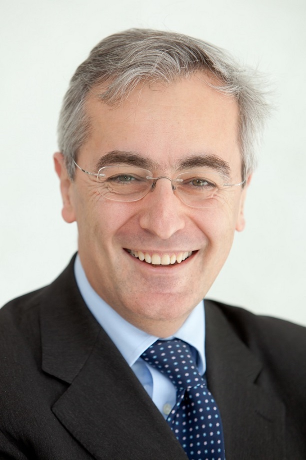 Angelo Auricchio合作论文数Faculty of Biomedical Sciences, Università della Svizzera;University Hospital in Magdeburg;Center for Computational Medicine in Cardiology, Università Della Svizzera Italiana in Lugano;Division of Cardiology, Istituto Cardiocentro Ticino23Sp Chough合作论文数22
 Richard Kobza合作论文数22
 Helge Servatius合作论文数22
 Haran Burri合作论文数Hopital Cantonal De Geneve22
 Etienne Pruvot合作论文数Arrhythmia Unit, Department of Heart and Vascular, Lausanne University Hospital;Department of Heart and Vascular, University of Lausanne22Rl Baker合作论文数21Diego Mannhart合作论文数21Meyre Pascal合作论文数21
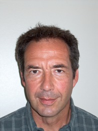 Jean-Marc Vesin合作论文数Electrical Engineering Section, School of Engineering, École Polytechnique Fédérale de Lausanne;ETH Zurich20Di Valentino Marcello合作论文数20
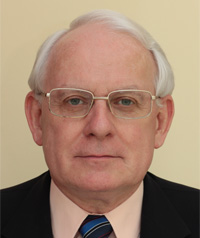 Marek Malik合作论文数Cardiac Electrophysiology at the University of London, Head of Non-Invasive Electrophysiology unit at St.20




