DNA damage and the elicited cellular response underlie the etiology of tumorigenesis and ageing. Yet, how this response integrates inputs from cells' environmental cues remains underexplored. Here we report that the BMP-Smad1 pathway, which is essential for embryonic development and tissue homeostasis, has an important role in the DNA damage response and oncogenesis. On genotoxic stress, Atm phosphorylates BMPs-activated Smad1 in the nucleus on S239, which disrupts Smad1 interaction with protein phosphatase PPM1A, leading to enhanced activation and upregulation of Smad1. Smad1 then interacts with p53 and inhibits Mdm2-mediated p53 ubiquitination and degradation to regulate cell proliferation and survival. Enhanced Smad1 S239 phosphorylation, and Smad1 mutations causing S239 substitution were detected in oesophageal and gastric cancer samples, respectively. These findings suggest that BMP-Smad1 signalling participates in the DNA damage response via the Atm-p53 pathway, thus providing a molecular mechanism whereby BMP-Smad1 loss-of-function leads to tumorigenesis, for example, juvenile polyposis and Cowden syndromes.
Background: Smad4 mutant embryos arrest shortly after implantation and display a characteristic shortened proximodistal axis, a significantly reduced epiblast, as well as a thickened visceral endoderm layer. Conditional rescue experiments demonstrate that bypassing the primary requirement for Smad4 in the extra-embryonic endoderm allows the epiblast to gastrulate. Smad4-independent TGF-beta signals are thus sufficient to promote mesoderm formation and patterning. To further analyse essential Smad4 activities contributed by the extra-embryonic tissues, and characterise Smad4 dependent pathways in the early embryo, here we performed transcriptional profiling of Smad4 null embryonic stem (ES) cells and day 4 embryoid bodies (EBs).Results: Transcripts from wild-type versus Smad4 null ES cells and day 4 EBs were analysed using Illumina arrays. In addition to several known TGF-beta/BMP target genes, we identified numerous Smad4-dependent transcripts that are mis-expressed in the mutants. As expected, mesodermal cell markers were dramatically down-regulated. We also observed an increase in non-canonical potency markers (Pramel7, Tbx3, Zscan4), germ cell markers (Aire, Tuba3a, Dnmt3l) as well as early endoderm markers (Dpp4, H19, Dcn). Additionally, expression of the extracellular matrix (ECM) remodelling enzymes Mmp14 and Mmp9 was decreased in Smad4 mutant ES and EB populations. These changes, in combination with increased levels of laminin alpha1, cause excessive basement membrane deposition. Similarly, in the context of the Smad4 null E6.5 embryos we observed an expanded basement membrane (BM) associated with the thickened endoderm layer.Conclusion: Smad4 functional loss results in a dramatic shift in gene expression patterns and in the endodermal cell lineage causes an excess deposition of, or an inability to breakdown and remodel, the underlying BM layer. These structural abnormalities probably disrupt reciprocal signalling between the epiblast and overlying visceral endoderm required for gastrulation.
Heterozygous disruption of the Men1 gene predisposes mice to the development of multiple endocrine tumors, accurately mimicking the human MEN1 cancer predisposition syndrome. Additionally, Men1 +/– mice frequently develop sex cord adenomas. The mechanism underlying the susceptibility of these mice to sex cord tumor development has not been fully determined, but data suggest it may involve transcriptional regulation of key growth promoting/repressing genes. To identify potential menin‐regulated genes that may be important for tumor suppression in sex cord cells, we compared the global gene expression profiles of testis and ovary adenomas with other endocrine tumors of the pancreas and pituitary from Men1 heterozygous mice and with control tissues. Gonadal tumors clustered separately from pancreas and pituitary tumors with only a few genes ( e.g., Cdkn2c ) commonly dysregulated in all tumor types. Testis and ovary tumors displayed a higher level of transcriptional similarity to each other than they did to their respective control tissues. Among genes that had decreased expression in tumors was significant over‐representation of genes associated with the TGF‐β, hedgehog and Wnt signaling, indicating that loss of menin function affects these pathways at the level of transcription. Aberrant protein expression in Leydig and granulosa cells of 2 transcriptionally dysregulated gene products, Gata6 and Csf1r were confirmed by immunohistochemistry. We propose that sex cord tumor susceptibility in Men1 +/– mice involves deregulated cell proliferation due to dysregulation of multiple cell growth regulating genes including: reduced Cdkn2c transcription, loss of TGF‐β pathway tumor suppressor function ( e.g., Gata6 ) and transcriptional activation of Csf1r. © 2008 Wiley‐Liss, Inc.
Although the identification of menin-interacting partners and other evidence support a role for menin, the multiple endocrine neoplasia type 1 gene (MEN1) product, in regulating gene expression, little is known about the cellular pathways dysregulated by menin loss during tumorigenesis. The mouse models of MEN1 accurately mimic the human syndrome and provide an opportunity to assess the transcriptional effects of Men1 deletion in different endocrine tumor types to identify common pathway aberrations underlying tumorigenesis in MEN1-affected tissues. We compared the global gene expression profiles of pituitary adenomas and pancreatic islet tumors with control tissues from wild-type littermates. Amongst the 551 differentially expressed genes was significant over-representation of genes associated with chromatin remodelling, transcription and cell cycling, including some genes known to encode menin-binding partners, e.g., Rhox5 and Mll1. Consistent with increased cell-cycle transition from G1 to S phase was an elevation of Cdc7 expression in the tumors, which was confirmed by qRT-PCR using independent samples. In support of previous findings in islet tumors, we found down-regulation of the cell-cycle regulator, p18, in both the pancreatic islet and pituitary adenomas, suggesting that reduced p18 levels may be important for Men1-related tumorigenesis in multiple tissues. Surprisingly, we identified increased p16 transcript in pancreatic islet and pituitary tumors. This was accompanied by increased cytoplasmic localization p16 protein in tumor cells. The specific genes and general pathways we have found to be commonly dysregulated in MEN1 tumors, provide a platform for determining their roles in endocrine tumorigenesis.
Multiple endocrine neoplasia type 1 (MEN1) is an inherited cancer predisposition syndrome typified by development of tumors in parathyroid, pituitary and endocrine pancreas, as well as less common sites including both endocrine and nonendocrine organs. Deletion or mutation of the tumor suppressor gene MEN1 on chromosome 11 has been identified in many cases of MEN1 as well as in sporadic tumors. The molecular biology of menin, the protein encoded by MEN1 , remains poorly understood. Here we describe a mouse model of MEN1 in which tumors were seen in pancreatic islets, pituitary, thyroid and parathyroid, adrenal glands, testes and ovaries. The observed tumor spectrum therefore includes types commonly seen in MEN1 patients and additional types. Pancreatic pathology was most common, evident in over 80% of animals, while other tumor types developed with lower frequency and generally later onset. Tumors of multiple endocrine organs were observed frequently, but progression to carcinoma and metastasis were not evident. Tumors in all sites showed loss of heterozygosity at the Men1 locus, though the frequency in testicular tumors was only 36%, indicating that a different molecular mechanism of tumorigenesis occurs in those Leydig tumors that do not show loss of the normal Men1 allele. Menin expression was below the level of detection in ovary, thyroid and testis, but loss of nuclear menin immunoreactivity was observed uniformly in all pancreatic islet adenomas and in some hyperplastic islet cells, suggesting that complete loss of Men1 is a critical point in islet tumor progression in this model. © 2006 Wiley‐Liss, Inc.
Smad4 in partnership with R-Smads (receptor-regulated Smads) activates TGF-beta (transforming growth factor-beta)-dependent signalling pathways essential for early mouse development. Smad4 null embryos die shortly after implantation due to severe defects in cell proliferation and visceral endoderm differentiation. In the basal state, Smad4 undergoes continuous shuttling between the cytoplasm and the nucleus due to the combined activities of an N-terminal NLS (nuclear localization signal) and an NES (nuclear export signal) located in its linker region. Cell culture experiments suggest that Smad4 nucleocytoplasmic shuttling plays an important role in TGF-beta signalling. In the present study we have investigated the role of Smad4 shuttling in vivo using gene targeting to engineer two independent mutations designed to eliminate Smad4 nuclear export. As predicted this results in increased levels of Smad4 in the nucleus of homozygous ES cells (embryonic stem cells) and primary keratinocytes, in the presence or absence of ligand. Neither mutation affects Smad4 expression levels nor its ability to mediate transcriptional activation in homozygous cell lines. Remarkably mouse mutants lacking the Smad4 NES develop normally. Smad4 NES mutants carrying one copy of a Smad4 null allele also fail to display developmental defects. The present study clearly demonstrates that Smad4 nucleocytoplasmic shuttling is not required for embryonic development or tissue homoeostasis in normal, healthy adult mice.
To identify possible genetic interactions between the mechanisms of tumor suppression of menin and pRb, we intercrossed mice with targeted deletions of Men1 and Rb1, and compared tumor development in cohorts of animals carrying single or dual mutations of these tumor-suppressor genes. In mice lacking one copy of Men1, pancreatic islet and anterior pituitary adenomas are common. In animals lacking one copy of Rb1, intermediate pituitary and thyroid tumors occur at high frequency, with less frequent development of pancreatic islet hyperplasia and parathyroid lesions. In mice heterozygous for both Men1 and Rb1, pancreatic hyperplasia and tumors of the intermediate pituitary and thyroid occurred at high frequency. Serum measurements of calcium and glucose did not vary significantly between genotypic groups. Loss of heterozygosity at the Rb1 locus was common in pituitary and thyroid tumors, whereas loss of menin was observed in pancreatic and parathyroid lesions. The tumor spectrum in the double heterozygotes was a combination of pathologies seen in each of the individual heterozygotes, without decrease in age of onset, indicating independent, non-additive effects of the two mutations. Together with the lack of increased tumor spectrum, this suggests that menin and pRb function in a common pathway of tumor suppression.
Mutations of the MEN1 gene, encoding the tumor suppressor menin, predispose individuals to the cancer syndrome multiple endocrine neoplasia type 1, characterized by the development of tumors of the endocrine pancreas and anterior pituitary and parathyroid glands. We have targeted the murine Men1 gene by using Cre recombinase-loxP technology to develop both total and tissue-specific knockouts of the gene. Conditional homozygous inactivation of the Men1 gene in the pituitary gland and endocrine pancreas bypasses the embryonic lethality associated with a constitutional Men1(-/-) genotype and leads to beta-cell hyperplasia in less than 4 months and insulinomas and prolactinomas starting at 9 months. The pituitary gland and pancreas develop normally in the conditional absence of menin, but loss of this transcriptional cofactor is sufficient to cause beta-cell hyperplasia in some islets; however, such loss is not sufficient to initiate pituitary gland tumorigenesis, suggesting that additional genetic events are necessary for the latter.
The cellular function of the menin tumor suppressor protein, product of the MEN1 gene mutated in familial multiple endocrine neoplasia type 1, has not been defined. We now show that menin is associated with a histone methyltransferase complex containing two trithorax family proteins, MLL2 and Ash2L, and other homologs of the yeast Set1 assembly. This menin-associated complex methylates histone H3 on lysine 4. A subset of tumor-derived menin mutants lacks the associated histone methyltransferase activity. In addition, menin is associated with RNA polymerase II whose large subunit carboxyl-terminal domain is phosphorylated on Ser 5. Men1 knockout embryos and cells show decreased expression of the homeobox genes Hoxc6 and Hoxc8. Chromatin immunoprecipitation experiments reveal that menin is bound to the Hoxc8 locus. These results suggest that menin activates the transcription of differentiation-regulating genes by covalent histone modification, and that this activity is related to tumor suppression by MEN1.
genesisVolume 32, Issue 2 p. 150-151 ArticleFree Access Targeting and conditional inactivation of the murine Men1 locus using the Cre recombinase: loxP System Christine Biondi, Christine Biondi Joint Experimental Oncology Program, The Queensland Institute of Medical Research and The University of Queensland, Brisbane, Queensland, AustraliaSearch for more papers by this authorMichael Gartside, Michael Gartside Joint Experimental Oncology Program, The Queensland Institute of Medical Research and The University of Queensland, Brisbane, Queensland, AustraliaSearch for more papers by this authorIan Tonks, Ian Tonks Joint Experimental Oncology Program, The Queensland Institute of Medical Research and The University of Queensland, Brisbane, Queensland, AustraliaSearch for more papers by this authorCarol Paterson, Carol Paterson Joint Experimental Oncology Program, The Queensland Institute of Medical Research and The University of Queensland, Brisbane, Queensland, AustraliaSearch for more papers by this authorNicholas K. Hayward, Nicholas K. Hayward Joint Experimental Oncology Program, The Queensland Institute of Medical Research and The University of Queensland, Brisbane, Queensland, AustraliaSearch for more papers by this authorGraham F. Kay, Corresponding Author Graham F. Kay [email protected] Joint Experimental Oncology Program, The Queensland Institute of Medical Research and The University of Queensland, Brisbane, Queensland, AustraliaThe Queensland Institute of Medical Research, Post Office Royal Brisbane Hospital, Queensland 4029, AustraliaSearch for more papers by this author Christine Biondi, Christine Biondi Joint Experimental Oncology Program, The Queensland Institute of Medical Research and The University of Queensland, Brisbane, Queensland, AustraliaSearch for more papers by this authorMichael Gartside, Michael Gartside Joint Experimental Oncology Program, The Queensland Institute of Medical Research and The University of Queensland, Brisbane, Queensland, AustraliaSearch for more papers by this authorIan Tonks, Ian Tonks Joint Experimental Oncology Program, The Queensland Institute of Medical Research and The University of Queensland, Brisbane, Queensland, AustraliaSearch for more papers by this authorCarol Paterson, Carol Paterson Joint Experimental Oncology Program, The Queensland Institute of Medical Research and The University of Queensland, Brisbane, Queensland, AustraliaSearch for more papers by this authorNicholas K. Hayward, Nicholas K. Hayward Joint Experimental Oncology Program, The Queensland Institute of Medical Research and The University of Queensland, Brisbane, Queensland, AustraliaSearch for more papers by this authorGraham F. Kay, Corresponding Author Graham F. Kay [email protected] Joint Experimental Oncology Program, The Queensland Institute of Medical Research and The University of Queensland, Brisbane, Queensland, AustraliaThe Queensland Institute of Medical Research, Post Office Royal Brisbane Hospital, Queensland 4029, AustraliaSearch for more papers by this author First published: 13 February 2002 https://doi.org/10.1002/gene.10061Citations: 15AboutPDF ToolsRequest permissionExport citationAdd to favoritesTrack citation ShareShare Give accessShare full text accessShare full-text accessPlease review our Terms and Conditions of Use and check box below to share full-text version of article.I have read and accept the Wiley Online Library Terms and Conditions of UseShareable LinkUse the link below to share a full-text version of this article with your friends and colleagues. Learn more.Copy URL No abstract is available for this article. LITERATURE CITED Chandrasekharappa SC, Guru SC, Manickam P, Olufemi SE, Collins FS, Emmert-Buck MR, Debelenko LV, Zhuang Z, Lubensky IA, Liotta LA, Crabtree JS, Wang Y, Roe BA, Weisemann J, Boguski MS, Agarwal SK, Kester MB, Kim YS, Heppner C, Dong Q, Spiegel AM, Burns AL, Marx SJ. 1997. Positional cloning of the gene for multiple endocrine neoplasia-type 1. Science 276: 404– 407. Heppner C, Kester MB, Agarwal SK, Debelenko LV, Emmert-Buck MR, Guru SC, Manickam P, Olufemi SE, Skarulis MC, Doppman JL, Alexander RH, Kim YS, Saggar SK, Lubensky IA, Zhuang Z, Liotta LA, Chandrasekharappa SC, Collins FS, Spiegel AM, Burns AL, Marx SJ. 1997. Somatic mutation of the MEN1 gene in parathyroid tumors. Nat Genet 16: 375– 378. Jonkers J, Meuwissen R, van der Gulden H, Peterse H, van der Valk M, Berns A. 2001. Synergistic tumor suppressor activity of BRCA2 and p53 in a conditional mouse model for breast cancer. Nat Genet 29: 418– 425. Lemmens I, Van de Ven WJ, Kas K, Zhang CX, Giraud S, Wautot V, Buisson N, De Witte K, Salandre J, Lenoir G, Pugeat M, Calender A, Parente F, Quincey D, Gaudray P, De Wit MJ, Lips CJ, Hoppener JW, Khodaei S, Grant AL, Weber G, Kytola S, Teh BT, Farnebo F, Thakker RV, et al. 1997. Identification of the multiple endocrine neoplasia type 1 (MEN1) gene. The European Consortium on MEN1. Hum Mol Gen 6: 1177– 1183. Stewart C, Parente F, Piehl F, Farnebo F, Quincey D, Silins G, Bergman L, Carle GF, Lemmens I, Grimmond S, Xian CZ, Khodei S, Teh BT, Lagercrantz J, Siggers P, Calender A, Van de Vem V, Kas K, Weber G, Hayward N, Gaudray P, Larsson C. 1998. Characterization of the mouse Men1 gene and its expression during development. Oncogene 17: 2485– 2493. Zhuang Z, Ezzat SZ, Vortmeyer AO, Weil R, Oldfield EH, Park WS, Pack S, Huang S, Agarwal SK, Guru SC, Manickam P, Debelenko LV, Kester MB, Olufemi SE, Heppner C, Crabtree JS, Burns AL, Spiegel AM, Marx SJ, Chandrasekharappa SC, Collins FS, Emmert-Buck MR, Liotta LA, Asa SL, Lubensky IA. 1997a. Mutations of the MEN1 tumor suppressor gene in pituitary tumors. Cancer Res 57: 5446– 5451. Zhuang Z, Vortmeyer AO, Pack S, Huang S, Pham TA, Wang C, Park WS, Agarwal SK, Debelenko LV, Kester M, Guru SC, Manickam P, Olufemi SE, Yu F, Heppner C, Crabtree JS Skarulis MC, Venzon DJ, Emmert-Buck MR, Spiegel AM, Chandrasekharappa SC, Collins FS, Burns AL, Marx SJ, Lubensky IA, et al. 1997b. Somatic mutations of the MEN1 tumor suppressor gene in sporadic gastrinomas and insulinomas. Cancer Res 57: 4682– 4686. Citing Literature Volume32, Issue2Special Issue: Conditional Alleles in MiceFebruary 2002Pages 150-151 ReferencesRelatedInformation
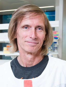 Nicholas Hayward合作论文数Oncogenomics Laboratory, QIMR Berghofer Medical Research Institute8
 Graham F. Kay合作论文数8Michael Gartside合作论文数5
 Kelly A. Loffler合作论文数Institute for Molecular Bioscience, The University of Queensland4Serewko-Auret Magdalena合作论文数3
 Arne Mould合作论文数Royal Brisbane Hospital, The University of Queensland3
 Elizabeth Robertson合作论文数Sir William Dunn School of Pathology, University of Oxford;Oxford Stem Cell Institute, University of Oxford3Russell Duncan合作论文数2Stuart S Levine合作论文数2KS Shanmugam合作论文数2
 Mitchell Stark合作论文数Dermatology Research Centre, Faculty of Health, Medicine and Behavioural Sciences, The University of Queensland;Frazer Institute, Faculty of Health, Medicine and Behavioural Sciences, The University of Queensland2Bikoff Elizabeth K合作论文数2Terry D. Copeland合作论文数2
 Maria Celia Hughes合作论文数2
 Thomas Milne合作论文数Radcliffe Department of Medicine, University of Oxford2J. C. Lee合作论文数2
 Paul Waring合作论文数Translational Pathology Consultation Services2
 Neil Hayes合作论文数Division of Med Hematology, Department of Medicine, College of Medicine, The University of Tennessee Health Science Center;Center for Cancer Research, The University of Tennessee Health Science Center;Heme / Onc, Regional One2Arindam Bhattacharjee合作论文数University College of Science, University of Calcutta2Jay L. Hess合作论文数2
 Matthew L. Meyerson合作论文数Harvard Medical School;Center for Cancer Genomics, Dana-Farber Cancer Institute2Orit Rozenblatt-Rosen合作论文数1I D Tonks合作论文数1Jennifer M Taylor合作论文数1Debipriya Das合作论文数1Gartside Michael合作论文数1xueying wang合作论文数1Zhongfeng Wang合作论文数1P Waring合作论文数1
 Lin He (贺林)合作论文数School of Life Sciences and Biotechnology, Shanghai Jiao Tong University;Institutes of Biomedical Sciences, Fudan University1Rozenblatt-Rosen Orit合作论文数1Tonks Ian D合作论文数1
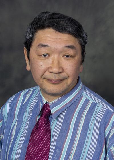 Yuji Mishina合作论文数Department of Biologic and Materials Sciences & Prosthodontics, School of Dentistry, University of Michigan1Leong Wai Fook合作论文数1
 Jia Hao (贾浩)合作论文数Shanghai Jiao Tong University College of Basic Medical Sciences1
 Mark A. Magnuson合作论文数Department of Molecular Physiology and Biophysics, School of Medicine, Vanderbilt University;Department of Cell and Developmental Biology, School of Medicine, Vanderbilt University1Au Bijin合作论文数1
 Huijuan Liu (刘慧娟)合作论文数School of Life Sciences and Biotechnology, Shanghai Jiao Tong University;Bio-X Instiutes, Shanghai Jiao Tong University1
 Yeguang Chen (陈晔光)合作论文数Nanchang University1
 Ball Keng Po Lai (黎鏡波)合作论文数Guilin Medical University1
 Dong Xie (谢东)合作论文数Shanghai Institutes for Biological Sciences, Chinese Academy of Sciences1
 Caroline Hill合作论文数Francis Crick Institute1
 Qiang Yu (于強)合作论文数Faculty of Health Sciences, University of Macau;Genome Institute of Singapore;Department of Physiology, National University of Singapore;Duke-NUS1
 Konrad Muller合作论文数1
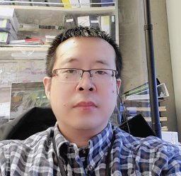 Deyong Jia合作论文数Shanghai Jiao Tong University, Shanghai Jiaotong Univ.1Ita Costello合作论文数Sir William Dunn School of Pathology, University of Oxford1Michael D. Howell合作论文数1
 Baojie Li (李保界)合作论文数School of Life Sciences and Biotechnology, Shanghai Jiao Tong University;Bio-X Institutes, Shanghai Jiao Tong University1H. K. Muller合作论文数1




