Abstract Introduction Rasal1, a GTPase-activating protein, is a novel component of the T-cell receptor (TCR) complex that associates with ZAP-70 and negatively regulates the p21ras/ERK pathway (Thaker et al., Nat. Commun., PMID: 31641113). Here, we demonstrate that Rasal1 selectively regulates mitochondrial oxidative phosphorylation (OXPHOS) metabolism, while cooperating with anti—PD—1 therapy to limit T-cell exhaustion. Methods Utilizing a knock-in mutant mouse expressing Rasal1 lacking the C2-1 domain (Rasal1c-mut), we analyzed glycolytic- and mitochondrial-driven metabolic programme by measuring the oxygen consumption rate (OCR), and the extracellular acidification rate (ECAR). In vivo studies were conducted in Rasal1c-mut mice using several immune checkpoint blockade (ICB)-resistant melanoma, colorectal, lymphoma, and lung carcinoma models in response to anti—PD—1 treatment. Results Primary T cells from mice expressing impaired Rasal1 exhibited a normal glycolytic shift following antigen stimulation but displayed a pronounced and selective increase in mitochondrial respiration. These findings indicate that Rasal1 fine-tunes TCR-dependent mitochondrial activity to sustain ATP generation and biosynthetic output during activation. Unexpectedly, inactive Rasal1–with elevated ZAP-70 and ERK signaling–synergized with anti—PD-1 blockade to reduce tumor-infiltrating lymphocyte exhaustion and promote the expansion of CD8+PD-1+TCF1+ stem-like progenitor cells, leading to enhanced tumor control. The same mechanism also increased the presence of cytolytic CD4+ TILs. Conclusion Collectively, our data reveal Rasal1 as a unique metabolic brake in T cells whose modulation offers a promising strategy to enhance the durability and breadth of immune checkpoint therapies. Funding Source Canadian Institutes of Health Research (159912) Topic Categories Tumor Immunology: Cellular Responses and Tumor Microevironment (TIME)
Severe COVID-19 can trigger a cytokine storm, leading to acute respiratory distress syndrome (ARDS) with similarities to superantigen-induced toxic shock syndrome. An outstanding question is whether SARS-CoV-2 protein sequences can directly induce inflammatory responses. In this study, we identify a region in the SARS-CoV-2 S2 spike protein with sequence homology to bacterial super-antigens (termed P3). Computational modeling predicts P3 binding to sites on MHC class I/II and the TCR that partially overlap with sites for the binding of staphylococcal enterotoxins B and H. Like SEB and SEH peptides, P3 stimulated 25-40% of human CD4+ and CD8+ T cells, increasing IFN-γ and granzyme B production. viSNE and SPADE profiling identified overlapping and distinct IFN-γ and GZMB subsets. The super-antigenic properties of P3 were further evident by its selective expansion of T cells expressing specific TCR Vα and Vβ chain repertoires. In vivo experiments in mice revealed that the administration of P3 led to a significant upregulation of proinflammatory cytokines IL-1β, IL-6, and TNF-α. While the clinical significance of P3 in COVID-19 remains unclear, its homology to other mammalian proteins suggests a potential role for this peptide family in human inflammation and autoimmunity.
While immune checkpoint blockade (ICB) has revolutionized cancer treatment, the key T-cell signaling pathways responsible for its potency remain unclear. GSK-3 is an inhibitory kinase that is most active in resting T-cells. In this study, we demonstrate that GSK-3 facilitates PD-1 blockade, an effect seen by modulating CD4 T-cell help for CD8+ CTL responses against ICB resistant tumors. We show that GSK-3 controls metabolic reprogramming towards glycolysis and synergizes with PD-1 to induce a transcriptional program that reduces suppressive CD4+ Treg numbers while generating super-armed effector-memory CD8+ CTLs that express an unprecedented 7/9 granzymes from the genome. Crucially, we found that GSK-3 cooperates with PD-1 blockade to determine the dependency of CD8+ CTLs on help from CD4+ T-cells. Our study unravels a novel cooperative PD-1 blockade-dependent signaling pathway that potentiates CTL responses against tumors, offering a new strategy to overcome immunotherapy resistance by modulating CD4+ helper and CD8+ cytotoxic functions. Significance:This study demonstrates for the first time that GSK-3 controls the crosstalk between CD4+ and CD8+ T cells, synergizing with anti-PD-1 therapy to overcome resistance to checkpoint blockade and to generate super-armed CD8+ effector cells in cancer immunotherapy. This newly uncovered GSK-3-dependent CD4-CD8 T-cell crosstalk mechanism presents a new approach to enhance anti-PD-1 immunotherapy.
T-cells constitute an essential component of the adaptive immune response, mount a protective response against foreign pathogens and are important regulators of anti-tumor immunotherapy. In this context, the activation of T-cells and chimeric antigen receptor (CAR)-expressing T-cells is orchestrated by various signaling pathways, involving the initiation of a protein tyrosine phosphorylation cascade. For T-cells, this involves initiation of the phosphorylation cascade via src-related protein-tyrosine kinase p56lck, which we show to associate with the co-receptors CD4 and CD8 for the induction of a phosphorylation cascade needed for the activation of T-cells. Likewise, p56lck phosphorylation of the antigen receptor immunoreceptor tyrosine-based activation motifs (ITAMs) and key CD28 tyrosine motifs ensures the functionality and the survival of CARs, while their phospho-targets are also inhibited by PD-1, a key component of the immune checkpoint blockade. This review covers historic and current elements of our knowledge of CD4/CD8–p56lck-induced activation events and their importance to the development of CAR T-cell immunotherapies.
COVID-19, caused by SARS-CoV-2, can lead to a severe inflammatory disease characterized by significant lymphopenia. However, the underlying cause for the depletion of T-cells in COVID-19 patients remains incompletely understood. In this study, we assessed the presence of different T-cell subsets in the progression of COVID-19 from mild to severe disease, with a focus on TCF1 expressing progenitor T-cells that are needed to replenish peripheral T-cells during infection. Our results showed a preferential decline in TCF1+ progenitor CD4 and CD8+ T-cells with disease severity. This decline was seen in various TCF1+ subsets including naive, memory and effector-memory cells, and surprisingly, was accompanied by a loss in cell division as seen by a marked decline in Ki67 expression. In addition, TCF1+ T-cells showed a reduction in pro-survival regulator, BcL2, and the appearance of a new population of TCF1 negative caspase-3 expressing cells in peripheral blood from patients with severe disease. The decline in TCF1+ T-cells was also seen in a subgroup of severe patients with vitamin D deficiency. Lastly, we found that sera from severe patients inhibited TCF1 transcription ex vivo which was attenuated by a blocking antibody against the cytokine, interleukin-12 (IL12). Collectively, our findings underscore the potential significance of TCF1+ progenitor T-cells in accounting for the loss of immunity in severe COVID-19 and outline an array of markers that could be used to identify disease progression. This study shows that severity of disease in COVID-19 patients is characterized by the preferential loss of TCF1+ progenitor T-cells needed to self-renew and replenish the immune system.
SummaryPD-1 immune checkpoint blockade (ICB) is now a promising first-line treatment for many cancers. While the steric blockade of PD-1 binding to its ligand plays a role, the role of internalisation in promoting the efficacy of ICB has not been explored. In this study, we show that PD-1 internalisation also contributes by unlocking the full cytolytic potential of ICB in cancer immunotherapy. We found that anti-mouse and human PD-1 downregulate a subset of PD-1 surface receptors on T-cells with high-density surface PD-1 leaving T-cells with intermediate expression resistant to further internalisation. Down regulation was seen on both CD4 and CD8 cells but was maximally effective on CD8 effector cells. In human T-cells, nivolumab outperformed pembrolizumab in terms of rate and efficacy. We also found that PD-1 internalisation depended on bivalent antibody (Ab)-induced crosslinking, while monovalent Ab sterically blocked PD-1 without inducing endocytosis. Immunologically, while both monovalent and bivalent Ab limited B16-PD-L1 tumor growth, bivalent Ab was significantly more effective. In molecular terms, while both antibodies increased granzyme B (GZMB) expression in CD8+ cytolytic T-cells, the induction of the second key cytolytic pore-forming mediator, perforin, was dependent on the blockade and internalisation mediated by bilavent anti-PD-1. Our findings unveil a novel mechanism in checkpoint blockade where steric blockade combined with the removal of PD-1 from the cell surface by endocytosis can complement and optimize therapy. The targeting of PD-1 internalisation holds promise for enhancing anti-tumor immunity and improving PD-1 checkpoint blockade therapy.Graphical AbstractIn briefBen Saad et al define the mechanism of PD-1 inhibitory endocytosis and show that the removal of surface PD-1 by endocytosis plays a role in complementing and optimizing checkpoint blockade. Targeting PD-1 internalisation holds promise for enhancing anti-tumor immunity and improving the efficacy of PD-1 checkpoint blockade therapy.
Cancer is a major global health concern, with immune suppression hindering treatment. Immunotherapy, specifically immune checkpoint blockage on T cells, has revolutionized cancer treatment. T-cell exhaustion is an abnormal activation state that develops when continuous exposure to antigens, like cancer. In this context, recent evidence suggests that parathyroid hormone-related protein (PTHrP) plays a previously underappreciated role in fostering an immunosuppressive tumor microenvironment. Further, blocking PTHrP activity reduces primary tumor growth, prevents metastasis, and prolongs survival in mice with various cancers. Here, we confirm that administration of anti-PTHrP monoclonal antibodies can reduce the growth of B16-PDL1 melanoma tumors and that although the therapy did not alter the presence of CD4+ and CD8+ TILs, we noted that all stages of T-cell exhaustion were reduced. Further, the expression of cytolytic proteins PERFORIN and GZMB also increased. By contrast, anti-PTHrP therapy increased the relative presence of pre-pro B cells with a decline in mature B cells in the bone marrow. Overall, our data indicates that anti-PTHrP therapy acts by reducing T-cell exhaustion and by affecting B-cell development. These provide further mechanistic evidence to support the application of anti-PTHrP blockade as an alternate therapeutic approach to boost anti-tumor immunity.
CD8+ T cells recognize tumor-associated antigens presented by major histocompatibility complex (MHC) class I molecules. How CD8+ T cells eliminate cancer cells deficient in MHC class I has been unclear. A study now shows that adaptive CD8+ T cell activation induces expression of the innate receptor NKG2D for the elimination of MHC class I–deficient tumors.
Introduction: Glycogen synthase kinase 3 (GSK-3) has been proposed as a novel cancer target due to its regulating role in both tumor and immune cells. However, the connection between GSK-3 and immunoevasive contexture, including tumor budding (TB) has not been previously examined.Methods: we investigated the expression levels of total GSK-3 as well as its isoforms (GSK-3 beta and GSK-3 alpha) and examined their potential correlation with TB grade and the programmed cell death-ligand 1 (PD-L1) in colorectal cancer (CRC) tumor samples. Additionally, we compared the efficacy of GSK-3-inhibition with PD-1/PD-L1 blockade in humanized patient-derived (PDXs) xenografts models of high-grade TB CRC.Results: we show that high-grade (BD3) TB CRC is associated with elevated expression levels of total GSK-3, specifically the GSK-3 beta isoform, along with increased expression of PD-L1 in tumor cells. Moreover, we define an improved risk stratification of CRC patients based on the presence of GSK-3+/PD-L1+/BD3 tumors, which are associated with a worse prognosis. Significantly, in contrast to the PD-L1/PD-1 blockade approach, the inhibition GSK-3 demonstrated a remarkable enhancement in the antitumor response. This was achieved through the reduction of tumor buds via necrosis and apoptosis pathways, along with a notable increase of activated tumor -infiltrating CD8+ T cells, NK cells, and CD4- CD8- T cells.Conclusions: our study provides compelling evidence for the clinical significance of GSK-3 expression and TB grade in risk stratification of CRC patients. Moreover, our findings strongly support GSK-3 inhibition as an effective therapy specifically targeting high-grade TB in CRC.
Acute myeloid leukemia (AML) is a cancer that originates from the bone marrow (BM). Under physiological conditions, the bone marrow supports the homeostasis of immune cells and hosts memory lymphoid cells. In this review, we summarize our present understanding of the role of the immune microenvironment on healthy bone marrow and on the development of AML, with a focus on T cells and other lymphoid cells. The types and function of different immune cells involved in the AML microenvironment as well as their putative role in the onset of disease and response to treatment are presented. We also describe how the immune context predicts the response to immunotherapy in AML and how these therapies modulate the immune status of the bone marrow. Finally, we focus on allogeneic stem cell transplantation and summarize the current understanding of the immune environment in the post-transplant bone marrow, the factors associated with immune escape and relevant strategies to prevent and treat relapse.
T-cell activation is a complex process involving a network of kinases and downstream molecular scaffolds or adaptors that integrate surface signals with effector functions. One key immune-specific adaptor is Src kinase-associated phosphoprotein 1 (SKAP1), which is also known as src kinase-associated protein of 55 kDa (SKAP55). This mini-review explains how SKAP1 plays multiple roles in regulating integrin activation, the "stop-signal", and the optimization of the cell cycling of proliferating T cells through interactions with various mediators, including the Polo-like kinase 1 (PLK1). Ongoing research on SKAP1 and its binding partners will likely provide important insights into the regulation of immune function and have implications for the development of new treatments for disease states such as cancer and autoimmunity.
Cancer remains the leading cause of death in the world despite the significant advancements made in anticancer drug discovery. This study is aimed to computationally evaluate the efficacy of 63 in-house synthesized pyrazole derivatives targeted to bind with prominent cancer targets namely EGFR, RSK1, RAF1, PARP2 and LIN28B known to be expressed, respectively, in lung, colon, skin, ovarian and pancreatic cancer cells. Initially, we perform the molecular docking investigations for all pyrazole compounds with a comparison to known standard drugs for each target. Docking studies have revealed that some pyrazole compounds possess better binding affinity scores than standard drug compounds. Thereafter, a long-range of 1 μs molecular dynamic (MD) simulation study for top ranked docked compounds with all respective proteins was carried out to assess the interaction stability in a dynamic environment. The results suggested that the top ranked complexes showed a stable interaction profile for a longer period of time. The outcome of this study suggests that pyrazole compounds, M33, M36, M76 and M77, are promising molecular candidates that can modulate the studied target proteins significantly in comparison to their known inhibitor based on their selective binding interactions profile. Furthermore, ADME-T profile has been explored to check for the drug-likeness and pharmacokinetics profiles and found that all proposed compounds exhibited acceptable values for being a potential drug-like candidate with non-toxic characteristics. Overall, extensive computational investigations indicate that the four proposed pyrazole inhibitors/modulators studied against each respective target protein will be helpful for future cancer therapeutic developments.Communicated by Ramaswamy H. Sarma.
In this study, a local quantitative structure-activity relationship (QSAR) models were developed for set of compounds tested for their inhibitory activity against six different cancer cell lines viz. A-549, MCF-7, HeLa, HepG-2, PaCa-2 and DLD-1. Two different statistical approaches Principal Component Analysis (PCA) and Partial Least Square (PLS) analyses were employed to developed QSAR models. Further, activity predictions were carried out for in-house synthesized 63 pyrazole derivatives. Prediction of pIC50 value of all 63 synthesized pyrazole derivatives were estimated based on the most significant QSAR model developed for each cancer cell line. Several statistical parameters such as correlation coefficient R2, RMSE, Cross validated R2, Cross validated RMSE, internal validation Q2 and the external validation R2 revealed that developed models showed a significant value for explaining an acceptable QSAR model. The results derived highlighted some important compounds for being the most promise lead candidate against the six-cancer cell line with a significant pIC50 value. Considering the contribution of most important descriptors, we have designed new molecules which found to have greater inhibitory potentiality than the reference compounds. Overall, the results suggest that the developed QSAR models might be useful as a theoretical reference for experimental studies and designing more potent anti-cancer therapeutic pyrazoles based compounds.
Intracellular signaling networks control every aspect of T cell biology. Proper signaling promotes T cell development, cellular differentiation, adaptive immune responses and maintenance of immune homeostasis. T cell signaling and activation mechanisms play a fundamental role in disease processes. Ligation of the T-cell receptor (TCR) alone by major histocompatibility complex (MHC)-peptide or cancer neo-antigens is needed to activate T-cells. Stimulation is induced by a tyrosine phosphorylation cascade initiated by the src kinase p56 and its binding to the co-receptors CD4 and CD8 (1–3). p56 phosphorylates immune tyrosine activation motifs (ITAMs) on the TCR associated CD3 and zeta chains that recruit and activate a second kinase, zeta-chain-associated protein kinase 70 (ZAP-70) (4). p56 and ZAP-70 in turn then phosphorylate adaptors to form complexes or molecular switches that amplify and integrate signals for specific functions. Adaptors LAT and SLP-76 facilitate phospholipase C phosphorylation and calcium mobilization, while ADAP and SKAP1 (aka SKAP55) activate integrins (5–7). Despite this, second and third sets of signals from positive and inhibitory co-receptors (IFs) determine the final outcome of activation. CD28 and ICOS potentiate activation, while inhibitory co-receptors such as CTLA-4, PD-1, LAG3, TIGIT, TIM-3 and others limit or terminate T-cell responses (8–10). They are also expressed on exhausted or dysfunctional Tcells which develop after repeated antigenic stimulation. CD28 can both amplify TCR signaling or generate independent signals (10, 11). Subsequent signals are provided by the CD40 pathways as well as an array of cytokines to bind to receptors leading to the full development of T-cell subsets and different levels of differentiation and effector function. This work has led to the development of chimeric antigen receptor modified T cells (CAR-T) to thwart diseases while further advances in our basic understanding of the signals proteins while further improve the efficacy of CAR therapy (12, 13). New technologies have also revolutionized our ability to better visualize the organization of signaling processes, characterize signaling cascades at the systems level and study the role of metabolites and signaling lipids in regulating T cell activation. As shown in this Research Topic, there is still much to learn about signaling in T cells and that a more in-depth basic understanding of signaling mechanisms could be leveraged to control T cell activity and rationally engineer T cellbased immunotherapies. O’Brien et al. detail work on the spontaneous differentiation of T follicular helper cells in mice carrying a mutant form of the LAT (linker for activation of T cells) adaptor on the PLCg1 binding site (Y136). LAT is an integral membrane adaptor protein that constitutes in T cells a major substrate of the ZAP-70 protein tyrosine kinase. They show that mice with a tyrosine to
Despite the decades of scientific studies for developing promising new therapies, cancer remains a major cause of illness and mortality, worldwide. Several cancer types are the major topic of research in drug discovery programs due to their global incidence cases and growing frequency. In the present study, using two different statistical approaches PCA (principal component analysis) and PLS (partial least squares), six 2D-QSAR (quantitative structure activity relationship) models have been developed for the set of compounds retrieved against seven cancer cell lines vizPC-3, B16F10, K562, MDA-MB-231, A2780, and ACHN. For the creation and validation of 2D-QSAR models, OECD (Organization for Economic Co-operation and Development) requirements have been strictly followed. All of the generated 2D-QSAR models produce a significant and high correlation coefficient value with several other statistical parameters. Moreover, developed 2D-QSAR models have been used for activity predictions of in-house synthesized 63 pyrazole derivatives compounds. Precisely, most statistically significant and accepted2D-QSAR model generated for each cancer cell line has been used to predict the pIC50 value (anti-cancer activity) of all 63 synthesized pyrazole derivatives. Furthermore, designing of novel pyrazole derivatives has been carried out by substituting the essential functional groups based on the best derived 2D-QSAR models for each cancer cell line, more precisely, based on the most significant molecular descriptors with enhanced anti-cancer activity. Finally, the prediction of the new designed molecules reveals higher pIC50 than the standard compounds.
 Helga Schneider合作论文数30
 Stuart F. Schlossman合作论文数Harvard Medical School25M Raab合作论文数24
 Monika Raab合作论文数19
 Hongyan Wang (王红艳)合作论文数Center for Excellence in Molecular Cell Science, Chinese Academy of Sciences18
 Alison Taylor合作论文数Department of Educational Studies, Faculty of Education, The University of British Columbia16KVS PRASAD合作论文数15
 Chikao Morimoto合作论文数Department of Therapy Development and Innovation for Immune Disorders and Cancers, Graduate School of Medicine, Juntendo University;The University of Tokyo13Janna Krueger合作论文数11Aj Dasilva合作论文数10
 Ottmar Janssen合作论文数10
 Bin Wei (魏滨)合作论文数School of Life Sciences, Shanghai University10
 Alexandra Kazanova合作论文数9
 Yuning Lu合作论文数9Hebin Liu合作论文数9D. Rothstein合作论文数8Xin Smith合作论文数8H Schneider合作论文数7
 Thaker Youg R合作论文数School of Biological Sciences, Nanyang Technological University7
 Walter Bodmer合作论文数Department of Oncology, Medical Sciences Division, University of Oxford;Cancer and Immunogenetics Laboratory, University of Oxford7Klaus Strebhardt合作论文数7Fatima Ezzahra Bennani合作论文数6
 Norman L. Letvin合作论文数Division of Viral Pathogenesis, Department of Medicine, Beth Israel Deaconess Medical Center;Harvard Medical School;Faculty of Medicine, Harvard University6Issa Mark E合作论文数6Y C Cai合作论文数6JG BODMER合作论文数5SD Dias合作论文数5M J Crumpton合作论文数5Jm Trevillyan合作论文数5
 Daniel M Davis合作论文数Division of Immunology, Immunity to Infection and Respiratory Medicine, School of Biological Sciences, University of Manchester/Manchester Institute for Collaborative Research on Ageing5
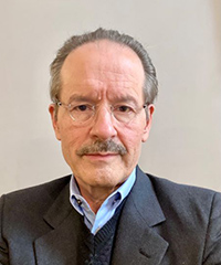 Lewis Cantley合作论文数Department of Cell Biology, Harvard Medical School;Department of Cell Biology, Dana-Farber Cancer Institute5
 Daniel Cefai合作论文数5Heinrich Repke合作论文数5Elke Valk合作论文数5E. Valk合作论文数4Lp Geng合作论文数4
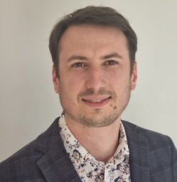 Francois Santinon合作论文数4
 Latifa Doudach合作论文数4
 Khalid Karrouchi合作论文数Faculty of Medicine and Pharmacy, Mohammed V University in Rabat4X Wu合作论文数4Nancy Hogg合作论文数4
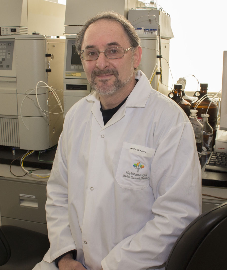 Uri Saragovi合作论文数Department of Pharmacology & Therapeutics, Faculty of Medicine and Health Sciences, McGill University4
 Richard Kremer合作论文数Division of Endocrinology and Metabolism, Department of Medicine, Faculty of Medicine and Health Sciences, McGill University4
 Andrew Smith合作论文数Eastman Dental Institute, Faculty of Medical Sciences, University College London4
 Tsutomu Takeuchi (竹内勤)合作论文数Research Center for Advanced Science and Technology, The University of Tokyo;Keio University;Saitama Medical University4Faouzi My El Abbes合作论文数Faculty of Medicine and Pharmacy, Rabat-Institute4
 John Ladbury合作论文数School of Molecular and Cellular Biology, Faculty of Biological Sciences, University of Leeds4Heon Kang合作论文数Department of Chemistry;Center for Integrated Molecular Systems, and;Pohang University of Science;Department of Chemistry|and Center for Integrated Molecular Systems, Pohang University of Science4
 Jonathan S. Duke-Cohan合作论文数The Lautenberg Center for General and Tumor Immunology, Hebrew University-Hadassah Hospital Medical School4




