Schistosomiasis is a major Neglected Tropical Disease, caused by the infection with blood flukes in the genus Schistosoma. To complete the life cycle, the parasite undergoes asexual and sexual reproduction within an intermediate snail host and a definitive mammalian host, respectively. The intra-molluscan phase provides a critical amplification step that ensures a successful transmission. However, the cellular and molecular mechanisms underlying the development of the intra-molluscan stages remain poorly understood. Here, single cell suspensions from S. mansoni mother sporocysts were produced and sequenced using the droplet-based 10X Genomics Chromium platform. Six cell clusters comprising two tegument, muscle, neuron, parenchyma and stem/germinal cell clusters were identified and validated by in situ hybridisation. Gene Ontology term analysis predicted key biological processes for each of the clusters, including three stem/germinal sub-clusters. Furthermore, putative transcription factors predicted for stem/germinal and tegument clusters may play key roles during parasite development and interaction with the intermediate host.
Although spectral flow cytometry has become a ubiquitous tool for cell analysis, the use of spectral cytometry on cell sorters requires additional considerations arising from the unique requirements of sorting workflows. Here, we show that care should be taken when ascertaining the purity of a sort on a spectral cell sorter, as the mismatch of buffers used for initial sample suspension and the buffers used for sort collection can affect the unmixing of the data, potentially giving rise to erroneous purity check results. ### Competing Interest Statement Laura Ferrer Font, Peter Mage, Aaron Tyznik, Xiaoshan Shi are employees of Becton Dickinson, Inc., the manufacturer of the BD FACSDiscover S8TM Cell Sorter used in these studies.
Although spectral flow cytometry has become a ubiquitous tool for cell analysis, the use of spectral cytometry on cell sorters requires additional considerations arising from the unique requirements of sorting workflows. Here, we show that care should be taken when ascertaining the purity of a sort on a spectral cell sorter, as the mismatch of buffers used for initial sample suspension and the buffers used for sort collection can affect the unmixing of the data, potentially giving rise to erroneous purity check results.
A homologue of binding immunoglobulin protein/BiP—IRL201805 alters the function of immune cells in pre-clinical in vivo and in vitro studies. The aim of the study was to select biomarkers that clearly delineate between RA patients who respond to IRL201805 and placebo patients and reveal the immunological mode of action of IRL201805 driving the extended pharmacodynamics observed in responding patients. Biomarkers that distinguished between responding patients and placebo patients included downregulation of serum interferon-γ and IL-1β; upregulation of anti-inflammatory mediators, serum soluble CTLA-4, and intracellular monocyte expression of IDO; and sustained increased CD39 expression on CD3+CD4+CD25hi CD127lo regulatory T cells. In the responding patients, selected biomarkers verified that the therapeutic effect could be continuous for at least 12 weeks post-infusion. In secondary co-culture, pre-infusion PBMCs cultured 1:1 with autologous PBMCs, isolated at later time-points during the trial, showed significantly inhibited IL-6 and IL-1β production upon anti-CD3/CD28 stimulation demonstrating IRL201805 alters the function of immune cells leading to prolonged pharmacodynamics confirmed by biomarker differences. IRL201805 may be the first of a new class of biologic drug providing long-term drug-free therapy in RA.
Cell sorting is a technique commonly used in academic and biotechnology laboratories in order to separate out cells or particles of interest from heterogeneous populations. Cell sorters use the same principles as flow cytometry analyzers, but instead of cell populations passing to the waste of the instrument, they can be collected for further studies including DNA sequencing as well as other genomic, in vitro and in vivo experiments. This chapter aims to give an overview of cell sorting, the different types of cell sorters, details on how a cell sorter works, as well as protocols that are useful when embarking on a journey with cell sorting.
With the increase in the number of parameters that can be detected at the single-cell level using flow and mass cytometry, there has been a paradigm shift when handling and analyzing data sets. Cytometry Shared Resource Laboratories (SRLs) already take on the responsibility of ensuring users have resources and training in experimental design and operation of instruments to promote high-quality data acquisition. However, the role of SRLs downstream, during data handling and analysis, is not as well defined and agreed upon. Best practices dictate a central role for SRLs in this process as they are in a pivotal position to support research in this context, but key considerations about how to effectively fill this role need to be addressed. Two surveys and one workshop at CYTO 2022 in Philadelphia, PA, were performed to gain insight into what strategies SRLs are successfully employing to support high-dimensional data analysis and where SRLs and their users see limitations and long-term challenges in this area. Recommendations for high-dimensional data analysis support provided by SRLs will be offered and discussed.
The purpose of this document is to provide guidance for establishing and maintaining growth and development of flow cytometry shared resource laboratories. While the best practices offered in this manuscript are not intended to be universal or exhaustive, they do outline key goals that should be prioritized to achieve operational excellence and meet the needs of the scientific community. Additionally, this document provides information on available technologies and software relevant to shared resource laboratories. This manuscript builds on the work of Barsky et al. 2016 published in Cytometry Part A and incorporates recent advancements in cytometric technology. A flow cytometer is a specialized piece of technology that require special care and consideration in its housing and operations. As with any scientific equipment, a thorough evaluation of the location, space requirements, auxiliary resources, and support is crucial for successful operation. This comprehensive resource has been written by past and present members of the International Society for Advancement of Cytometry (ISAC) Shared Resource Laboratory (SRL) Emerging Leaders Program https://isac-net.org/general/custom.asp?page=SRL-Emerging-Leaders with extensive expertise in managing flow cytometry SRLs from around the world in different settings including academia and industry. It is intended to assist in establishing a new flow cytometry SRL, re-purposing an existing space into such a facility, or adding a flow cytometer to an individual lab in academia or industry. This resource reviews the available cytometry technologies, the operational requirements, and best practices in SRL staffing and management.
With the recent growth in spectral flow cytometry many laboratories are investing in new spectral flow cytometers in order to maximise the information gathered about every cell. This study hypothesised that traditional cytometers already within many laboratories may be used as spectral cytometers and have shown using a range of different cytometers that data acquired may be unmixed after acquisition.
Over 250 million people suffer from schistosomiasis, a tropical disease caused by parasitic flatworms known as schistosomes. Humans become infected by free-swimming, water-borne larvae, which penetrate the skin. The earliest intra-mammalian stage, called the schistosomulum, undergoes a series of developmental transitions. These changes are critical for the parasite to adapt to its new environment as it navigates through host tissues to reach its niche, where it will grow to reproductive maturity. Unravelling the mechanisms that drive intra-mammalian development requires knowledge of the spatial organisation and transcriptional dynamics of different cell types that comprise the schistomulum body. To fill these important knowledge gaps, we perform single-cell RNA sequencing on two-day old schistosomula of Schistosoma mansoni . We identify likely gene expression profiles for muscle, nervous system, tegument, oesophageal gland, parenchymal/primordial gut cells, and stem cells. In addition, we validate cell markers for all these clusters by in situ hybridisation in schistosomula and adult parasites. Taken together, this study provides a comprehensive cell-type atlas for the early intra-mammalian stage of this devastating metazoan parasite.
Shared resource laboratories (SRLs) offer instrumentation, training, and support to investigators and play an important role in the progress and development of science. To facilitate daily tasks and to provide an effective service, we have made use of computer scripts; a list of computer commands that are processed sequentially, to automate tasks in our flow cytometry facility. Using Python and an application programming interface (API), we automate user communication and produce a daily schedule display screen. We exploit the accessible nature of open standards to use R and Python to analyze and backup data from the BD Influx cell sorter. Finally, we show that through simple scripting, we can add value to an existing service by producing sort statistics from the Beckman Coulter XDP cell sorter. With these five examples, we demonstrate and wish to inspire other SRLs that the use of scripts helps to improve work efficiency, can solve problems, and can enhance the service provided by the SRL. © 2019 The Authors. Cytometry Part A published by Wiley Periodicals, Inc. on behalf of International Society for Advancement of Cytometry.
This article reports on a conference workshop conducted at CYTO 2019. This workshop centered on an online directory for non-commercial cytometry innovations called CYTO Lab Hacks. The CYTO Lab Hacks website is being developed to become a curated platform to collate and to promote cytometry related materials developed by the wider scientific community. The website will present brief summaries and links to repositories with experimental protocols, descriptions of hardware changes, document templates, software code, and other innovations. The workshop outcomes, summarized in this manuscript, cover the topics of the website functionality and user experience, organization of the volunteer task force, and understanding the needs of the cytometry community in respect to sharing innovations.
Kamila Czechowska,* Joanne Lannigan, Lili Wang, Judith Arcidiacono, Thomas M. Ashhurst, Ruth M. Barnard, Steven Bauer, Cláudia Bispo, Diana L. Bonilla, Ryan R. Brinkman, Maciej Cabanski, Hyun-Dong Chang, Lina Chakrabarti, Grace Chojnowski, Bunny Cotleur, Heba Degheidy, Gelo V. Dela Cruz, Steven Eck, John Elliott, Rachel Errington, Andy Filby, Dominic Gagnon, Rui Gardner, Cherie Green, Michael Gregory, Christopher J. Groves, Christopher Hall, Frederik Hammes, Michael Hedrick, Robert Hoffman, Jaroslav Icha, Johanna Ivaska, Dominic C. Jenner, Derek Jones, Frederiek-Maarten Kerckhof, Christian Kukat, David Lanham, Silas Leavesley, Michael Lee, Sheng Lin-Gibson, Virginia Litwin, Yanli Liu, Jenny Molloy, Jonni S. Moore, Susann Müller, Jakub Nedbal, Raluca Niesner, Nao Nitta, Betsy Ohlsson-Wilhelm, Nicole E. Paul, Stephen Perfetto, Ziv Portat, Ruben Props, Stefan Radtke, Radhika Rayanki, Aja Rieger, Samson Rogers, Peter Rubbens, Robert Salomon, Matthias Schiemann, John Sharpe, Soren Ulrik Sonder, Jennifer J. Stewart, Yongliang Sun, Henning Ulrich, Gert Van Isterdael, Alessandra Vitaliti, Caryn van Vreden, Michael Weber, Jakob Zimmermann, Giacomo Vacca, Paul Wallace, Attila Tárnok
Kamila Czechowska,* Joanne Lannigan, Nima Aghaeepour, Jessica B. Back, Julfa Begum, Greg Behbehani, Cláudia Bispo, Daniel Bitoun, Alfonso Blanco Fernández, Samuel Tony Boova, Ryan Remy Brinkman, Christopher O. Ciccolella, Bunny Cotleur, Derek Davies, Gelo Victoriano Dela Cruz, Roxana Del Rio-Guerra, Ann Marie Des Lauriers-Cox, Iyadh Douagi, Claudia Dumrese, Diana L. Bonilla Escobar, Jose Estevam, Christina Ewald, Anna Fossum, Brice Gaudillière, Cherie Green, Christopher Groves, Christopher Hall, Yasmin Haque, Michael Nathan Hedrick, Karen Hogg, Elena W.Y. Hsieh, Jonathan Irish, James Lederer, Michael Leipold, Laura J. Lewis-Tuffin, Virginia Litwin, Peter Lopez, Ines Nasdala, Jakub Nedbal, Betsy M. Ohlsson-Wilhelm, Kylie M. Price, Adeeb H. Rahman, Radhika Rayanki, Aja M. Rieger, J. Paul Robinson, Howard Shapiro, Yongliang Steve Sun, Vera A. Tang, Lydia Tesfa, William G. Telford, Rachael Walker, Joshua A. Welsh, Paul Wheeler, Attila Tárnok
ObjectiveThe association between inflammation and dysregulated bone remodeling is apparent in rheumatoid arthritis and is recapitulated in the human tumor necrosis factor transgenic (hTNFtg) mouse model. We investigated whether extracellular binding immunoglobulin protein (BiP) would protect the hTNFtg mouse from both inflammatory arthritis as well as extensive systemic bone loss and whether BiP had direct antiosteoclast properties in vitro.MethodshTNFtg mice received a single intraperitoneal administration of BiP at onset of arthritis. Clinical disease parameters were measured weekly. Bone analysis was performed by microcomputed tomography and histomorphometry. Mouse bone marrow macrophage and human peripheral blood monocyte precursors were used to study the direct effect of BiP on osteoclast differentiation and function in vitro. Monocyte and osteoclast signaling was analyzed by Western blotting, flow cytometry, and imaging flow cytometry.ResultsBiP‐treated mice showed reduced inflammation and cartilage destruction, and histomorphometric analysis revealed a decrease in osteoclast number with protection from systemic bone loss. Abrogation of osteoclast function was also observed in an ex vivo murine calvarial model. BiP inhibited differentiation of osteoclast precursors and prevented bone resorption by mature osteoclasts in vitro. BiP also induced downregulation of CD115/c‐Fms and Receptor Activator of NF‐κB (RANK) messenger RNA and protein, causing reduced phosphorylation of the p38 mitogen–activated protein kinases, extracellular signal–regulated kinases 1/2 and p38, with suppression of essential osteoclast transcription factors, c‐Fos and NFATc1. BiP directly inhibited TNF‐α– or Receptor Activator of NF–κB Ligand (RANKL)–induced NF‐κB nuclear translocation in THP‐1 monocytic cells and preosteoclasts by the canonical and noncanonical pathways.ConclusionBiP combines an anti‐inflammatory function with antiosteoclast activity, which establishes it as a potential novel therapeutic for inflammatory disorders associated with bone loss.
The use of the DNA dyes Hoechst (HO) and chromomycin A3 (CA3) has become the preferred combination for the bivariate analysis of chromosomes from both human and animals. This analysis requires a flow cytometer equipped with lasers of specific wavelength and of higher power than is typical on a conventional bench top flow cytometer. In this study, we have investigated the resolution of chromosome peaks in a human cell line with normal flow karyotype using different combinations of DNA dyes on a number of flow cytometers available in a flow cytometry core facility. Chromosomes were prepared from the human cell line using a modified polyamine isolation buffer. The bivariate flow karyotypes of different DNA dyes combination; 4'-6-diamidino-2-phenylindole (DAPI) or Hoechst with propidium iodide (PI), obtained from different flow cytometers were compared to the reference flow karyotype of DAPI or Hoechst with chromomycin A3, generated from a Mo-Flo cell sorter using laser power settings of 300 mW each of UV and 457 nm. Good chromosome separation was observed in most of the flow cytometers used in the study. This study demonstrates that chromosome analysis and sorting can also be performed on benchtop flow cytometers equipped with the standard solid state 488 and 355 nm lasers, using a DNA dye combination of DAPI or Hoechst with PI. © 2018 The Authors. Cytometry Part A published by Wiley Periodicals, Inc. on behalf of International Society for Advancement of Cytometry.
Summary The programmed cell death 1 (PD-1) receptor plays a major role in regulating T cell activation. Our aim was to determine how inflammation influences PD-1-mediated T cell suppression. Flow cytometry analysis of rheumatoid arthritis (RA) and psoriatic arthritis (PsA) synovial fluid (SF) mononuclear cells showed an increase in the percentage of PD-1+ cells within the CD4+ and CD8+ T cell compartment compared to paired peripheral blood (PB). Upon in-vitro T cell receptor (TCR) stimulation of healthy control (HC) CD4+ T cells in the presence of plate-bound PD-L1fc chimera, significantly decreased proliferation and interferon (IFN)-γ secretion was observed. In contrast, CD4+ T cells from RA and PsA PB and SF appeared resistant to such PD-1-mediated inhibition. Addition of the proinflammatory cytokines tumour necrosis factor (TNF)α, interleukin (IL)-6 and IL-1β, which were increased in RA and PsA SF compared to osteoarthritis (OA) SF, consistently abrogated PD-1-mediated suppression in HC CD4+ T cell cultures. This effect was reversed by inhibitors of these cytokines. Soluble PD-1 (sPD-1) levels were increased in cell culture supernatants from TNFα and IL-6-stimulated cultures compared to untreated controls, and also in RA and PsA, but not in OA, serum and SF. Functionally, addition of sPD-1fc counteracted PD-1-mediated suppression of HC CD4+ T cells, and increased T cell proliferation in HC CD4+ T cell/monocyte co-cultures. These in-vitro findings indicate that CD4+ T cells from patients with RA and PsA show increased resistance to PD-1-mediated suppression, which may be explained in part by the presence of soluble PD-1 in the inflammatory environment.
Background The Programmed Cell Death 1 (PD-1) receptor plays a major role in maintaining self-tolerance. Engagement of PD-1 with its ligand PD-L1 on CD4+ T cells is reported to downregulate inflammatory responses through inhibition of cell proliferation and cytokine production. In inflammatory arthritis (IA), PD-1 is described as overexpressed on synovial fluid (SF) CD4+ T cells compared to peripheral blood (PB) CD4+ T cells while conflicting evidence exists regarding the expression and functionality of PD-L1 during chronic inflammation. The aim of this study was to determine (i) PD-1 and PD-L1 expression on IA SF CD4+ T cells and (ii) the effects of the pro-inflammatory cytokines TNFα and IL-6 on PD-1 ligation in healthy control (HC) CD4+T cells. Materials and methods CD4+ T cells were isolated from PB of HC or patients with IA and cultured in anti-CD3 or anti-CD3/PD-L1fc chimaera coated plates. IL-6 (10 ng/ml) +/- anti-IL-6R (tocilizumab; 1 ug/ml), or TNFα (10 ng/ml) or acellular SF (0.5%) +/- anti-TNFα (adalimumab; 1ug/ml) were added to the cultures. Proliferation was assessed at day 5 by 3H-thymidine uptake. Day 5 supernatants were analysed by ELISA for soluble PD-1 (sPD-1). Results IA SF samples were enriched for PD-1+CD4+ T cells compared to PB (n = 6, p = 0.03) while no significant difference was observed in the percentage of PD-L1+ T cells. CD4+ T cells from HC showed a significant decrease in proliferation upon PD-1 ligation (n = 8; p < 0.05) while CD4+ T cells from IA patients appeared more resistant (n = 8; p > 0.05). In HC CD4+ T cell cultures, addition of TNFα or IL-6 completely abrogated PD-L1fc activity (n = 10; p > 0.05). In these cultures, the TNFα and IL-6 effect was neutralised by adalimumab (n = 9) and tocilizumab (n = 6) (p < 0.05). Addition of a-cellular SF (0.5%) to HC CD4+ cell cultures (n = 3) also reduced the effects of PD-1 crosslinking. ELISA analysis showed that both TNFα or IL-6 induced sPD-1 production (n = 7; p < 0.05) and that adalimumab and tocilizumab successfully blocked this effect. Conclusion We hypothesise that the pro-inflammatory cytokines TNFα and IL-6 modulate CD4+ T cells reactivity to PD-L1 by inducing the release of a soluble form of PD-1 which might interfere with PD-1/PD-L1 signalling.
OBJECTIVES Binding immunoglobulin protein (BiP) is a human endoplasmic reticulum-resident stress protein. In pre-clinical studies it has anti-inflammatory properties due to the induction of regulatory cells. This randomized placebo-controlled, dose ascending double blind phase I/IIA trial of BiP in patients with active RA, who had failed accepted therapies, had the primary objective of safety. Potential efficacy was measured by DAS28-ESR and changes in biomarkers. METHODS Twenty-four patients with active RA who had failed one or more DMARDs were sequentially assigned to three groups each of eight patients randomly allocated to receive placebo (two patients) or BiP (six patients), 1, 5 or 15 mg. Patients received a single i.v. infusion over 1 h and were observed as inpatients overnight. A 12-week follow-up for clinical, rheumatological and laboratory assessments for safety, efficacy (DAS28-ESR) and biomarker analysis was performed. RESULTS No infusion reactions or serious adverse drug reactions were noted. Adverse events were evenly distributed between placebo and BiP groups with no BiP-related toxicities. Haematological, renal and metabolic parameters showed no drug-related toxicities. Remission was only achieved by patients in the 5 and 15 mg groups, and not patients who received placebo or 1 mg BiP. Good DAS28-ESR responses were achieved in all treatment groups. The BiP responding patients showed significantly lower serum concentrations of CRP, 2 weeks post-infusion compared with pre-infusion levels, and of VEGF and IL-8 from the placebo group. CONCLUSION BiP (⩽15 mg) is safe in patients with active RA. Some patients had clinical and biological improvements in RA activity. BiP merits further study. TRIAL REGISTRATION ISRCTN registry, http://isrctn.com, ISRCTN22288225 and EudraCT, https://eudract.ema.europa.eu, 2011-005831-19.
 Valerie M. Corrigall合作论文数Guy's Hospital, King's College London School of Medicine11
 Rachael Walker合作论文数6
 Davide Bommarito合作论文数Ctr Inflammat Biol & Canc Immunol, Kings Coll London4
 Laura Ferrer Font合作论文数4Kirkham Bruce W合作论文数4Rieger Aja M合作论文数4
 Bispo Claudia合作论文数4Litwin Virginia合作论文数4
 Leonie Taams合作论文数School of Immunology & Microbial Sciences, Faculty of Life Sciences & Medicine, King’s College London4Betsy Ohlsson-Wilhelm合作论文数3Bunny Cotleur合作论文数3Sam Thompson合作论文数3
 Gert Van Isterdael合作论文数Dept Plant Syst Biol, Univ Ghent VIB3
 Nedbal Jakub合作论文数Richard Dimbleby Cancer Research Laboratory, King's College London3
 Ernest Choy合作论文数School of Medicine, Cardiff University3Bee Ling Ng合作论文数3Gabriel S. Panayi合作论文数3
 Jonathan Michael Irish合作论文数2Gelo Victoriano Dela Cruz合作论文数2Kamila Czechowska合作论文数2Sabrina Ceeraz合作论文数2Jennifer Graham合作论文数2patrik granberg合作论文数2jonatan skyllberg合作论文数2Christopher J Groves合作论文数2
 Carmen Diaz Soria合作论文数2Cherie Green合作论文数Genentech, Inc2
 Toby Prevost合作论文数Cicely Saunders Institute of Palliative Care, Policy & Rehabilitation, Florence Nightingale Faculty of Nursing, Midwifery & Palliative Care, King’s College London2
 Agi Grigoriadis合作论文数Faculty of Dentistry, Oral & Craniofacial Sciences, King’s College London2
 Anna C Belkina合作论文数Cancer Research Center, Boston University School of Medicine2
 Aaron Tyznik合作论文数2
 David M Gravano合作论文数2Chaabo Khaldoun合作论文数2
 Zhigang Lu合作论文数2Back Jessica B合作论文数2
 Ryan Brinkman合作论文数Terry Fox Laboratory, British Columbia Cancer Research Institute;Cytapex Bioinformatics Inc;Faculty of Medicine, University of British Columbia2
 Vera Tang合作论文数2
 Matthew Berriman合作论文数School of Infection & Immunity, University of Glasgow;Wellcome Trust Sanger Institute2
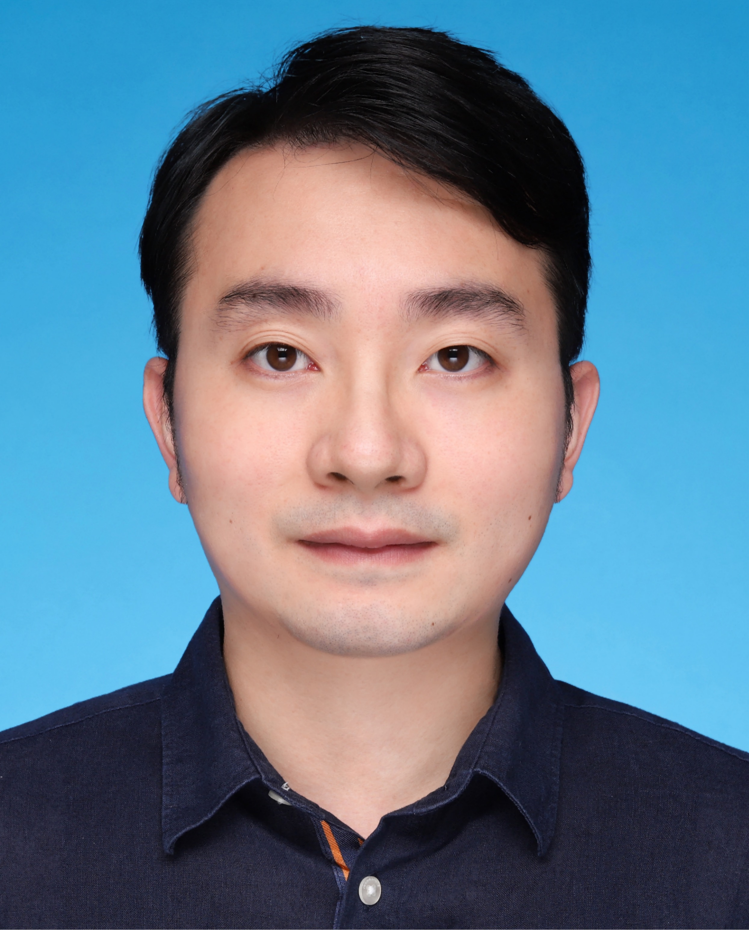 Xiaoshan Shi (施小山)合作论文数University of Chinese Academy of Sciences;Shenzhen Institutes of Advanced Technology, Chinese Academy of Sciences2Lili Wang合作论文数2
 Charlotte Christie Petersen合作论文数2
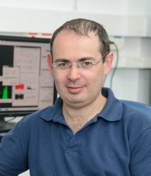 Ziv Porat合作论文数Department of Molecular Genetics, The Weizmann Institute of Science2Attila Tarnok合作论文数2Toby Garrood合作论文数2
 Derek Davies合作论文数Cancer Research UK FACS Laboratory, London Research Institute London2
 Diana L. Bonilla合作论文数2
 Joanne A. Lannigan合作论文数Flow Cytometry Support Services, LLC2
 Rui Gardner合作论文数Instituto Gulbenkian de Ciência2
 Gabriel Rinaldi合作论文数UDELAR, Universidad de la República2




