Microfluidic-spun hydrogel fibers are appealing for tissue engineering and cell transplantation applications, as they give access to fiber-shaped tissues that mimic blood vessels, muscle fibers, or neural networks in vivo. Alginates are overwhelmingly employed as their base material, as they allow for simple processing, despite their poor cell adherence and weak cell-matrix interactions. Alginates also require crosslinking, and thus can leach ions and lose integrity under physiological conditions. To overcome these limitations, we are reporting herein the first synthesis of pure chitosan fibers by microfluidic wet spinning, avoiding the use of crosslinking agents, and achieving excellent cell viability of 85%. These fibers exhibit higher mechanical strength (695 MPa) than alginate counterparts (2-4 MPa). Our system can also accommodate a core of chitin nanocrystals to modulate mechanical properties or serve as a reservoir of bioactive molecules, such as methylene blue. As chitosan is a natural biopolymer, this work addresses the United Nations Sustainable Development Goals (UN SDGs) 6 and 14. The resulting pure chitosan and chitin/chitosan composite fibers exhibit high processability and can be woven into a variety of structures. Finally, these microstructured chitosan fibers have the potential to be used as templates to create fiber-shaped tissues or to develop into live building blocks for the assembly of very complex artificial tissues.
Physical forces direct cell behaviors such as differentiation, migration, and division. Cell fusion is no exception. In addition to protein-based machinery, mechanical stresses, pressure differentials, and electric fields have been suggested to also regulate fusion. Decades of research have identified numerous fusogenic membrane proteins, yet the precise mechanisms of their action remain unclear. Here, we outline a biophysical framework that links the physics of model membranes to fusion in living cells. We describe how surface free energy, membrane curvature, tension, pressure, and electric fields can play a role in driving fusion, drawing on insights from both model membranes and cellular systems. Finally, we highlight how these biophysical factors contribute to fusion in the context of placental development and how they may inform future work in skeletal muscle, bone tissue, and tumors. By integrating molecular and biophysical perspectives, we provide a starting point to uncover conserved principles of cell fusion and advance a more integrated view of fusion as both a biochemical and biophysical process.
The yield stress at which biomaterials undergo plastic deformation limits the stresses that can be developed in encapsulated growing tissues. While matrix mechanical properties such as stiffness and viscoelasticity have a profound effect on cells, the role of yield stress has remained challenging to define. Here, we design a granular hydrogel platform with supramolecular host-guest dynamic crosslinkers to precisely and quantitatively tune the stress at which the matrix repeatedly yields and reconfigures around tissues as they grow. Designed to provide similar mechanical constraints as a mesh stress ball, matrix yield stresses can be tuned between 12 and 370 Pa, while maintaining a storage modulus below ∼0.1 kPa. Our study suggests that this range of yield stress is sufficient to promote or limit peripheral shedding in a model of non-adhesive cancer migration, and that early development of midbrain organoids is exquisitely sensitive to these matrix mechanics. Yield stresses of only 25 Pa promoted bud-like protrusions and large, luminized neural rosettes, while variations as small as 10 Pa limited these phenotypes. These studies indicate that morphogenesis and tissue organization can be controlled via the material's yield stress, suggesting a new mechanical parameter to target in designing biomaterials for disease modeling and regenerative medicine.
ABSTRACT The mechanical mutability of sea cucumbers is a source of bioinspiration for different stimuli‐responsive materials for biomedical applications, soft robotics, self‐healing hydrogels, and tunable scaffolds. However, using the source material itself has rarely been explored. Here, the structure‐function relationships of decellularized mutable collagenous tissue (MCT) from sea cucumbers are investigated, for their potential use as a tissue scaffold material. Given the harsh decellularization process, this necessitated an in‐depth investigation of decellularized MCT (dMCT) vis‐à‐vis native MCT (nMCT). Thus, a cross‐disciplinary hierarchical investigation was performed, utilizing multi‐length scale techniques to study the molecular, fibrillar, and bulk tissue composition and structure within the native and decellularized MCTs. Results highlight a similar composition and structure within 2D sections of both tissues; however, a 3D analysis of fibrillar orientation suggests an increase in the overall percentage of ordered fibrils of the dMCT. Moreover, a dramatic increase in bulk tissue stiffness was observed via rheology, supporting a previously described protein‐based mechanism of mechanical mutability. Finally, cell biocompatibility studies demonstrate that decellularized MCT is not toxic to living cells. Given the retention of native structure, cytocompatibility, and demonstration of modified mechanics following decellularization, MCT shows exceptional promise as an adaptable scaffold for tissue engineering applications.
Antibacterial membranes are often proposed for applications in which the membranes are in contact with the human body or in contact with food or drink, and hence their designs must minimize unintended toxicity. Zinc oxide (ZnO) is a well-known antibacterial agent and is relatively nontoxic, making it a promising material for the design of antibacterial membranes. Needle-like ZnO nanomaterials are believed to be additionally capable of a cell puncturing mechanism when they are agitated in suspension with bacteria. It is unclear, however, whether the puncturing mechanism is effective when the needle-like nanomaterials are immobilized as surface coatings. In this study, we assessed the antibacterial performance of two types of ZnO coatings synthesized on nylon membranes. One ZnO coating possessed no distinct hierarchical structure whereas the second consisted of ZnO microflowers each comprised of numerous ZnO nanoneedles, collectively forming a nanoneedle topography on the membrane surface. For antibacterial assessment of these coatings, we used several conventional assays and a variation of a recently developed bacterial bioluminescence monitoring assay. The conventional assays evaluated the antibacterial effects of zinc released from the membranes. The bioluminescence monitoring assay uniquely captured antibacterial effects of cell-surface contact between bacteria and the ZnO nanoneedle topography such as the puncture mechanism in question in real time without disturbing ongoing cell-surface interactions throughout incubation. Bioluminescent Staphylococcus aureus and bioluminescent Pseudomonas aeruginosa exposed to the ZnO nanoneedle topography exhibited loss and recovery of bioluminescence comparable to bacteria that were exposed to the ZnO coating without nanoneedle topography. We conclude that the nanotextured topography therefore did not further enhance antibacterial performance of the ZnO-coated membranes. S. aureus and P. aeruginosa were able to survive, recover, and proliferate directly atop the nanotextured ZnO coating.
Microscale engineering technologies are reshaping strategies for pancreatic islet replacement therapies. This review surveys three broad domains where microscale approaches are demonstrating considerable promise. In biomanufacturing applications, microscale platforms enhance control over oxygenation, nutrient delivery, hydrodynamic forces, and aggregate size, addressing longstanding challenges in the scalable and consistent production of stem cell-derived islets. In preclinical processing of therapeutic replacement and augmentation devices, we review recent advances in micro- and macroencapsulation, with emphasis on how microscale material design, membrane architecture, and oxygen-management strategies collectively aim to balance immunoprotection with adequate mass transport, an essential requirement for long-term graft survival. Finally, we also examine the expanding toolkit for functional assessment across the implantation lifecycle. Integrated microsensors and microfluidic testing systems enable localized, real-time monitoring of metabolic behavior before and during implantation, while transport-defined ex vivo platforms improve failure-mode analysis after graft retrieval. Together, these categories provide a coherent framework illustrating how microscale systems are being leveraged to improve the robustness, interpretability, and translational readiness of next-generation islet replacement therapies.
Reliabl electrophysiological sensing in brain organoids is limited by the high interfacial impedance and noise of microscale electrodes, particularly when scalable and reusable platforms are required. In this work, we introduce PEDOT-coated printed circuit board (PCB) electrodes as a biosensing platform for low-noise recording of organoid electrical activity. Conducting polymer coatings were electrodeposited directly onto PCB-integrated electrodes and systematically optimized to maximize interfacial capacitance while preserving coating adhesion and durability. The optimized PEDOT interfaces reduced electrode impedance to 3-5 kΩ at 1 kHz, corresponding to about three-order-of-magnitude decrease compared to Au-coated electrodes. The low-impedance response was retained after repeated autoclave sterilization and sonication, demonstrating robustness under conditions relevant to routine biological use. When applied to human brain organoids, the PEDOT-coated electrodes exhibited markedly reduced background noise and significantly enhanced signal-to-noise ratios (SNR), enabling reliable detection of extracellular spikes and synchronized burst activity across multiple channels. Relative to gold electrodes, the PEDOT-modified PCB platform recorded higher-amplitude signals and increased spike counts, indicating improved electrode-tissue coupling. These results establish PEDOT-coated PCB electrodes as a scalable and reusable biosensing interface for electrophysiological interrogation of 3D neural tissues.
Cancer associated fibroblasts (CAFs) play a critically important role in facilitating tumour cell invasion during metastasis. They also modulate local biophysical features of the tumour microenvironment through the formation of fibrotic foci, which have been correlated with breast cancer aggression. However, the impact of the evolving three-dimensional biophysical tumour microenvironment on CAF function remains undefined. Here, by isolating CAFs from primary human triple-negative breast cancer tissue at the time of surgery, we find that their ability to remodel the local microenvironment and invade into a three-dimensional matrix correlates with disease state. We then engineered culture models to systematically deconstruct and recreate mechanical tissue features of early breast cancer fibrotic foci; and demonstrate that invasion is mechanically-activated only in CAFs from patients with no detectable pre-existing metastases, but is independent of mechanical cues in CAFs isolated from patients with later-stage axillary lymph node metastases. By comparing the differential transcriptional response of these cells to microenvironmental tissue stiffness, we identify the aryl hydrocarbon receptor (AhR) as being significantly upregulated in invasive sub-populations of both mechanically-activated and mechanically-insensitive CAFs. Increasing AhR expression in CAFs induced invasion, while suppressing AhR significantly reduced invasion in both mechanically-activated and mechanically-insensitive CAF populations, even on stiffnesses that recapitulate late-stage disease. This work therefore uses mechanobiological analyses to identify AhR as a mediator of CAF invasion, providing a potential stratification marker to identify those patients who might respond to future mechanics-based prophylactic therapies, and provides a targetable mechanism to limit CAF-associated metastatic disease progression in triple-negative breast cancer patients. STATEMENT OF SIGNIFICANCE: By designing a mechanically-tunable tissue-engineered model of fibroblastic foci, and using this to culture patient-derived cancer-associated fibroblasts, we demonstrate that these cells are differentially mechanosensitive, depending on disease stage of the patient. While comparing transcriptomic profiles of patient-derived cells produces too many pathways to screen, identifying the pathways activated by local tissue mechanics that were common across each patient allowed us to identify a specific target to limit fibroblast invasion. This broad discovery strategy may be useful across a variety of biomaterials-based tissue engineered models; and these specific findings suggest (1) a strategy to identify patients who might respond to CAF- or matrix-targeting therapies, and (2) a specific actionable target to limit CAF-associated metastatic disease progression.
Generating microgels is of critical importance in developing granular biomaterials, which have diverse emerging applications in regenerative medicine and tissue engineering. However, producing large volumes of microgels while maintaining a reasonably low population of polydispersity remains a challenge. Here, we introduce the Turbinator, a device that can be added on to the commercially available Shirasu Porous Glass (SPG) microdroplet production system to provide precise control of the local shear stresses around the porous glass droplet production head. In addition to reducing the polydispersity of droplet sizes produced using the SPG, this system allows for continuous production of droplets in inexpensive and massively scalable kerosene oil baths for industrial manufacturing applications. To validate the device, we develop finite element models to understand the local shear stresses applied and characterize the droplets produced under various operating conditions. Finally, we confirmed that this production method supports biological activity via viability and spreading assays of fibroblast cells and invasion assays in a model cancer spheroid system.
Heart-on-a-chip platforms aim to recapitulate cardiac tissue structure and function in vitro. Traditionally, microfabricated pillars are used to estimate contractile forces based on pillar deflection. However, this approach measures only global forces at the pillar interface and lacks the spatial resolution needed to capture local mechanical stresses. In this study, we present a non-destructive optical method for continuous, multi-scale stress mapping using ultrasoft edge-labeled micro-spherical stress gauges (eMSGs). These embedded mechanosensors visibly deform in response to cellular and extracellular matrix (ECM)-generated stresses, enabling real-time measurements at cell and tissue scales. Our platform features dual cell-seeding chambers with flexible polydimethylsiloxane pillars, into which neonatal rat cardiomyocytes are seeded within a fibrin/Geltrex hydrogel containing eMSGs. Over time, tissues compacted, aligned, and exhibited spontaneous contractions and calcium transients. By modulating ECM composition, we found that reduced fibrin concentration enhanced contractile frequency, regularity, and force generation. Analysis of eMSG deformation enabled calculation of lateral and longitudinal stresses, revealing the impact of compaction and contraction on local mechanics. Finally, drug testing was performed using norepinephrine, which enhanced contractile force, and blebbistatin, which inhibited contraction, demonstrating robust pharmacological responsiveness. This platform provides a powerful tool for real-time biomechanical analysis and drug testing in engineered cardiac tissues.
Mechanical features of tissues have been recognised as key drivers of disease progression and are increasingly investigated as diagnostic and therapeutic targets. Engineered tissue models with integrated embedded biomechanical sensors have recently uncovered complex mechanical behaviors across micro- and nanoscale environments, offering novel insights into developmental and disease mechanisms. This short opinion synthesizes emerging mechanical signatures that have been identified at high measurement sensitivities and spatial resolutions by embedding customized biomechanical sensors into engineered tissues, particularly for soft tissue pathologies like cancer and fibrosis. We then describe the challenges of achieving these increased resolutions in clinical practice, and these gaps. If successful, these improved biomechanical measurement systems could open new pathways for improving diagnostics and patient outcomes.
Measuring the transport dynamics of soluble molecules such as nutrients, growth factors, and therapeutics within cell aggregates is essential to understand the transport-limiting effects of 3D cell culture models. Traditional methods to study molecular transport within engineered tissues often face challenges related to access for delivery and sampling and require sacrificing the culture. Here, we introduce an accessible, device-innovation platform that allows spatially defined delivery into a living cell aggregate. By integrating a highly perfusable, hollow-core agarose hydrogel microtube into a polyacrylamide microwell, our system allows local access to the aggregate's interior without advanced microfabrication or complex vascularization. Transport out of the hydrogel tube is limited to a specific spatial region within the aggregate by the deliberate placement and fracturing of a glass capillary sheath. We demonstrate an application of this system in an engineered model of placental trophoblast plugs, which arise temporarily during the first trimester of pregnancy to protect the placenta during development. Partial cell fusion occurs within these plugs, which would alter local transport characteristics, whether this morphological feature affects transport in a 3D tissue remains undefined. To address this, we created cylindrical plug-like aggregates and applied quantitative fluorescence measurements coupled with finite element modeling to determine that patterns of diffusivity are heterogeneous, with enhanced transport at the aggregate core compared to the periphery. This specific application highlights the platform's potential to study molecular transport dynamics within cell aggregates and provides a foundation for further exploration of diffusion-limited processes in 3D culture systems.
Stem-cell-derived islets (SC-islets) offer an alternative to cadaveric islets for Type 1 diabetes treatment. Controlling aggregate size during differentiation can improve reproducibility and performance, but current protocols are challenging to scale while maintaining control over this important parameter. We first establish that due to stochastic fusion, SC-islet sizes produced with conventional protocols can create chronic oxygen limitations during differentiation. We then propose and demonstrate the use of a micropocket hydrogel platform to facilitate the handling and long-term culture of separated cellular aggregates during SC-islet production. Micropockets are formed with an overhanging lip-and-funnel geometry, which allows single cells to easily enter the aggregation chamber while preventing aggregates from escaping during media exchanges. We design and fabricate these micropockets in inert polyacrylamide hydrogels and demonstrate that this design protects the aggregates from shear stress (∼50× reduction) during media exchanges. We also compare pyramidal, conical, and spherical micropocket chamber geometries to optimize the aggregate formation. Pancreatic progenitor cells were aggregated and differentiated into SC-islets over 23 days, during which aggregate sizes consistent with human islets were maintained (136 μm, ±31 μm SD). In contrast, aggregates produced in suspension culture increased in size throughout differentiation (from 114 μm ± 8 μm SD to 275 μm ± 62 μm SD) due to stochastic aggregate fusion. Furthermore, no aggregate losses during daily media exchanges were incurred in micropockets, suggesting a highly scalable approach to produce appropriately sized islets while maintaining precise control over microenvironmental conditions. Overall, this work demonstrates the utility of a single-step culture system to form and maintain aggregates during lengthy differentiation processes, which can ultimately be scaled for therapeutic production applications.
Heart-on-a-chip platforms aim to miniaturize and replicate the complex structure and function of cardiac tissue. Traditionally, microfabricated pillar pairs have been employed in these systems to provide tissue anchorage and determine contractility parameters based on pillar deflection. However, this approach lacks the spatial resolution required to capture local cell- and tissue-scale mechanical stresses. In this study, we established a non-destructive optical method for continuous micro- and macro-scale contractile force measurements. We utilized our previously developed edge-labeled micro-spherical stress gauges (eMSGs) to map the stresses within a heart-on-a-chip. These ultrasoft mechanosensors visibly deform in response to stresses generated by cells and the extracellular matrix (ECM). The chip consisted of two cell-seeding chambers, each containing flexible silicone pillar pairs to support tissue formation and compaction. Neonatal rat cardiomyocytes (CMs) were encapsulated in a fibrin/Geltrex hydrogel mixture containing eMSGs and seeded into each chamber. Over time, the tissue compacted and began beating spontaneously, demonstrating structural alignment and functional cardiac hallmarks, such as calcium transients and tissue-scale beating. The effects of ECM composition on tissue function were examined, revealing that lower fibrin concentrations significantly enhanced contractile frequency, regularity, and stress generation. Local cell- and ECM-scale mechanics were further investigated by analyzing the shape changes of the dispersible sensors. Lateral and longitudinal stresses were calculated for each sensor, highlighting the critical role of tissue compaction and contraction in cell-generated forces. Finally, the platform was validated using two known drug candidates, with their effects on contractility clearly demonstrated. ### Competing Interest Statement M.A. is a co-founder of eNUVIO Inc. All other authors declare no competing financial interest. Natural Sciences and Engineering Research Council of Canada (NSERC), RGPIN-2021-03960, DGECR-2021-00337, RGPIN-2022-05165 Fonds de Recherche du Québec - Santé, https://ror.org/02eqrsj93, Chercheurs-boursiers J1 (313837), Establishment of Young Investigators (324277) Montréal TransMedTech Institute (iTMT), X
Organoids have emerged as valuable tools for the study of development and disease. Assembloids are formed by integrating multiple organoid types to create more complex models. However, the process by which organoids integrate to form assembloids remains unclear and may play an important role in the resulting organoid structure. Here, a microfluidic platform is developed that allows separate culture of distinct organoid types and provides the capacity to partially control the geometry of the resulting organoid surfaces. Removal of a microfabricated barrier then allows the shaped and positioned organoids to interact and form an assembloid. When midbrain and unguided brain organoids were allowed to assemble with a defined spacing between them, axonal projections from midbrain organoids and cell migration out of unguided organoids were observed and quantitatively measured as the two types of organoids fused together. Axonal projection directions were statistically biased toward other midbrain organoids, and unguided organoid surface geometry was found to affect cell invasion. This platform provides a tool to observe cellular interactions between organoid surfaces that are spaced apart in a controlled manner, and may ultimately have value in exploring neuronal migration, axon targeting, and assembloid formation mechanisms.
The syncytiotrophoblast is a multinucleated structure that arises from fusion of mononucleated cytotrophoblasts, to sheath the placental villi and regulate transport across the maternal-fetal interface. Here, we ask whether the dynamic mechanical forces that must arise during villous development might influence fusion, and explore this question using in vitro choriocarcinoma trophoblast models. We demonstrate that mechanical stress patterns arise around sites of localized fusion in cell monolayers, in patterns that match computational predictions of villous morphogenesis. We then externally apply these mechanical stress patterns to cell monolayers and demonstrate that equibiaxial compressive stresses (but not uniaxial or equibiaxial tensile stresses) enhance expression of the syndecan-1 and loss of E-cadherin as markers of fusion. These findings suggest that the mechanical stresses that contribute towards sculpting the placental villi may also impact fusion in the developing tissue. We then extend this concept towards 3D cultures and demonstrate that fusion can be enhanced by applying low isometric compressive stresses to spheroid models, even in the absence of an inducing agent. These results indicate that mechanical stimulation is a potent activator of cellular fusion, suggesting novel avenues to improve experimental reproductive modelling, placental tissue engineering, and understanding disorders of pregnancy development.
Congenital and acquired valvular heart diseases (VHDs) are significant causes of mortality worldwide. With valve replacement being the primary solution for VHD, current options display shortcomings, including calcification, thrombogenicity, and hemodynamic alteration, leading to repetitive surgeries. Tissue engineering, however, has shown great potential for fabricating heart valves (HVs) with fewer complications. Here, a series of inks are developed, combining poly(vinyl alcohol), gelatin, and carrageenan for 3D printing of tissue‐engineered heart valves (TEHVs). The inks/hydrogels are investigated to characterize their physico‐chemical, morphological, mechanical, and rheological characteristics. In vitro and in vivo biocompatibility, immune response, hemolysis, and thrombogenicity of the inks/hydrogels are also evaluated. Moreover, in vitro hydrodynamics of the TEHVs under physiological conditions are reported. Inks demonstrate mechanical characteristics comparable to native leaflets. Subcutaneous implantation reveals that the hydrogels do not induce chronic inflammation and can undergo remodeling. In vitro hemocompatibility assessments of the hydrogels show minimal hemolysis with low thrombogenicity. Different sizes and types of HVs are successfully printed with high fidelity in the air. In vitro hydrodynamic assessment confirms that the TEHVs can withstand aortic conditions. Altogether, the 3D‐printed TEHVs can be a promising alternative for valve replacement to solve the problems associated with the current options.
Three-dimensional (3D) bioprinting is an advanced fabrication technique to build biomimetic constructs for different biomedical applications. Here, we developed composite bioinks based on photocrosslinkable natural polymers, gelatin methacryloyl (GelMA) and alginate methacrylate (AlgMA), and electroconductive reduced graphene oxide (rGO) nanomaterials. The photocrosslinked hydrogels indicated interconnected porous microarchitecture with detectable rGO nanosheets on the pore walls. The addition of AlgMA and rGO to GelMA bioinks significantly enhanced tensile and compressive mechanical properties, improved the rheological properties and printability of inks, and controlled the degradation profile of the constructs. The optimization was done based on the suitable electromechanical properties for cardiac tissue engineering applications. The ring-shaped cardiac constructs were further 3D bioprinted with different cardiac cell types (neonatal rat cardiomyocytes, cardiac fibroblasts, and HL-1 cells). The optimized bioink supported a high level of cardiac cell viability, proliferation, spreading, elongation, and alignment with functional hallmarks (e.g., calcium transient and cellular spontaneous beating). Proof-of-concept validation was demonstrated for utilizing this platform as a 3D bioprinted heart-on-a-chip model with automated high-throughput in the 12-well plates. The developed bioprinted cardiac model could be used for different applications, including drug screening and disease modeling.
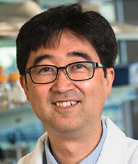 Shuichi Takayama合作论文数Wallace H. Coulter Department of Biomedical Engineering, College of Engineering, Georgia Institute of Technology20
 Yu Sun (孙钰)合作论文数Department of Mechanical and Industrial Engineering, University of Toronto;University of Toronto Robotics Institute20
 Craig A. Simmons合作论文数Simmons Lab for Cellular Mechanobiology, Department of Mechanical & Industrial Engineering, Faculty of Applied Science and Engineering, University of Toronto;Boutiq Solutions Inc.;Ted Rogers Centre for Heart Research19
 Stephanie Mok合作论文数Department of Chemical Engineering, McGill University11
 Wontae Lee合作论文数McGill Univ, Dept Chem Engn, Montreal, PQ, Canada10
 Nikita Kalashnikov合作论文数McGill Univ, Dept Chem Engn, Montreal, PQ H3A 0C5, Canada10
 Nathalie Tufenkji合作论文数Biocolloids and Surfaces Laboratory, Department of Chemical Engineering, McGill University10
 Prabu Karthick Parameshwar合作论文数Dept Biol & Biomed Engn, McGill Univ9
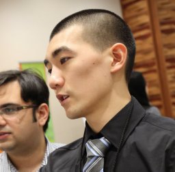 Nicholas Lin合作论文数Dept Chem Engn, McGill Univ9
 Raymond Tran合作论文数Dept Chem Engn, McGill Univ8
 Cathy Vaillancourt合作论文数INRS – Institut Armand-Frappier, Université du Québec8
 Arvind Chandrasekaran合作论文数CAVE Research Center, CON;Optical Microsystems Laboratory;Dept . of Mechanical Engineering;CONCORDIA University;W;CONCAVE Research Center, CONCORDIA University7
 Richard L Leask合作论文数Department of Biomedical Engineering, Faculty of Medicine and Health Sciences, McGill University6
 Luke McCaffrey合作论文数Rosalind and Morris Goodman Cancer Institute, Faculty of Medicine and Health Sciences, McGill University/Department of Medicine, Division of Experimental Medicine, Faculty of Medicine and Health Sciences, McGill University6
 Michael Thouless合作论文数Department of Materials Science and Engineering, University of Michigan;Department of Mechanical Engineering, University of Michigan6
 Brendan M. Leung合作论文数University of Toronto Institute of Biomaterials and Biomedical Engineering6Zhenwei Ma合作论文数5Lucas Sagrillo-Fagundes合作论文数5
 Dao Nguyen合作论文数Division of Experimental Medicine, Department of Medicine, Faculty of Medicine and Health Sciences, McGill University5
 Corinne Hoesli合作论文数Department of Chemical Engineering, Faculty of Engineering, McGill University5
 Sasha Cai Lesher-Perez合作论文数5Kim Byoung Choul合作论文数University of Michigan, University of Michigan5Gregor Andelfinger合作论文数5Geoffrey A. Mckay合作论文数5
 Houman Savoji合作论文数Chemical & Petroleum Engineering Department, Sharif University of Technology5
 Bogdan Beca合作论文数4Toshiki Matsuoka合作论文数4Christina-Marie Boghdady合作论文数4
 Thomas Durcan合作论文数Department of Neurology and Neurosurgery, Faculty of Medicine and Health Sciences, McGill University4
 Jonathan Spicer合作论文数Division of Experimental Medicine, Department of Medicine, Faculty of Medicine and Health Sciences, McGill University4
 Amin Valiei合作论文数Department of Chemical and Material Engineering, University of Alberta.4
 Labuz Joseph M合作论文数Department of Biomedical Engineering, University of Michigan4
 Morag Park合作论文数Department of Biochemistry, School of Biomedical Sciences, Faculty of Medicine and Health Sciences, McGill University;Rosalind and Morris Goodman Cancer Institute, Faculty of Medicine and Health Sciences, McGill University4
 Roni F. Rayes合作论文数Florida State University4
 Zhe Lu合作论文数Bluewrist Inc.4Benjamin E Campbell合作论文数3Jiexi Huang合作论文数3Nicholas Wong合作论文数3Camille Cassel de Camps合作论文数3
 Paula Lepine合作论文数3Naimeh Rafatian合作论文数3Sonya Kouthouridis合作论文数3Claire Wang合作论文数3Ali Mousavi合作论文数University of Montreal, University of Montreal3
 Katherine N MacDonald合作论文数Michael Smith Laboratories, University of British Columbia, Vancouver, British Columbia V6T 1Z4, Canada.3
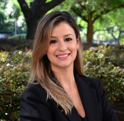 Nathalia Azevedo Portilho合作论文数Inst Mol Med, Univ Texas Hlth Sci Ctr Houston3
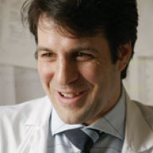 Lorenzo Ferri合作论文数Gerald Bronfman Department of Oncology, Faculty of Medicine and Health Sciences, McGill University;Division of General Surgery, Department of Surgery, Faculty of Medicine and Health Sciences, McGill University;Division of Thoracic Surgery, Department of Surgery, Faculty of Medicine and Health Sciences, McGill University3
 Sidong Huang合作论文数Department of Biochemistry, Faculty of Medicine and Health Sciences, McGill University3
 William Pastor合作论文数Department of Biochemistry, Faculty of Medicine and Health Sciences, McGill University3




