Background: While nasal epistaxis balloons are generally seen as safe and routinely utilized by both surgical and nonsurgical providers, the complication profile related to this type of device has not been well defined. Objective: The objective of this study was to utilize the FDA MAUDE (Manufacturer and User Facility Device Experience) database to better assess adverse events (AE) related to use of nasal epistaxis balloons. Reports were individually tabulated and events were categorized with special attention to AEs. Methods: The FDA MAUDE database was queried for all medical device reports (MDR) related to nasal epistaxis balloon devices from January 2012 to November 2022. Results: 19 MDRs met inclusion criteria. 5 MDRs were classified as device related (26.3 %); two events were reported for balloon leak and deflation, two events were reported for device breakage, and one device related event was unknown. 14 MDRs (73.7 %) were classified as patient related. Two documented MDRs were patient deaths due to exsanguination. Additional serious AEs included balloon ingestion and subsequent small bowel perforation (n = 1), cerebrospinal fluid leak (n = 1), skull base violation and intracranial placement of the device (n = 1), and respiratory distress (n = 3). Conclusion: Though epistaxis control with nasal balloons is generally seen as a safe procedure, there have been several concerning AEs reported. While two reports of death due to exsanguination were the most severe AEs, multiple other life-threatening AEs were also documented. Increased awareness of associated complications can be used to better counsel patients during the informed consent process as well as providers in their clinical decision making.
SUMMARY:Virtual surgical planning (VSP) has been applied to many aspects of head and neck reconstruction. The authors describe the use of VSP to create auricular templates in addition to cartilage cutting and suturing guides for microtia repair in 2 patients with unilateral and bilateral grade 3 microtia. Both patients had satisfactory aesthetic results. This technique allows for increased precision, may decrease operative time, and has good cosmetic outcomes.
Objectives Endoscopic endonasal anterior skull base surgery has expanding use in the pediatric population, but the anatomy of pediatric patients can lead to limitations. This study aims to characterize the important anatomical implications of the pediatric skull base using computed tomography (CT) scans. Design This study is designed as retrospective analysis. Setting The study setting comprises of tertiary academic medical center. Participants In total, 506 patients aged 0 to 18 who had undergone maxillofacial and or head CTs between 2009 to 2016 were involved. Methods Measurements included piriform aperture width, nare to sella distance (NSD), sphenoid pneumatization, olfactory fossa depth, lateral lamella cribriform plate angles, and intercarotid distances (ICD) at the superior clivus and cavernous sinus. These patients were then subdivided into three age groups adjusting for sex. Analysis of covariance (ANCOVA) models were fit comparing between all age groups and by sex. Results Piriform aperture width, NSD, sphenoid sinus pneumatization as measured using lateral aeration and anterior sellar wall thickness, olfactory fossa depth, and ICD at the cavernous sinus were significantly different among all age groups ( p <0.0001). Our results show that mean piriform aperture width increased with each age group. The mean olfactory fossa depth also had consistent age dependent growth. In addition, ICD at the cavernous sinus showed age dependent changes. When comparing by sexes, females consistently showed smaller measurements. Conclusion The process of skull base development is age and sex dependent. During preoperative evaluation of pediatric patients for skull base surgery piriform aperture width, sphenoid pneumatization in both the anterior posterior and lateral directions, and ICD at the cavernous sinus should be carefully reviewed.
A myriad of techniques have been described to correct brow ptosis via a blepharoplasty incision, mid-forehead incision, endoscopic approach, pretrichial incision, coronal approach or direct brow lift. There are varying risks and results for each technique including paresthesia, facial nerve injury, alopecia, and unsightly scars. In this article we describe the mini-direct browlift technique that utilizes bone anchored sutures and allows for good cosmesis and control of brow height with minimal morbidity. A myriad of techniques have been described to correct brow ptosis via a blepharoplasty incision, mid-forehead incision, endoscopic approach, pretrichial incision, coronal approach or direct brow lift. There are varying risks and results for each technique including paresthesia, facial nerve injury, alopecia, and unsightly scars. In this article we describe the mini-direct browlift technique that utilizes bone anchored sutures and allows for good cosmesis and control of brow height with minimal morbidity. A myriad of techniques have been described to correct brow ptosis via a blepharoplasty incision, mid-forehead incision, endoscopic approach, pretrichial incision, coronal approach or direct brow lift. There are varying risks and results for each technique including paresthesia, facial nerve injury, alopecia, and unsightly scars. In this article we describe the mini-direct browlift technique that utilizes bone anchored sutures and allows for good cosmesis and control of brow height with minimal morbidity. A myriad of techniques have been described to correct brow ptosis via a blepharoplasty incision, mid-forehead incision, endoscopic approach, pretrichial incision, coronal approach or direct brow lift. There are varying risks and results for each technique including paresthesia, facial nerve injury, alopecia, and unsightly scars. In this article we describe the mini-direct browlift technique that utilizes bone anchored sutures and allows for good cosmesis and control of brow height with minimal morbidity.
Background Eustachian tube balloon dilation (ETBD) has been Food and Drug Administration (FDA) approved for refractory Eustachian tube dysfunction since 2016. While ETBD is generally seen as safe, the complication profile has not been well defined.Objective The objective of this study was to utilize the FDA manufacturer and user facility device experience (MAUDE) database to better assess adverse events (AE) related to ETBD.Methods This is a study of a multiinstitutional database maintained by the U.S. FDA. A database analysis was performed via the collaboration of multiple clinicians at tertiary referral centers. The FDA MAUDE database was queried for all medical device reports (MDR) related to ETBD devices from January 2012 to November 2022. Eighty-eight unique MDR were identified, 16 of which met inclusion criteria.Results Three MDRs were classified as device-related (18.8%); none resulted in an AE. Thirteen MDRs (81.3%) were patient-related; all were classified as AEs. The most common AE was postoperative subcutaneous emphysema (n = 6, 46.2%). Of the patients with subcutaneous emphysema, there was a wide range of severity. The most severe AE (n = 1, 6.3%) was postoperative stroke secondary to carotid artery dissection.Conclusion Though ETBD is generally seen as a safe procedure, there have been several concerning AEs reported to date. Increased awareness of ETBD complications can serve as a primer for improved patient education and counseling during the informed consent process and aid surgeons in clinical decision-making. Future studies with standardized reporting protocols are warranted to create a central registry for ETBD.
Skull base defects following resection of anterior cranial fossa and sinonasal tumors are not uncommon. Advances in endoscopic techniques have allowed for entirely endonasal resection and reconstruction of these tumors. This article discusses techniques in the evaluation and management of anterior skull base defects.
KEY POINTS:Eustachian tube recanalization is a feasible procedure but additional studies are needed to determine its safety. Eustachian tube closure can result from different etiologies and can cause severe symptoms. Ureteral stents have appropriate shape and pliability for placement and long-term healing. Multidisciplinary team approach allows for simultaneous endonasal and otologic approaches.
The LaryngoscopeEarly View Triological Society Best Practice When is the Optimal Time to Reduce Pediatric Nasal Bone Fractures? Sheng Zhou MD, Sheng Zhou MD USC Caruso Department of Otolaryngology-Head and Neck Surgery, Los Angeles, California, USASearch for more papers by this authorMarta Kulich MD, Marta Kulich MD USC Caruso Department of Otolaryngology-Head and Neck Surgery, Los Angeles, California, USASearch for more papers by this authorChristopher Pool MD, Christopher Pool MD Kaiser Permanente Orange County, Irvine, California, USASearch for more papers by this authorJavan Nation MD, Javan Nation MD Division of Pediatric Otolaryngology, Rady Children's Hospital, San Diego, California, USA Department of Otolaryngology—Head and Neck Surgery, University of California San Diego, La Jolla, California, USASearch for more papers by this authorShelby Leuin MD, Shelby Leuin MD Division of Pediatric Otolaryngology, Rady Children's Hospital, San Diego, California, USA Department of Otolaryngology—Head and Neck Surgery, University of California San Diego, La Jolla, California, USASearch for more papers by this authorJeffrey A. Koempel MD, MBA, Jeffrey A. Koempel MD, MBA Division of Pediatric Otolaryngology, Children's Hospital Los Angeles, Los Angeles, California, USASearch for more papers by this authorVijay A. Patel MD, Corresponding Author Vijay A. Patel MD [email protected] orcid.org/0000-0002-8145-1721 Division of Pediatric Otolaryngology, Rady Children's Hospital, San Diego, California, USA Department of Otolaryngology—Head and Neck Surgery, University of California San Diego, La Jolla, California, USA Send correspondence to Vijay Patel, Rhinology and Cranial Base Surgery, Complex Pediatric Otolaryngology, Rady Children's Hospital, Department of Otolaryngology – Head & Neck Surgery, University of California San Diego, La Jolla, California, USA. Email: [email protected]Search for more papers by this author Sheng Zhou MD, Sheng Zhou MD USC Caruso Department of Otolaryngology-Head and Neck Surgery, Los Angeles, California, USASearch for more papers by this authorMarta Kulich MD, Marta Kulich MD USC Caruso Department of Otolaryngology-Head and Neck Surgery, Los Angeles, California, USASearch for more papers by this authorChristopher Pool MD, Christopher Pool MD Kaiser Permanente Orange County, Irvine, California, USASearch for more papers by this authorJavan Nation MD, Javan Nation MD Division of Pediatric Otolaryngology, Rady Children's Hospital, San Diego, California, USA Department of Otolaryngology—Head and Neck Surgery, University of California San Diego, La Jolla, California, USASearch for more papers by this authorShelby Leuin MD, Shelby Leuin MD Division of Pediatric Otolaryngology, Rady Children's Hospital, San Diego, California, USA Department of Otolaryngology—Head and Neck Surgery, University of California San Diego, La Jolla, California, USASearch for more papers by this authorJeffrey A. Koempel MD, MBA, Jeffrey A. Koempel MD, MBA Division of Pediatric Otolaryngology, Children's Hospital Los Angeles, Los Angeles, California, USASearch for more papers by this authorVijay A. Patel MD, Corresponding Author Vijay A. Patel MD [email protected] orcid.org/0000-0002-8145-1721 Division of Pediatric Otolaryngology, Rady Children's Hospital, San Diego, California, USA Department of Otolaryngology—Head and Neck Surgery, University of California San Diego, La Jolla, California, USA Send correspondence to Vijay Patel, Rhinology and Cranial Base Surgery, Complex Pediatric Otolaryngology, Rady Children's Hospital, Department of Otolaryngology – Head & Neck Surgery, University of California San Diego, La Jolla, California, USA. Email: [email protected]Search for more papers by this author First published: 07 June 2023 https://doi.org/10.1002/lary.30794 Editor's Note: This Manuscript was accepted for publication on May 16, 2023. The authors have no funding, financial relationships, or conflicts of interest to disclose. Read the full textAboutPDF ToolsRequest permissionExport citationAdd to favoritesTrack citation ShareShare Give accessShare full text accessShare full-text accessPlease review our Terms and Conditions of Use and check box below to share full-text version of article.I have read and accept the Wiley Online Library Terms and Conditions of UseShareable LinkUse the link below to share a full-text version of this article with your friends and colleagues. Learn more.Copy URL No abstract is available for this article. BIBLIOGRAPHY 1Vyas RM, Dickinson BP, Wasson KL, Roostaeian J, Bradley JP. Pediatric facial fractures: current national incidence, distribution, and health care resource use. J Craniofac Surg. 2008; 19(2): 339- 349; discussion 350. https://doi.org/10.1097/SCS.0b013e31814fb5e3. 2Yabe T, Tsuda T, Hirose S, Ozawa T. Comparison of pediatric and adult nasal fractures. J Craniofac Surg. 2012; 23(5): 1364- 1366. https://doi.org/10.1097/SCS.0b013e31824dfb7b. 3Lee DH, Jang YJ. Pediatric nasal bone fractures: does delayed treatment really lead to adverse outcomes? Int J Pediatr Otorhinolaryngol. 2013; 77(5): 726- 731. https://doi.org/10.1016/j.ijporl.2013.01.027. 4Yilmaz MS, Guven M, Kayabasoglu G, Varli AF. Efficacy of closed reduction for nasal fractures in children. Br J Oral Maxillofac Surg. 2013; 51(8): e256- e258. https://doi.org/10.1016/j.bjoms.2013.07.008. 5Kang WK, Han DG, Kim SE, Lee YJ, Shim JS. Comparison of postoperative outcomes between early and delayed surgery for pediatric nasal fractures. Arch Craniofacial Surg. 2021; 22(2): 93- 98. https://doi.org/10.7181/acfs.2021.00122. Early ViewOnline Version of Record before inclusion in an issue ReferencesRelatedInformation
Abstract Objective To determine if otolaryngologists and audiologists of the American Academy of Otolaryngology – Head and Neck Surgery have noticed an increase in the incidence of sudden sensorineural hearing loss during the coronavirus disease 2019 pandemic. Methods A questionnaire was developed for the purpose of providing a cross-sectional descriptive analysis of perceived association between the coronavirus disease 2019 pandemic and an increase in the incidence of sudden sensorineural hearing loss. Results Of respondents, 63.0 per cent did not notice an increase in sudden sensorineural hearing loss during the coronavirus disease 2019 pandemic. There was a weak positive correlation between patients identified with sudden sensorineural hearing loss and the percentage of coronavirus disease 2019 positive patients reported by each medical care provider (Spearman correlation = 0.20, 95 per cent confidence interval = 0.05–0.33). There was no association between geographical location and perceived increase in sudden sensorineural hearing loss (p = 0.38). Conclusion The majority of respondents did not perceive an increase in the incidence of sudden sensorineural hearing loss during the coronavirus pandemic, regardless of geographical region.
Abstract Background The risk of skull base injury during choanal atresia repair can be mitigated via thorough understanding of skull base anatomy. There is a paucity of data describing differences in skull base anatomy between patients with coloboma, heart defects, atresia choanae, growth retardation, genital abnormalities, and ear abnormalities (CHARGE) syndrome and those without. Objectives The aim of this study was to measure nasal and skull base anatomy in patients with isolated bilateral choanal atresia (BCA), CHARGE syndrome, and other syndromic congenital anomalies. Methods Retrospective chart review of patients with bilateral choanal atresia and computed tomography of the face between 2001 and 2019 were evaluated. Choanal width, height, mid-nasal height, and skull base slope were measured radiographically. Differences in anatomy between healthy patients, those with CHARGE syndrome, and those with other congenital anomalies were compared. Results Twenty-one patients with BCA and relevant imaging were identified: 7 with isolated BCA, 6 with CHARGE syndrome, and 8 with other congenital anomalies. A t-test indicated insignificant difference in skull base slope, choanal height, choanal width, or mid-nasal skull base height between isolate BCA cases and patients with any congenital anomaly. When comparing CHARGE to isolated BCA cases, mid-nasal height was shorter in CHARGE patients (p = 0.03). There were no differences in measurements between patients with congenital anomalies excluding CHARGE (p > 0.05). Two patients in the congenital anomaly group were found to have bony skull base defects preoperatively. Conclusion This study represents the largest description of skull base and nasal anatomy in patients with CHARGE syndrome and BCA. Surgeons should be aware of the lower skull base in CHARGE patients to avoid inadvertent skull base injury.
Background: Sinocutaneous fistulae (SCF) are abnormal communications between the paranasal sinuses and the overlying skin. They may be difficult to manage due to facial geometry, scar contraction, and poor tissue vascularity. We describe a novel use of the buccal flap and review the literature to examine management options for this disease process. Methods: A PubMed/MEDLINE literature search was performed for studies published between January 1, 1950 and April 29, 2020 that describe management strategies for SCF. The clinical record, imaging, and operative reports were reviewed of the case in which the buccal fat flap was used in reconstruction. Results: A total of 359 articles were retrieved. After removing duplicate articles, non-English studies, animal studies, duplicate articles and studies that mentioned SCF without specific mention of management strategies, 51 articles were reviewed. Management paradigms throughout the articles include (1) removal of infection, (2) ensuring patency of sinus outflow tracts, (3) tensionless multilayered closure using well vascularized tissue, and (4) prevention or minimization of future risk factors for fistula formation. Conclusion: This article informs surgeons on reconstructive options for sinocutaneous fistulae including a novel description of the buccal fat flap.
Facial Plastic Surgery & Aesthetic MedicineVol. 24, No. 6 Research LettersCost Analysis of Cadaveric Versus Autologous Costal Cartilage Grafting in SeptorhinoplastyGhazal S. Daher, Kasra Ziai, Robert A. Saadi, Christopher Pool, and Jessyka G. LighthallGhazal S. Daherhttps://orcid.org/0000-0001-5013-3555Department of Otolaryngology—Head and Neck Surgery, College of Medicine, Pennsylvania State University, Hershey, Pennsylvania, USA.Search for more papers by this author, Kasra ZiaiDepartment of Otolaryngology—Head and Neck Surgery, College of Medicine, Pennsylvania State University, Hershey, Pennsylvania, USA.Search for more papers by this author, Robert A. SaadiDepartment of Otolaryngology—Head and Neck Surgery, College of Medicine, Pennsylvania State University, Hershey, Pennsylvania, USA.Search for more papers by this author, Christopher PoolDepartment of Otolaryngology—Head and Neck Surgery, College of Medicine, Pennsylvania State University, Hershey, Pennsylvania, USA.Search for more papers by this author, and Jessyka G. Lighthall*Address correspondence to: Jessyka G. Lighthall, MD, FACS, Facial Plastic and Reconstructive Surgery, Department of Otolaryngology-Head and Neck Surgery, College of Medicine, The Pennsylvania State University, 500 University Drive, H091, Hershey, PA 17033-0850, USA, E-mail Address: jlighthall@pennstatehealth.psu.eduhttps://orcid.org/0000-0002-8500-5059Facial Plastic and Reconstructive Surgery, Department of Otolaryngology-Head and Neck Surgery, College of Medicine, The Pennsylvania State University, Hershey, Pennsylvania, USA.Search for more papers by this authorPublished Online:8 Nov 2022https://doi.org/10.1089/fpsam.2021.0249AboutSectionsView articleView Full TextPDF/EPUB Permissions & CitationsPermissionsDownload CitationsTrack CitationsAdd to favorites Back To Publication ShareShare onFacebookTwitterLinked InRedditEmail View article"Cost Analysis of Cadaveric Versus Autologous Costal Cartilage Grafting in Septorhinoplasty." Facial Plastic Surgery & Aesthetic Medicine, 24(6), pp. 489–490FiguresReferencesRelatedDetails Volume 24Issue 6Dec 2022 InformationCopyright 2022, American Academy of Facial Plastic and Reconstructive Surgery, Inc.To cite this article:Ghazal S. Daher, Kasra Ziai, Robert A. Saadi, Christopher Pool, and Jessyka G. Lighthall.Cost Analysis of Cadaveric Versus Autologous Costal Cartilage Grafting in Septorhinoplasty.Facial Plastic Surgery & Aesthetic Medicine.Dec 2022.489-490.http://doi.org/10.1089/fpsam.2021.0249Published in Volume: 24 Issue 6: November 8, 2022Online Ahead of Print:April 4, 2022PDF download
Objective: To evaluate the safety profile, including adverse drug reactions and operative complications, of liposomal bupivacaine (LB) use for local anesthesia during functional septorhinoplasty. Study Design: Retrospective review. Subject and Methods: A database query was conducted for patients >18 years of age who underwent septorhinoplasty from January 1, 2019, to August 1, 2020. Adverse drug reactions and postoperative outcomes were compared between patients who received locally administered LB and patients who received standard local anesthetic at the completion of the surgery. Results: A total of 95 cases were included in our data analysis. No significant differences were found in adverse reactions overall (6.3% vs. 3.1%, p = 0.51) or complications, including rate of infection (3.2% vs. 3.1%, p = 1), cartilage warping (1.6% vs. 0%, p = 1), graft resorption (0% vs. 0%, p = 1), septal hematoma (0% vs. 0%, p = 1), and need for revision surgery (4.8% vs. 3.1%, p = 1) between LB and control groups. Conclusion: LB demonstrated an acceptable safety profile when compared with standard local anesthetics during septorhinoplasty, with no significant difference in systemic or local adverse drug reactions or postoperative complications.
BACKGROUND:The color change of topical intranasal fluorescein has been used to confirm the presence of cerebrospinal fluid (CSF) during endoscopic endonasal surgery. We aimed to validate the use of topical intranasal fluorescein for CSF detection. METHODS:Blood, CSF, saliva, and normal saline were combined with decreasing fluorescein concentrations (from 10% to 0.1%). The solutions were photographed in high definition on nasal pledgets and in 1.5-mL Eppendorf tubes. The color difference (ΔE) was objectively measured via the International Commission on Illumination coordinates. Four otolaryngologists who were unaware of the study parameters also evaluated the samples for perceptible color differences. The human eye cannot detect color differences at an International Commission on Illumination ΔE of <5. RESULTS:All otolaryngologists agreed a color difference could be seen with blood across all fluorescein concentrations. However, a perceptible color difference between the experimental samples that excluded blood was not appreciable. Objectively, the ΔE was <5 on average for all nonblood samples when mixed with 5% and 10% fluorescein in the Eppendorf experiment. The ΔE for the nonblood samples was >5 for the remaining tested. Similarly, the average ΔE for the nonblood samples in the pledget experiment was >5 across all fluorescein concentrations. The blood ΔE was consistently >50 throughout all fluorescein concentrations in the Eppendorf experiment and >20 throughout the pledget experiment, correlating with the subjective ease of discernment between blood and the control sample in both groups. CONCLUSIONS:Color change alone is not sufficient to determine a difference between CSF, saliva, and saline. Blood, however, is readily identified using this method. Adjunct characteristics, in addition to the color change, are necessary to properly identify an active CSF leak.
BACKGROUND:Juvenile nasopharyngeal angiofibroma (JNA) is a locally aggressive benign vascular tumor that typically afflicts young adolescent males. Historically removed via open approaches, these tumors are now being removed endoscopically. As the modern healthcare setting emphasizes value, efficient utilization of resources may lead to decreased cost while maintaining or improving patient outcomes. OBJECTIVE:The objective of this study was to investigate how perioperative management of juvenile nasopharyngeal angiofibromas (JNAs) influence overall cost. We specifically investigate the effect of approach type (open, endoscopic, or combined) with regards to cost and length of stay. We also delineated practice patterns, analyzed safety profiles, and characterize clinical outcomes. METHODS:The 2016 Healthcare Cost and Utilization Project Kids' Inpatient Database (HCUP-KID) was queried to identify males aged <21 years with an ICD-10-CM diagnosis code of D10.6 (benign neoplasm of nasopharynx) and ICD-10-PCS codes to determine whether an open, endoscopic or combined approach was performed. Univariate statistical analysis and multivariable logistic regression were performed to examine the effects of demographics, patient characteristics, procedure type, and complications on length of stay (LOS) and cost. RESULTS:A total of 89 male patients were analyzed with a mean age of 14.8 years (range 8-20 years). Mean LOS was 3.4 days. Mean total charges were $128,780. Comparing open (n = 16), endoscopic (n = 65), and combined (n = 8) approaches, there was a significant difference in the need for fresh frozen plasma (p = 0.02) and packed red blood cell (pRBC) (p = 0.03) transfusion but no difference in preoperative embolization (p > 0.05) between approach types. LOS was associated with age (p = 0.02), pRBC transfusion (p = 0.04) and septal deviation (p = 0.03). Charges varied with LOS (p < 0.001) on linear regression analysis but not with other variables in this dataset. CONCLUSION:Approach type for JNA appears to be unrelated to LOS or charges in this multi-site, population-based analysis. However, septal deviation, pRBC transfusion, and young age are associated with increased LOS in patients undergoing JNA resection.
Objectives/Hypothesis The goal of head and neck cancer surgery is the complete resection of tumor with a cuff of healthy tissue. A 5‐mm margin is optimal but not always achievable in the oropharynx. We aimed to identify a consensus of definition and management of close margins for human papilloma virus (HPV)‐associated oropharyngeal cancer without other risk factors. Study Design Descriptive survey. Methods A survey of the American Head and Neck Society (AHNS) was conducted to evaluate the abovementioned objectives by presenting hypothetical scenarios and asking questions regarding management. Results One‐hundred fifty‐five AHNS members completed the survey (18% response rate). Close margins were defined as <5 mm, <3 mm, and <1 mm by 27.7%, 32.3%, and 32.3% of respondents. There was no significant difference in margin determination with experience level ( P = .186). In an HPV‐positive tumor with close margins, 51% chose postoperative observation. The remainder chose adjuvant radiation (22.6%), chemoradiation (1.9%), or re‐excision of the wound bed (19.4%). There was no association between postoperative close margin management and experience level ( P = .80). Conclusion Heterogeneity exists in the definition and management of close margins in HPV‐mediated oropharyngeal carcinoma (OPSCC). Establishing a standard regarding close margins in HPV‐mediated OPSCC may allow for the optimization of outcomes and help define best practices. Level of Evidence 5 Laryngoscope , 131:E2650–E2654, 2021
Juvenile nasopharyngeal angiofibromas (JNAs) are a rare, highly vascular benign tumor affecting adolescent males. Recent advances in endoscopic visualization and technique have allowed for complete extirpation of these tumors endoscopically. Proponents of this approach find benefit in the lack of facial incisions, decreased intraoperative blood loss, and excellent tumor control. This chapter describes the endoscopic approach to JNAs. Juvenile nasopharyngeal angiofibromas (JNAs) are a rare, highly vascular benign tumor affecting adolescent males. Recent advances in endoscopic visualization and technique have allowed for complete extirpation of these tumors endoscopically. Proponents of this approach find benefit in the lack of facial incisions, decreased intraoperative blood loss, and excellent tumor control. This chapter describes the endoscopic approach to JNAs.
Background The direct costs associated with different diagnostic algorithms to localize cerebrospinal fluid (CSF) rhinorrhea have not been described. Methods A decision-tree analysis of imaging modalities used to localize CSF rhinorrhea was performed to compare associated direct costs. The primary outcome was cost, which was determined based on reimbursement data published by the Centers for Medicare and Medicaid Services in 2018. The model was parameterized after a literature review of published studies was performed from 1990 to 2018 to estimate the sensitivity CSF rhinorrhea localization of the following radiographic modalities: high-resolution computed tomography (HRCT), magnetic resonance cisternography (MRC), and CT cisternography (CTC). In addition to base case analysis, 1-way sensitivity analyses were also performed to evaluate the robustness of results to changes in model parameters. Results Among patients with a high suspicion for CSF rhinorrhea, use of HRCT followed by exploration in the operating room if preliminary HRCT was negative was found to be the optimal localization modality from a cost perspective ($172.25). The next least costly algorithm was HRCT followed by MRC ($294.10). Imaging algorithms beginning with CTC were the next least costly modality ($727.37). Sensitivity analyses generally supported HRCT to be the optimal initial radiographic strategy over a wide range of parameter values. Conclusion This work advocates HRCT as first-line modality to localize CSF rhinorrhea from a cost perspective. Although algorithms beginning with MRC were on average $35 more expensive than those starting with CTC, associated risks of CTC were not modeled and may play a role in decision making.
OBJECTIVE:The risk of expansile hematoma and airway compromise following neck surgery have been used to validate overnight observation. We investigated the outcomes of pediatric patients undergoing a Sistrunk procedure via either same day surgery or overnight observation. METHODS:A retrospective review of patients undergoing Sistrunk procedures between January 1, 2008 to January 1, 2019 was performed. 76 cases were identified for review. Bivariate and multivariable analyses were performed to determine predictive factors for overnight admission as well as associations between overnight observation and adverse outcomes (hematoma, seroma, airway compromise, infection). Factors evaluated for analysis included ASA class, surgeon type, history of pre-operative infection, recurrent case, operation >90 min, pharyngeal violation, intraoperative cyst rupture, cyst size, and drain placement. RESULTS:No patients had life-threatening adverse events. There was no difference in complication rates between same day discharge (17%) and overnight observation (23%, p = 0.47). Otolaryngologists were more likely to admit patients overnight (88% vs. 14%, p = 0.042) as well as place a drain (97% vs. 24%, p < 0.001) when compared to pediatric surgeons. Drain placement was associated with overnight observation (73% vs. 3%, p < 0.001). Multivariable logistic regression demonstrated drain placement (OR 21.9, 95%CI (2.5-189.7), p = 0.005) and otolaryngologist as operative surgeon (OR 11.7, 95%CI (2.8-48.2), p < 0.001) as strong predictive variables for overnight observation. There was no association between other investigated variables and adverse events or overnight stay. CONCLUSION:Same day Sistrunk operations are safe in select healthy patients. Overnight observation appears to be driven by drain placement and surgeon practice patterns.
 Lighthall Jessyka G合作论文数7
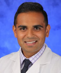 Patel Vijay A合作论文数Penn State Hershey Medical Center7
 Neerav Goyal合作论文数Department of Surgical Gastroenterology, Sir Ganga Ram Hospital7David Goldenberg合作论文数5
 Darrin Bann合作论文数Penn State College of Medicine5Meghan N. Wilson合作论文数5
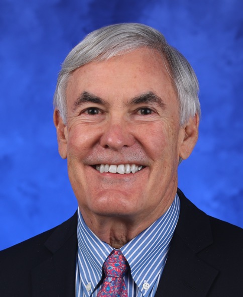 James R. Broach合作论文数Penn State Cancer Institute, College of Medicine, The Pennsylvania State University;Department of Biochemistry and Molecular Biology, College of Medicine, The Pennsylvania State University;Princeton University5
 Feng Yue合作论文数Department of Biochemistry and Molecular Genetics, Feinberg School of Medicine, Northwestern University4Saadi Robert A合作论文数4
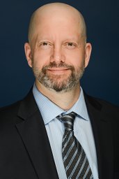 Brett A. Miles合作论文数4Eric M. Genden合作论文数4
 Sheetij Dutta合作论文数Department of Immunology, Walter Reed Army Institute of Research4
 Andrea Crisanti合作论文数3Max Hennessy合作论文数3
 Roberta Spaccapelo合作论文数Department of Medicine and Surgery, University of Perugia3Ockenhouse Christian F合作论文数3Teng Marita合作论文数3Murphy Jittawadee合作论文数3Khan Mohemmed N合作论文数3Brooke A. Bozick合作论文数3
 Henry Crist合作论文数2Lisa Dlugosz合作论文数2Jennifer Carter合作论文数2Sriranga Iyyanki合作论文数2Adrian H Batchelor合作论文数2Nicole Williams合作论文数2Tyler B. Merrill合作论文数2Junjia Zhu合作论文数2Victoria T Le合作论文数2Christopher Ehret合作论文数2
 Abhijit Chakraborty合作论文数La Jolla Institute for Allergy and Immunology;2
 Elizabeth Cottrill合作论文数Dept Otolaryngol Head & Neck Surg, Thomas Jefferson Univ2Kristen Lee合作论文数2
 Jie Xu合作论文数Univ Penn, Sch Vet Med, Dept Pathobiol, Philadelphia, PA 19104 USA2
 Yanli Wang合作论文数Penn State Univ, Dept Biol, University Pk, PA 16802 USA2Tonya S. King合作论文数2
 Hongbo Yang合作论文数Department of Biochemistry and Molecular Genetics, Feinberg School of Medicine, Northwestern University2
 Michael Cappello合作论文数Council on African Studies, Yale Macmillan Center, Yale University2
 Vishnu Dileep合作论文数Department of Biological Science, Florida State University2
 Duncan Odom合作论文数Division of Regulatory Genomics and Cancer Evolution, German Cancer Research Center;Ruprecht Karls Universität Heidelberg2
 David M Gilbert合作论文数Laboratory of Chromosome Replication and Epigenome Regulation, San Diego Biomedical Research Institute;DNA DAVE LLC2
 Jesse Dixon合作论文数Gene Expression Laboratory, Salk Institute for Biological Studies2Clark Royden A合作论文数2Sasaki Takayo合作论文数2
 Jon J Vermeire合作论文数2
 Joshua I Warrick合作论文数2
 Zhan Ye合作论文数2Morgan Diegel合作论文数2
 Einat Slonimsky合作论文数Sackler Faculty of Medicine, Tel Aviv University2




