Recently, a link was shown between the dystrophin-glycoprotein complex (DGC) protein sarcospan (SSPN) and development and progression of arrhythmias. Variants in the SSPN gene have been reported as a potential cause of atrial fibrillation (Afib) and studies in SSPN-deficient mice have demonstrated increased susceptibility to pre-ventricular contractions and stress-induced arrhythmias. To examine potential sources of arrhythmogenesis in SSPN-deficient hearts it was assessed whether loss of SSPN expression affected the abundance or localization of connexin 43, an important gap junction protein connexin 43. It was found that SSPN-deficient hearts exhibit increased connexin 43 abundance and changes in localization at the cardiac sarcolemma. To address the hypothesis that this disease develops early in development, neonatal mouse ventricular cardiomyocytes (NMVC) from SSPN-deficient and wild-type mice and immunofluorescence are used to assess changes in SSPN, connexin, and other gap junction proteins expression patterns in response to isoproterenol and other arrhythmia-inducing agents. Fluorescence microscopy will be performed to detect alterations in localization of connexin isoforms and other important gap junction proteins when SSPN-deficient mice. In WT, NMVC co-localization will be assessed for SSPN and other proteins important in cardiac conduction pathways. Immunoblotting and pull-down assays will also be utilized to determine endogenous interactions between SSPN and other key cardiac conduction proteins including connexin 40 43, and 45. Overall, these studies will be expanded to include adult cardiomyocytes and resolve novel SSPN interaction partners aside from the canonical adhesion complex proteins in the heart.
Many postdoctoral fellows and scholars who hope to secure tenure-track faculty positions in the United States apply to the National Institutes of Health (NIH) for a Pathway to Independence Award. This award has two phases (K99 and R00) and provides funding for up to 5 years. Using NIH data for the period 2006–2022, we report that ~230 K99 awards were made every year, representing up to ~$250 million annual investment. About 40% of K99 awardees were women and ~89% of K99 awardees went on to receive an R00 award annually. Institutions with the most NIH funding produced the most recipients of K99 awards and recruited the most recipients of R00 awards. The time between a researcher starting an R00 award and receiving a major NIH award (such as an R01) ranged between 4.6 and 7.4 years, and was significantly longer for women, for those who remained at their home institution, and for those hired by an institution that was not one of the 25 institutions with the most NIH funding. Shockingly, there has yet to be a K99 awardee at a historically Black college or university. We go on to show how K99 awardees flow to faculty positions, and to identify various factors that influence the future success of individual researchers and, therefore, also influence the composition of biomedical faculty at universities in the United States.
Regulation of myocardial mass is key for maintaining cardiovascular health. This review highlights the complex and regulatory relationship between mechanosignaling and myocardial mass, influenced by many internal and external factors including hemodynamic and microgravity, respectively. The heart is a dynamic organ constantly adapting to changes in workload (preload and afterload) and mechanical stress exerted on the myocardium, influencing both physiological adaptations and pathological remodeling. Mechanosignaling pathways, such as the mitogen-activated protein kinases (MAPKs) and the phosphoinositide 3-kinases and serine/threonine kinase (PI3K/Akt) pathways, mediate downstream effects on gene expression and play key roles in transducing mechanical cues into biochemical signals, thereby modulating cellular processes, including control of myocardial mass. Dysregulation of these processes can lead to pathological cardiac remodeling, such as hypertrophic cardiomyopathy. Furthermore, recent studies have highlighted the importance of protein quality control mechanisms, such as the ubiquitin-proteasome system, in settings of extreme physiological conditions that alter the heart workload such as pregnancy and microgravity. Overall, this review provides a thorough insight into how mechanical signals are converted into chemical signals to regulate myocardial mass in both healthy and diseased conditions.
The nonphysiological nutrient levels found in traditional culture media have been shown to affect numerous aspects of cancer cell physiology, including how cells respond to certain therapeutic agents. Here, we comprehensively evaluated how physiological nutrient levels affect therapeutic response by performing drug screening in human plasma-like medium. We observed dramatic nutrient-dependent changes in sensitivity to a variety of FDA-approved and clinically trialed compounds, including rigosertib, an experimental cancer therapeutic that recently failed in phase III clinical trials. Mechanistically, we found that the ability of rigosertib to destabilize microtubules is strongly inhibited by the purine metabolism end product uric acid, which is uniquely abundant in humans relative to traditional in vitro and in vivo cancer models. These results demonstrate the broad and dramatic effects nutrient levels can have on drug response and how incorporation of human-specific physiological nutrient medium might help identify compounds whose efficacy could be influenced in humans.
While P21-activated kinase-1 (PAK1) has been extensively studied in relation to cardiovascular health and glucose metabolism, its roles within adipose tissue and cardiometabolic diseases are less understood. In this study, we explored the effects of PAK1 deletion on energy balance, adipose tissue homeostasis, and cardiac function utilizing a whole-body PAK1 knockout (PAK1−/−) mouse model. Our findings revealed that body weight differences between PAK1−/− and WT mice emerged at 9 weeks of age, with further increases observed at 12 weeks. Furthermore, PAK1−/− mice displayed increased fat mass and decreased lean mass at 12 weeks, indicating a shift towards adiposity. In conjunction with the increased body weight, PAK1−/− mice had increased food intake and reduced energy expenditure. At a mechanistic level, PAK1 deletion boosted the expression of lipogenic markers while diminishing thermogenic markers expression in adipose tissues, contributing to reduced energy expenditure and the overall obesogenic phenotype. Moreover, our findings highlighted a significant impact on cardiac function following PAK1 deletion, including alterations in calcium kinetics and compromised systolic and lusitropy functions. In summary, our study emphasizes the significant role of PAK1 in weight regulation and cardiac function, enriching our comprehension of heart health and metabolism. These findings could potentially facilitate the identification of novel therapeutic targets in cardiometabolic diseases.
Loss of myocardial mass in a neonatal rat cardiomyocyte culture is studied to determine whether there is a distinguishable cellular response based on the origin of mechano‐signals. The approach herein compares the sarcomeric assembly and disassembly processes in heart cells by imposing mechano‐signals at the interface with the extracellular matrix (extrinsic) and at the level of the myofilaments (intrinsic). Experiments compared the effects of imposed internal (inside/out) and external (outside/in) loading and unloading on modifications in neonatal rat cardiomyocytes. Unloading of the cellular substrate by myosin inhibition (1 μ m mavacamten), or cessation of cyclic strain (1 Hz, 10% strain) after preconditioning, led to significant disassembly of sarcomeric α‐actinin by 6 h. In myosin inhibition, this was accompanied by redistribution of intracellular poly‐ubiquitin K48 to the cellular periphery relative to the poly‐ubiquitin K48 reservoir at the I‐band. Moreover, loading and unloading of the cellular substrate led to a three‐fold increase in post‐translational modifications (PTMs) when compared to the myosin‐specific activation or inhibition. Specifically, phosphorylation increased with loading while ubiquitination increased with unloading, which may involve extracellular signal‐regulated kinase 1/2 and focal adhesion kinase activation. The identified PTMs, including ubiquitination, acetylation, and phosphorylation, are proposed to modify internal domains in α‐actinin to increase its propensity to bind F‐actin. These results demonstrate a link between mechanical feedback and sarcomere protein homeostasis via PTMs of α‐actinin that exemplify how cardiomyocytes exhibit differential responses to the origin of force. The implications of sarcomere regulation governed by PTMs of α‐actinin are discussed with respect to cardiac atrophy and heart failure.
The hypothesis tested is that unloading of mechanical forces affects acetylation, ubiquitination, and phosphorylation of the actin-binding protein α-actinin located in the Z-disc. We applied targeted proteomics to interrogate the post-translational modification changes during loading and unloading. Cell morphology and post-translational modifications were determined in a mechanical intervention consisting of 1 Hz cyclic strain for 24 hr (loaded) followed by 6 hr rest (unloaded) of cultured neonatal rat ventricular myocytes (NRVMs). This was compared to a chemical intervention consisting of treating NRVMs with the myosin inhibitor Mavacamten (1 μM, 6hr) or activator Omecamtiv Mecarbil (0.5 μM, 6hr). Quantitative immunofluorescence showed both chemical and mechanical loading increased α-actinin content while Mavacamten decreased the α-actinin content. Mass spectrometry analysis of affinity-purified α-actinin revealed that the mechanical intervention led to increased levels of post-translational modifications compared with the chemical interventions. Specifically, α-actinin ubiquitination increased with mechanical loading-unloading; acetylation decreased with mechanical loading-unloading and increased with mechanical loading; and phosphorylation remained unchanged with mechanical loading-unloading but increased with mechanical loading. Fluorescence recovery after photobleaching (FRAP) experiments demonstrated that Mavacamten increased the dynamics of overexpressed YFP-tagged α-actinin and a GFP-tagged CapZ in NRVMs when compared to Omecamtiv Mecarbil treatments and controls. Overall, the results suggest a link between sarcomere homeostasis and mechanical forces via mechanisms involving acetylation, phosphorylation and ubiquitination of α-actinin and a second Z-disc protein, CapZ. These findings could have consequences for cardiac heart disease with abnormal sarcomeric proteostasis. Funded by NIH grants HL151825 (CS) and HL62426 (RJS, BR, and CMW).
In the heart, alternative splicing of the igf-I gene produces two isoforms: IGF-IEa and IGF-IEc, (Mechano-growth factor, MGF). The sequence divergence between their E-domain regions suggests differential isoform function. To define the biological actions of MGF’s E-domain, we performed in silico analysis of the unique C-terminal sequence and identified a phosphorylation consensus site residing within a putative 14-3-3 binding motif. To test the functional significance of Ser 18 phosphorylation, phospho-mimetic (S/E18) and phospho-null (S/A18) peptides were delivered to mice at different doses for 2 weeks. Cardiovascular function was measured using echocardiography and a pressure-volume catheter. At the lowest (2.25 mg/kg/day) and highest (9 mg/kg/day) doses, the peptides produced a depression in systolic and diastolic parameters. However, at 4.5 mg/kg/day the peptides produced opposing effects on cardiac function. Fractional shortening analysis also showed a similar trend, but with no significant change in cardiac geometry. Microarray analysis discovered 21 genes (FDR p < 0.01), that were expressed accordant with the opposing effects on contractile function at 4.5 mg/kg/day, with the nuclear receptor subfamily 4 group A member 2 (Nr4a2) identified as a potential target of peptide regulation. Testing the regulation of the Nr4a family, showed the E-domain peptides modulate Nr4a gene expression following membrane depolarization with KCl in vitro. To determine the potential role of 14-3-3 proteins, we examined 14-3-3 isoform expression and distribution. 14-3-3γ localized to the myofilaments in neonatal cardiac myocytes, the cardiac myocytes and myofilament extracts from the adult heart. Thermal shift analysis of recombinant 14-3-3γ protein showed the S/A18 peptide destabilized 14-3-3γ folding. Also, the S/A18 peptide significantly inhibited 14-3-3γ’s ability to interact with myosin binding protein C (MYPC3) and phospholamban (PLN) in heart lysates from dobutamine injected mice. Conversely, the S/E18 peptide showed no effect on 14-3-3γ stability, did not inhibit 14-3-3γ’s interaction with PLN but did inhibit the interaction with MYPC3. Replacing the glutamic acid with a phosphate group on Ser 18 (pSer18), significantly increased 14-3-3γ protein stability. We conclude that the state of Ser 18 phosphorylation within the 14-3-3 binding motif of MGF’s E-domain, modulates protein-protein interactions within the 14-3-3γ interactome, which includes proteins involved in the regulation of contractile function.
Age-related wild-type transthyretin amyloidosis (wtATTR) is characterized by systemic deposition of amyloidogenic fibrils of misfolded transthyretin (TTR) in the connective tissue of many organs. In the heart, this leads to age-related heart failure with preserved ejection fraction (HFpEF). The hypothesis tested is that TTR deposited in vitro disrupts cardiac myocyte cell-to-cell and cell-to-matrix adhesion complexes, resulting in altered calcium handling, force generation, and sarcomeric disorganization. Human iPSC-derived cardiomyocytes and neonatal rat ventricular myocytes (NRVMs), when grown on TTR-coated polymeric substrata mimicking the stiffness of the healthy human myocardium (10 kPa), had decreased contraction and relaxation velocities as well as decreased force production measured using traction force microscopy. Both NRVMs and adult mouse atrial cardiomyocytes had altered calcium kinetics with prolonged transients when cultured on TTR fibril-coated substrates. Furthermore, NRVMs grown on stiff (~GPa), flat or microgrooved substrates coated with TTR fibrils exhibited significantly decreased intercellular electrical coupling as shown by FRAP dynamics of cells loaded with the gap junction-permeable dye calcein-AM, along with decreased gap junction content as determined by quantitative connexin 43 staining. Significant sarcomeric disorganization and loss of sarcomere content, with increased ubiquitin localization to the sarcomere, were seen in NRVMs on various TTR fibril-coated substrata. TTR presence decreased intercellular mechanical junctions as evidenced by quantitative immunofluorescence staining of N-cadherin and vinculin. Current therapies for wtATTR are cost-prohibitive and only slow the disease progression; therefore, better understanding of cardiomyocyte maladaptation induced by TTR amyloid may identify novel therapeutic targets.
This Commentary is a call for submissions for the upcoming Issue Focus that will highlight some of the scientific topics discussed during the 2nd Costa Rica Biophysics Symposium.
All cells sense force and build their cytoskeleton to optimize function. How is this achieved? Two major systems are involved. The first is that load deforms specific protein structures in a proportional and orientation-dependent manner. The second is post-translational modification of proteins as a consequence of signaling pathway activation. These two processes work together in a complex way so that local subcellular assembly as well as overall cell function are controlled. This review discusses many cell types but focuses on striated muscle. Detailed information is provided on how load deforms the structure of proteins in the focal adhesions and filaments, using α-actinin, vinculin, talin, focal adhesion kinase, LIM domain-containing proteins, filamin, myosin, titin, and telethonin as examples. Second messenger signals arising from external triggers are distributed throughout the cell causing post-translational or chemical modifications of protein structures, with the actin capping protein CapZ and troponin as examples. There are numerous unanswered questions of how mechanical and chemical signals are integrated by muscle proteins to regulate sarcomere structure and function yet to be studied. Therefore, more research is needed to see how external triggers are integrated with local tension generated within the cell. Nonetheless, maintenance of tension in the sarcomere is the essential and dominant mechanism, leading to the well-known phrase in exercise physiology: "use it or lose it."
A transduced mechanical signal arriving at its destination in muscle alters sarcomeric structure and function. A major question addressed is how muscle mass and tension generation are optimized to match actual performance demands so that little energy is wasted. Three cases for improved energy efficiency are examined: the troponin complex for tuning force production, control of the myosin heads in a resting state, and the Z-disc proteins for sarcomere assembly. On arrival, the regulation of protein complexes is often controlled by post-translational modification (PTM), of which the most common are phosphorylation by kinases, deacetylation by histone deacetylases and ubiquitination by E3 ligases. Another branch of signals acts not through peptide covalent bonding but via ligand interactions (e.g. Ca2+ and phosphoinositide binding). The myosin head and the regulation of its binding to actin by the troponin complex is the best and earliest example of signal destinations that modify myofibrillar contractility. PTMs in the troponin complex regulate both the efficiency of the contractile function to match physiologic demand for work, and muscle mass via protein degradation. The regulation of sarcomere assembly by integration of incoming signaling pathways causing the same PTMs or ligand binding are discussed in response to mechanical loading and unloading by the Z-disc proteins CapZ, α-actinin, telethonin, titin N-termini, and others. Many human mutations that lead to cardiomyopathy and heart disease occur in the proteins discussed above, which often occur at their PTM or ligand binding sites.
Our review focuses on sarcomere regulatory mechanisms with a discussion of cardiac-specific modifications to the three-state model of thin filament activation from a blocked to closed to open state. We discuss modulation of these thin filament transitions by Ca2+, by crossbridge interactions, and by thick filament-associated proteins, cardiac myosin-binding protein C (cMyBP-C), cardiac regulatory light chain (cRLC), and titin. Emerging evidence supports the idea that the cooperative activation of the thin filaments despite a single Ca2+ triggering regulatory site on troponin C (cTnC) cannot be considered in isolation of other functional domains of the sarcomere. We discuss long- and short-range interactions among these domains with the regulatory units of thin filaments, including proteins at the barbed end at the Z-disc and the pointed end near the M-band. Important to these discussions is the ever-increasing understanding of the role of cMyBP-C, cRLC, and titin filaments. Detailed knowledge of these control processes is critical to the understanding of mechanisms sustaining physiological cardiac state with varying hemodynamic load, to better defining genetic and acquired cardiac disorders, and to developing targets for therapies at the level of the sarcomeres.
In cardiac muscle, binding of troponin (Tn) and tropomyosin (Tpm) to filamentous (F)‐actin forms thin filaments capable of Ca2+‐dependent regulation of contraction. Tpm binds to F‐actin in a head‐to‐tail fashion, while Tn stabilizes these linkages. Valuable structural and functional information has come from biochemical, X‐ray, and electron microscopy data. However, the use of fluorescence microscopy to study thin filament assembly remains relatively underdeveloped. Here, triple fluorescent labeling of Tn, Tpm, and F‐actin allowed us to track thin filament assembly by fluorescence microscopy. It is shown here that Tn and Tpm molecules self‐organize on actin filaments and give rise to decorated and undecorated regions. Binding curves based on colocalization of Tn and Tpm on F‐actin exhibit cooperative binding with a dissociation constant Kd of ~ 0.5 µm that is independent of the Ca2+ concentration. Binding isotherms based on the intensity profile of fluorescently labeled Tn and Tpm on F‐actin show that binding of Tn is less cooperative relative to Tpm. Computational modeling of Tn‐Tpm binding to F‐actin suggests two equilibrium steps involving the binding of an initial Tn‐Tpm unit (nucleation) and subsequent recruitment of adjacent Tn‐Tpm units (elongation) that stabilize the assembly. The results presented here highlight the utility of employing fluorescence microscopy to study supramolecular protein assemblies.
Scientists in developing countries face several challenges, including limited funding and a smaller and less connected scientific community. One opportunity of growth is to host scientific meetings in these countries to highlight the importance of scientific research in society. As early career investigators, we organized a biophysics symposium in Costa Rica, a developing country, with the goal of increasing the awareness of and interest in biophysics and biomedical research. In this report, we discuss our experience organizing this event to serve as a practical guide with actionable points to organize meetings of this kind in developing countries.Developed nations have benefited from the scientia potentia est or “knowledge is power” economy, in which pioneering work in the sciences has led to crucial new technologies and further economic growth. Developing countries, on the other hand, face challenges that limit investment in fundamental science that may not produce immediate tangible benefits but has significant potential to provide long-term returns. Hence, smaller scientific communities in developing nations must take advantage of the larger scientific network already in place after decades of investment in research in high-income countries. Some advances have been observed in this area. Chile, for example, has boosted its scientific productivity by establishing strong international collaborations (1). Overall, internationally coauthored articles in developing countries rose from 10% in 1990 to 25% in 2010 (2). Nonetheless, further actions must be implemented to bridge the gap between the developing and developed nations.Beyond increasing research funding, other actions can substantially benefit scientific progress. Experienced investigators who have built prestigious careers in developing nations highlight the need for national and international collaborations, as discussed above, but further emphasize the importance of participating in meetings and visiting other institutions as vital factors for success (3). Face-to-face encounters at scientific meetings are the standard to spur collaborations between investigators who otherwise would have difficulties connecting (4). Scientific and networking events in developing countries can promote fruitful professional relationships, while allowing scientists from these nations to better use their already limited resources.Costa Rica has a middle-income economy, which according to the World Bank invested approximately 0.5% of its gross domestic product in research and development in 2016. For comparison, Israel invested 4.58% in 2017 (5). Government-supported research in Costa Rica is varied, with internationally renowned groups such as the Clodomiro Picado Institute, a powerhouse of snake venom research tasked with the production of snakebite antidotes for human and veterinary use. Our field of interest, biophysics, does not have a strong presence in the Costa Rican scientific community. We organized the first Costa Rican Biophysics Symposium to create a space where biophysics could be introduced to a mixed audience of scientists, students, and the general public. We found it extremely important to build a strong organizing team. Organizing such a specialized networking event for the first time, while living in different countries, can be a tremendous a challenge.Biophysical research relies on particularly expensive, cutting-edge techniques and instrumentation that may not be available in most institutions. A strong network of collaborators is then key to promote effective resource sharing for biophysicists in developing countries. Furthermore, such collaborations are beneficial for national and international funding applications. We therefore surmised the event would be an excellent networking opportunity to promote such collaborations, relying on a mixture of local attendees and Costa Ricans performing research abroad. In addition, this would be an opportunity to bring awareness about the large umbrella that biophysics casts across multiple disciplines.Since the inception of this idea, we set the event close to the end-of-year holiday season, given that many Costa Rican expatriates return home during this time. Timing of the event is particularly important, because incentives are few for prospective speakers to invest time and resources in attending a small, first-of-its-kind meeting instead of an established conference. Speakers should be invited well in advance (more than a year of anticipation, if possible) to allow them to plan their schedules accordingly.To identify potential speakers, we took advantage of freely available databases. First, we used the Red Ticotal from the National Academy of Science of Costa Rica (ANC by its Spanish abbreviation; 6), which encompasses scientists and engineers who study or work abroad. Another resource was the “Find a Biophysicist” database from the Biophysical Society, which includes a country-specific search (7). Professional social media sites, such as LinkedIn or ResearchGate, helped make a broad search among the organizers' networks. Speaker selection for this type of meeting needs realistic expectations, and some flexibility was necessary in the research topics presented, given that the number of speakers determines the program. We recruited 8 speakers, including the 3 organizers, and organized the symposium as a half-day event. The first talk was an overview of what is encompassed by biophysical research, also highlighting prominent female and Latin American scientists.The venue is a major expense for a professional meeting. Our recommendation is to work with local universities or institutions that oversee the scientific enterprise to host the event in their facilities. We organized the symposium at the National Academy of Sciences of Costa Rica, which generously offered its auditorium for the event at no cost. This arrangement is also advantageous from a logistics and advertising perspective: it is in the best interest of these institutions to promote novel scientific events through their networks.We aimed to promote attendance by making the event free to the public. Procuring funding sources for a first-time event can be challenging, especially given the already limited resources in developing countries. Professional societies can provide a substantial help in this matter. The Biophysical Society kindly supported our event through a Networking mini-grant, which was enough to cover all expenses of the meeting, including refreshments and printed materials. By creating a precedent, sponsor recruitment may become easier in future editions (one of the authors has organized unrelated symposia and found that repetitions of an event increase confidence of prospective sponsors). In some instances, it may be unavoidable to ask for a registration fee.Recruiting attendees starts with the flyer. It does not need to have all the details in the first iteration. We sent flyers to local universities, relevant governmental institutions, and the organizers' professional networks. We promoted the event with the Costa Rican College of Physicians, the ANC's database, and Red Ticotal. We emphasized the role of the basic sciences as the starting point of many important translational discoveries in medicine. In fact, some medical doctors attended this event. Making a web page so anyone who is interested could follow updates such as changes in the schedule, the venue, or even the date of the event was also useful.The standard method to get feedback is to ask attendees to fill evaluation forms. To improve turnout, we provided printed forms and emphasized their importance throughout the event. Other alternatives are digital feedback surveys if attendees' email addresses are available. An online registration tool becomes helpful because email information can be collected and a head count can be determined for logistic purposes.We hope that these lessons and recommendations may encourage others to develop scientific conferences in other developing countries. No agenda, venue, or theme for a meeting is ideal. The best advice we can give is to have a very committed team and to be eager to adapt to any situation that arises. In the end, this professional experience is rewarding, and you will have the chance to make an unprecedented scientific impact. As research becomes more competitive and funding rates decrease even in developed nations, networking events such as this are crucial to stimulate scientific research and form the best collaborative teams.This work was supported by The Biophysical Society Networking Event mini-grant (CS, JAR, FJA). We thank Dayana Mora, Lizeth Lázaro, Melania Odio, and the ANC for their generous support and hosting of the symposium. The authors declare no competing financial interests.
Calcium binding to troponin C (TnC) is insufficient for full activation of myosin ATPase activity by actin-tropomyosin-troponin. Previous attempts to investigate full activation utilized ATP-free myosin or chemically modified myosin to stabilize the active state of regulated actin. We utilized the Δ14-TnT and the A8V-TnC mutants to stabilize the activated state at saturating Ca2+ and to eliminate one of the inactive states at low Ca2+. The observed effects differed in solution studies and in the more ordered in vitro motility assay and in skinned cardiac muscle preparations. At saturating Ca2+, full activation with Δ14-TnT·A8V-TnC decreased the apparent KM for actin-activated ATPase activity compared to bare actin filaments. Rates of in vitro motility increased at both high and low Ca2+ with Δ14-TnT; the maximum shortening speed at high Ca2+ increased 1.8-fold. Cardiac muscle preparations exhibited increased Ca2+ sensitivity and large increases in resting force with either Δ14-TnT or Δ14-TnT·A8V-TnC. We also observed a significant increase in the maximal rate of tension redevelopment. The results of full activation with Ca2+ and Δ14-TnT·A8V-TnC confirmed and extended several earlier observations using other means of reaching full activation. Furthermore, at low Ca2+, elimination of the first inactive state led to partial activation. This work also confirms, in three distinct experimental systems, that troponin is able to stabilize the active state of actin-tropomyosin-troponin without the need for high-affinity myosin binding. The results are relevant to the reason for two inactive states and for the role of force producing myosin in regulation.
Cardiac function mainly depends on the total myocyte mass in the ventricles. Assembly and disassembly of sarcomeres occurs to adjust this mass to altered mechanical demand. In the heart, hypertrophic cardiomyopathy results from myofibrillar assembly controlled by post-translational modification of proteins directed by signaling pathways. More is known about assembly on loading than disassembly on unloading. Here, the hypothesis tested is that unloading of mechanical forces affects acetylation (Ac) and ubiquitination (Ub) of the actin-binding proteins, α-actinin and CapZ. Omecamtiv mecarbil (0.5 μM) and mavacamten (1 μM) were used to increase (load) and decrease (unload) cardiomyocyte tension, respectively, via their action on myosin ATPase. Mavacamten decreased myocyte contractility in rat ventricular myocytes (NRVMs) and caused significant sarcomere disassembly by 6 h and 70% atrophy by 24 h. Assembly was preserved with omecamtiv mecarbil (0.5 μM) over the 24 h time period. Post-translational modification was determined in loaded and unloaded NRVMs at 6 h of drug treatment. Bottom-up mass spectrometry analysis showed single residues in α-actinin and CapZ that were acetylated or ubiquitinated. Acetylation levels appeared to increase in the mavacamten-treated samples while these levels are preserved in untreated and omecamtiv mecarbil-treated samples. Ac and Ub in the Z-discs were quantified on immunofluorescent images. The Z-discs colocalized oligo-Ub (K-48 oligo-Ub linkage) and Ac in untreated samples; this Z-disc localization of Ub and Ac was diminished with unloading. Fluorescence recovery after photobleaching (FRAP) measurements of the dynamics of α-actinin and CapZ after reduced cell tension with mavacamten (1 μM) and omecamtiv mercabil (0.5 μM) are ongoing. Overall, results suggest sarcomere assembly is regulated by mechanical forces through a mechanism involving Ac and Ub of myofibrillar proteins. These findings could have consequences for cardiac heart disease with abnormal sarcomeric proteostasis.
Muscle adaptation is a response to physiological demand elicited by changes in mechanical load, hormones, or metabolic stress. Cytoskeletal remodeling processes in many cell types are thought to be primarily regulated by thin filament formation due to actin-binding accessory proteins, such as the actin-capping protein. Here, we hypothesize that in muscle, the actin-capping protein (named CapZ) integrates signaling by a variety of pathways, including phosphorylation and phosphatidylinositol 4,5-bisphosphate (PIP2) binding, to regulate muscle fiber growth in response to mechanical load. To test this hypothesis, we assess mechanotransduction signaling that regulates muscle growth using neonatal rat ventricular myocytes cultured on substrates with the stiffness of the healthy myocardium (10 kPa), fibrotic myocardium (100 kPa), or glass. We investigate how PIP2 signaling affects CapZ using the PIP2 sequestering agent neomycin and the effect of PKC-mediated CapZ phosphorylation using the PKC-activating drug phorbol 12-myristate 13-acetate (PMA). Molecular simulations suggest that close interactions between PIP2 and the β-tentacle of CapZ are modified by phosphorylation at T267. Fluorescence recovery after photobleaching (FRAP) demonstrates that the kinetic binding constant of CapZ to sarcomeric thin filaments in living muscle cells increases with stiffness or PMA treatment but is diminished by PIP2 reduction. Furthermore, CapZ with a deletion of the β-tentacle that lacks the phosphorylation site T267 shows increased FRAP kinetics with lack of sensitivity to PMA treatment or PIP2 reduction. Förster resonance energy transfer (FRET) probes the molecular interactions between PIP2 and CapZ, which are decreased by PIP2 availability or by the β-tentacle truncation. These data suggest that CapZ is bound to actin tightly in the idle, locked state, with little phosphorylation or PIP2 binding. However, this tight binding is loosened in growth states triggered by mechanical stimuli such as substrate stiffness, which may have relevance to fibrotic heart disease.
En el músculo cardiaco, la activación de los filamentos delgados por medio de Ca2+permite la unión fuerte de miosina a la actina la cual genera el efecto de bazo palanca responsable de la conversión de energía química del ATP en energía mecánica. Esta activación es inhibida por la proteína Troponina (Tn) que es sensible al Ca2+. Empero, el nivel de detalle del mecanismo actual de puentes cruzados es insuficiente en describir lo que ocurre con Tn durante este ciclo. El presente estudio trata de estudiar los cambios conformacionales en Tn en función de Ca2+y miosina mediante el uso de microscopía de florescencia de tiempo de vida combinada con resonancia de transferencia de energía Förster (FLIM-FRET). Moléculas fluorescentes localizadas en la terminal C de TnC y TnI, respectivamente, reportan el estado de activación de Tn. Bajo las condiciones probadas, Ca2+es responsable de la mayor parte de la activación de Tn. Miosina en estado rigor (unión fuerte) activa Tn tanto en estados saturados como sub-saturados de Ca2+, pero éste último estado no supera el estado saturado con Ca2+. ATP-γ-S (ATP no hidrolizable) no afecta la activación de Tn significativamente; sin embargo, blebbistatin (induce unión débil en miosina) indujo activación de Tn significativamente en estados sub-saturados de Ca2+. La relación entre el grado de activación de Tn y el de su flexibilidad conformacional sugieren que estados activos e inactivos coexisten en diferentes proporciones que dependen de la combinación de los efectores Ca2+y miosina. Estos resultados satisfacen un modelo de activación alostérica en filamentos delgados.
 John Robinson合作论文数Department of Geography and Planning, Faculty of Arts and Science, University of Toronto11
 Maria E. Moutsoglou合作论文数10
 Giho Harry Kim合作论文数Department of Physics, University of Rhode Island10
 Brenda Russell合作论文数Department of Physiology and Biophysics, College of Medicine, University of Illinois at Chicago;Center for CardioVascular Research, University of Illinois at Chicago;Cell Habitats Inc10
 R. John Solaro合作论文数Department of Physiology & Biophysics, College of Medicine, University of Illinois at Chicago5
 Michael Mkrtschjan合作论文数4Chad Warren合作论文数Department of Physiology and Biophysics, University of Illinois at Chicago4Kyle Dittloff合作论文数3Elisabeth DiNello合作论文数2
 Aida Rahimi Kahmini合作论文数2
 Jose Rodriguez Corrales合作论文数Escuela de Química, Universidad Nacional2
 Carmen Mora-Aparicio合作论文数Inst Costarricense Acueductos & Alcantarillados2
 Tauras Vilgalys合作论文数Sect Genet Med, Univ Chicago2
 Maicon Landim-Vieira合作论文数Florida State Univ, Biomed Sci, Tallahassee, FL 32306 USA2
 Nicole Woitowich合作论文数Department of Medical Social Sciences, Feinberg School of Medicine, Northwestern University2
 Sarah R. Hengel合作论文数Department of Biochemistry, University of Iowa;Department of Biochemistry, University of Iowa2
 Tyrrell Daniel J合作论文数Department of Internal Medicine, Wake Forest School of Medicine2Le Long合作论文数2
 Babdor Joël合作论文数2
 Tejal A Desai合作论文数School of Engineering, Brown University;School of Pharmacy, University of California, San Francisco2Alex Murillo合作论文数University of Costa Rica2Pieter deTombe合作论文数1Koreena Rafael-Clyke合作论文数1Kelly Conger合作论文数1Sagar Dommaraju合作论文数1Paloma González-Sánchez合作论文数1José Á. Rodríguez-Corrales合作论文数1Admasu Y Wondmagegn合作论文数1
 Patrick R. DeLear合作论文数1
 Conrad Safranek合作论文数1Maximilian McCann合作论文数Department of Ophthalmology & Visual Sciences, University of Illinois at Chicago1tanya a baldwin合作论文数1
 Jooman Park合作论文数Department of Physiology and Biophysics, University of Illinois at Chicago1
 Wentao Dong合作论文数Department of Chemical Engineering, Stanford University;Department of Genetics, Stanford University;Stanford University, Stanford University1Priya Mohindra合作论文数Graduate Program in Bioengineering, UC Berkeley-UCSF1Jennifer E Endress合作论文数1Mete Emir Ozgurses合作论文数Canc Ctr, Univ Illinois1
 Marcos Munoz合作论文数1
 Gustavo Chaves合作论文数1Fang Qizhi合作论文数1Li Zhu合作论文数1
 Yuwei Jiang合作论文数College of Medicine, University of Illinois at Chicago1
 Dylan Johnson合作论文数Dept Biochem & Mol Biol, East Carolina Univ1
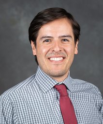 Francisco J Alvarado合作论文数Univ Michigan, Dept Mol & Integrat Physiol, Ann Arbor, MI 48109 USA1
 David Sept合作论文数Department of Biomedical Engineering, College of Engineering, University of Michigan-Ann Arbor1
 Paul H Goldspink合作论文数1
 Isaac S. Harris合作论文数Ontario Cancer Institute, University Health Network;Ontario Cancer Institute, University Health Network1
 Vipin Rawat合作论文数1
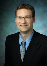 Ranek Mark J合作论文数Division of Basic Biomedical Sciences, University of South Dakota1




