Background: Continuous glucose monitors (CGMs) enable people with diabetes to proactively manage their blood glucose and reduce the risk of hypoglycemia. Commercially available CGMs utilize percutaneous electrodes that, after days to weeks of implantation, are subjected to the foreign body response that severely reduces sensor accuracy. The previous work demonstrated the use of hydrogels containing a glucose-responsive viologen that quenches a nearby fluorophore. Here, we investigate the immobilization of this sensing motif onto a nanoparticle surface and optimize local surface concentrations for optical glucose sensing. Methods: A viologen quencher-fluorescent dye system was incorporated into poly(2-hydroethyl methacrylate) hydrogels in varying quantities to assess the effect of quencher-fluorophore concentration on glucose responsiveness. The sensing motif was then immobilized onto silica nanoparticles by carbodiimide chemistry. Nanosensors with a range of dye and quencher concentrations were challenged for glucose responsiveness to determine the optimal sensor formulation. Results: When incorporated into a hydrogel, high concentrations of viologen quencher and fluorophore were required to permit electron transfer between the two components and yield a detectable glucose response. Immobilization of this glucose-responsive system onto a silica nanoparticle facilitated this electron transfer to yield detectable responses at even low concentrations. Increasing quencher concentration on the nanoparticle, relative to the fluorophore, resulted in the greatest apparent glucose response. Conclusion: The nanoparticle sensors demonstrated excellent glucose response in the physiological range and are a promising tool for real-time glucose tracking.
Cells interact intimately with complex microdomains in their extracellular matrix (ECM) and maintain a delicate balance of mechanical forces through mechanosensitive cellular components. Tissue injury results in acute degradation of the ECM and disruption of cell-ECM contacts, manifesting in loss of cytoskeletal tension, leading to pathological cell transformation and the onset of disease. Recently, microscale hydrogel constructs have been developed to provide cells with microdomains to form focal adhesion binding sites, which enable restoration of cytoskeletal tension. These synthetic anchors can recapitulate the complex 3D architecture of the native ECM to provide microtopographical cues. The mechanical deformation of proteins at the cell surface can activate signaling cascades to modulate downstream gene-level transcription, making this a unique materials-based approach for reprogramming cell behavior. An overview of the mechanisms underlying these mechanosensitive interactions in fibroblasts, stem and other cell types is provided to review their effects on cellular reprogramming. Recent investigations on the fabrication, functionalization and implementation of these materials and microtopographical features for drug testing and therapeutic applications are discussed.
Repairing cardiac tissue after myocardial infarction (MI) is one of the most challenging goals in tissue engineering. Following ischemic injury, significant matrix remodeling and the formation of avascular scar tissue significantly impairs cell engraftment and survival in the damaged myocardium. This limits the efficacy of cell replacement therapies, demanding strategies that reduce pathological scarring to create a suitable microenvironment for healthy tissue regeneration. Here, we demonstrate the successful fabrication of discrete hyaluronic acid (HA)-based microrods to provide local biochemical and biomechanical signals to reprogram cells and attenuate cardiac fibrosis. HA microrods were produced in a range of physiological stiffness and shown to degrade in the presence of hyaluronidase. Additionally, we show that fibroblasts interact with these microrods in vitro, leading to significant changes in proliferation, collagen expression and other markers of a myofibroblast phenotype. When injected into the myocardium of an adult rat MI model, HA microrods prevented left ventricular wall thinning and improved cardiac function at 6 weeks post infarct.
Cell migration is regulated by several mechanotransduction pathways, which consist of sensing and converting mechanical microenvironmental cues to internal biochemical cellular signals, such as protein phosphorylation and lipid signaling. While there has been significant progress in understanding protein changes in the context of mechanotransduction, lipid signaling is more difficult to investigate. In this study, physical cues of stiffness (10, 100, 400 kPa, and glass), and microrod or micropost topography were manipulated in order to reprogram primary fibroblasts and assess the effects of lipid signaling on the actin cytoskeleton. In an in vitro wound closure assay, primary cardiac fibroblast migration velocity was significantly higher on soft polymeric substrata. Modulation of PIP2 availability through neomycin treatment nearly doubled migration velocity on 10 kPa substrata, with significant increases on all stiffnesses. The distance between focal adhesions and the lamellar membrane (using wortmannin treatment to increase PIP2 via PI3K inhibition) was significantly shortest compared to untreated fibroblasts grown on the same surface. PIP2 localized to the leading edge of migrating fibroblasts more prominently in neomycin‐treated cells. The membrane‐bound protein, lamellipodin, did not vary under any condition. Additionally, fifteen micron‐high micropost topography, which blocks migration, concentrates PIP2 near to the post. Actin dynamics within stress fibers, measured by fluorescence recovery after photobleaching, was not significantly different with stiffness, microtopography, nor with drug treatment. PIP2‐modulating drugs delivered from microrod structures also affected migration velocity. Thus, manipulation of the microenvironment and lipid signaling regulatory drugs might be beneficial in improving therapeutics geared toward wound healing.
Recovery of live cells from three-dimensional (3D) culture would improve analysis of cell behaviors in tissue engineered microenvironments. In this work, we developed a temperature responsive hydrogel to enable transient 3D culture of human glioblastoma (GBM) cells. N-isopropylacrylamide was copolymerized with hydrophilic grafts and functionalized with the cell adhesion peptide RGD to yield the novel copolymer poly(N-isopropylacrylamide-co-Jeffamine(®) M-1000 acrylamide-co-hydroxyethylmethacrylate-RGD), or PNJ-RGD. This copolymer reversibly gels in aqueous solutions when heated under normal cell culture conditions (37°C). Moreover, these gels redissolve within 70 s when cooled to room temperature without the addition of any agents to degrade the synthetic scaffold, thereby enabling rapid recollection of viable cells after 3D culture. We tested the efficiency of cell recovery following extended 3D culture and were able to recover more than 50% of viable GBM cells after up to 7 days in culture. These data demonstrate the utility of physically crosslinked PNJ-RGD hydrogels as a platform for culture and recollection of cells in 3D.
The invasion of malignant glioblastoma (GBM) cells into healthy brain is a primary cause of tumor recurrence and associated morbidity. Here, we describe a high-throughput method for quantitative measurement of GBM proliferation and invasion in three-dimensional (3D) culture. Optically clear hydrogels composed of thiolated hyaluronic acid and gelatin were chemically crosslinked with thiol-reactive poly(ethylene glycol) polymers to form an artificial 3D tumor microenvironment. Characterization of the viscoelasticity and aqueous stability indicated the hydrogels were mechanically tunable with stiffness ranging from 18 Pa to 18.2 kPa and were resistant to hydrolysis for at least 30 days. The proliferation, dissemination and subsequent invasion of U118 and U87R GBM spheroids cultured on the hydrogels were tracked in situ with repeated fluorescence confocal microscopy. Using custom automated image processing, cells were identified and quantified through 500 µm of gel over 14 days. Proliferative and invasive behaviors were observed to be contingent on cell type, gel stiffness, and hepatocyte growth factor availability. These measurements highlight the utility of this platform for performing quantitative, fluorescence imaging analysis of the behavior of malignant cells within an artificial, 3D tumor microenvironment.
The oral route of drug administration is most preferred due to its ease of use, low cost, and high patient compliance. However, the oral uptake of many small molecule drugs and biotherapeutics is limited by various physiological barriers, and, as a result, drugs suffer from issues with low solubility, low permeability, and degradation following oral administration. The flexibility of micro- and nanofabrication techniques has been used to create drug delivery platforms designed to address these barriers to oral drug uptake. Specifically, micro/nanofabricated devices have been designed with planar, asymmetric geometries to promote device adhesion and unidirectional drug release toward epithelial tissue, thereby prolonging drug exposure and increasing drug permeation. Furthermore, surface functionalization, nanotopography, responsive drug release, motion-based responses, and permeation enhancers have been incorporated into such platforms to further enhance drug uptake. This review will outline the application of micro/nanotechnology to specifically address the physiological barriers to oral drug delivery and highlight technologies that may be incorporated into these oral drug delivery systems to further enhance drug uptake.
Here, efforts toward the development of a microneedle-based glucose sensor or "smart patch" for intradermal glucose sensing are described. Metallic microneedle array electrodes, conducting polymers, and glucose oxidase form the sensor platform. This work represents the first steps toward the development of painless, transdermal-sensing devices for continuous glucose monitoring.
Skeletal muscle injury resulting in tissue loss poses unique challenges for surgical repair. Despite the regenerative potential of skeletal muscle, if a significant amount of tissue is lost, skeletal myofibers will not grow to fill the injured area completely. Prior work in our lab has shown the potential to fill the void with an extracellular matrix (ECM) scaffold, resulting in restoration of morphology, but not functional recovery. To improve the functional outcome of the injured muscle, a muscle-derived ECM was implanted into a 1 x 1 cm(2), full-thickness defect in the lateral gastrocnemius (LGAS) of Lewis rats. Seven days later, bone-marrow-derived mesenchymal stem cells (MSCs) were injected directly into the implanted ECM. Partial functional recovery occurred over the course of 42 days when the LGAS was repaired with an MSC-seeded ECM producing 85.4 +/- 3.6% of the contralateral LGAS. This was significantly higher than earlier recovery time points (p < 0.05). The specific tension returned to 94 +/- 9% of the contralateral limb. The implanted MSC-seeded ECM had more blood vessels and regenerating skeletal myofibers than the ECM without cells (p < 0.05). The data suggest that the repair of a skeletal muscle defect injury by the implantation of a muscle-derived ECM seeded with MSCs can improve functional recovery after 42 days.
 Tejal A Desai合作论文数School of Engineering, Brown University;School of Pharmacy, University of California, San Francisco6
 Michael Mkrtschjan合作论文数3
 Brenda Russell合作论文数Department of Physiology and Biophysics, College of Medicine, University of Illinois at Chicago;Center for CardioVascular Research, University of Illinois at Chicago;Cell Habitats Inc3
 Christopher Solís合作论文数Dept Physiol & Biophys, Univ Illinois2
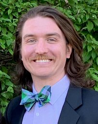 John Heffernan合作论文数Barrow Brain Tumor Research Center, Barrow Neurological Institute2Derek J. Overstreet合作论文数2
 Brent L Vernon合作论文数Center for Interventional Biomaterials, Arizona State University2
 Rachael Weiss Sirianni合作论文数Dept. of Biomedical Engineering, Yale University2Soya Gamsey合作论文数1Sagar Dommaraju合作论文数1
 Natalie Wisniewski合作论文数1cameron l nemeth合作论文数1Tae J. Song合作论文数1Tianlu Wang合作论文数1
 Conrad Safranek合作论文数1David W. Hammers合作论文数1Rohit Gokhale合作论文数1Gauree S Chendke合作论文数1Priya Mohindra合作论文数Graduate Program in Bioengineering, UC Berkeley-UCSF1
 Matthew Timothy Tierney合作论文数Sanford Burnham Prebys Med Discovery Inst1Fang Qizhi合作论文数1
 Apurva Sarathy合作论文数1
 Daniel G. Anderson合作论文数Koch Institute for Integrative Cancer Research, Massachusetts Institute of Technology;Department of Chemical Engineering, School of Engineering, Massachusetts Institute of Technology1Cannon Megan V合作论文数1Kim Jean合作论文数1Fox Cade B合作论文数Department of Bioengineering and Therapeutic Sciences, University of California1
 Richard E Sievers合作论文数1
 Randall Lee合作论文数School of Medicine, University of California, San Francisco1
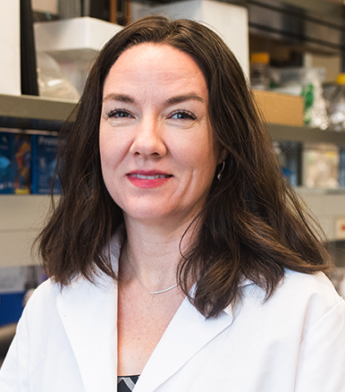 Laura Suggs合作论文数Department of Biomedical Engineering, Cockrell School of Engineering, University of Texas at Austin1
 Hariharasudhan D. Chirra合作论文数Department of Chemical and Materials Engineering, University of Kentucky1
 Edward K. Merritt合作论文数1Michael A. Invernale合作论文数Department of Chemistry and the Polymer Program, University of Connecticut1
 Daniel Bikle合作论文数School of Medicine, University of California, San Francisco;Departments of Medicine and Dermatology, University of California, San Francisco;University of California San Francisco Helen Diller Family Comprehensive Cancer Center;VA Medical Center1Roger P. Farrar合作论文数1
 Benjamin C. Tang合作论文数Department of Chemical and Biomolecular Engineering, The Johns Hopkins University1Yongmei Wang合作论文数1




