Introduction: Bulky disease has been identified as an independent predictor of poor outcomes following CAR-T therapy. Although lymphodepletion (LD) critically affects CAR-T cell functionality and treatment response, its clinical significance remains undervalued, with no established protocols for standardized regimens. While bendamustine has gained traction as a standalone LD approach in commercial CAR-T products, its mechanistic basis and ideal combinatorial applications need further investigation. In this study, we investigated the combination of bendamustine with the standard fludarabine/cyclophosphamide (FC) regimen in patients exhibiting high tumor burden, particularly those with extranodal involvement or bulky disease, to assessed the clinical efficacy and safety . Methods A total of 38 adult patients with R/R BCL undergoing CAR-T therapy were enrolled. The LD regimen comprised bendamustine (70 mg/m²) on day -6 and FC (fludarabine 25 mg/m², cyclophosphamide 250 mg/m² daily for 3 days, days -5 to -3) before CAR-T cell infusion (day 0). Clinical outcomes were retrospectively analyzed. Primary endpoints included median absolute lymphocyte count (ALC) reduction from pre-LD to CAR-T infusion, complete response (CR) rate, 1-year overall survival (OS), progression-free survival (PFS), and adverse events (AEs) such as cytokine release syndrome (CRS), immune effector cell-associated neurotoxicity syndrome (ICANS), and hematologic toxicity. Results: From March 2022 to March 2025, 38 patients were enrolled, with a median age of 56 years (range: 30–79). Among them, 35 (92.11%) had bulky disease (>5 cm) and/or extranodal involvement, 42.11% (16/38) were aged ≥60 years, 73.68% (28/38) had a CAR-HEMATOTOX score ≥3, and 50.0% (19/38) had an International Prognostic Index (IPI) score ≥3. The median ALC at CAR-T infusion was 0.04 × 10⁹/L (range: 0–0.23), with a median ALC reduction of -0.53 × 10⁹/L (range: -2.18 to 0.07) from pre-LD to infusion. The best response was evaluated at 1 to 6 months after CAR-T cell infusion. CR was achieved in 55.26% (21/38) of patients, and PR was observed in 26.31% (10/38), yielding an overall response rate (ORR) of 81.57%. With a median follow-up of 21.5 months (note: some patients had shorter enrollment durations), the median OS was not reached, while the median PFS was 10.3 months. Survival analysis demonstrated 12-month, 24-month, and 36-month OS rates of 79.14%, 67.04%, and 54.93%, respectively. The 12-month PFS rate was 52.37%. Safety assessment showed CRS of any grade/≥grade 3 in 63.1%/5.3% of patients, ICANS of any grade/≥grade 3 in 5.3%/2.6%, and hematologic toxicity including neutropenia (60.53%, 23/38) and grade 4 thrombocytopenia (28.95%, 11/38). Conclusion The B-FC regimen demonstrates promising efficacy and safety in elderly patients with high tumor burden and elevated CAR-HEMATOTOX scores. Further investigation is warranted to elucidate the mechanistic impact of B-FC on CAR-T cell kinetics.
Topic: 25. Gene therapy, cellular immunotherapy and vaccination - Clinical Background: Anti-CD19 chimeric antigen receptor (CAR) T-cell treatment has showed about 60-80% of response rate in relapsed or refractory (R/R) B-cell lymphoma. However, approximately 40% of patients (PTs) achieved long-term remissions and CD19 antigen target downregulation or loss has been proposed as a common mechanism. CD37, widely expressed on B cells, is involved in lymphoma pathogenesis and immune regulation. Therefore, we generated a bispecific CAR-T cell targeting CD19 and CD37. Preclinical studies found that bispecific CD19/CD37 CAR-T cells were effective against B-cell lymphoma (Cancers 2021,13, 981). Aims: An open-label single arm clinical trial (ChiCTR2100049827) was designed to explore the safety and efficacy of CAR19/37 T-cell therapy for PTs with R/R aggressive B-cell lymphoma. Methods: In this trial, bispecific CAR19/37 was comprised of a murine anti-CD19 scFv (clone FMC63) and humanized anti-CD37 scFv (based on clone 2B8D12F2D4) in loop, and a 4-1BB costimulatory molecule (Figure A). Enrolled PTs had measurable disease with CD19, CD37 or CD19/CD37 expression (Figure B) and have received ≥2 prior lines of therapy. All PTs received cyclophosphamide (500 mg/m2, d-5 to d-4) and fludarabine (30 mg/m2, d-5 to d-3) daily, followed by CAR-T cell infusion at a dose of 0.5x10⁶, 1×10⁶, or 2×10⁶ cells/kg on day 0. The primary endpoint was to characterize the safety. Adverse effects (AEs) were graded using CTCAE v5.0, except that cytokine release syndrome (CRS) and neurotoxicity were graded by ASCTC criteria. Secondary endpoints were pharmacokinetics and efficacy. Lugano criteria (2014) were used for response assessment. Results: As of Feb 22, 2023, 10 PTs s received CD19/CD37 CAR-T cells with a median age of 53 (range 42-67) years old and a median of 3 (range 2-6) prior lines of therapy. Of 10 PTs, 1 was Burkitt lymphoma, 1 was chronic lymphocytic leukemia (CLL) with Ritcher’s transformation, 2 were follicular lymphoma, and the rest were diffuse large B-cell lymphoma. Besides, 6 out of PTs had high-risk cytogenetic profiles, and 2PTs were relapsed after previous CD19 CAR-T cell therapy. Most frequent AEs of grade 3-4 were neutropenia (100%), leukopenia (90%), and thrombocytopenia (30%). 4 PTs (40%) experienced CRS, of which grade 3 CRS occurred once and lasted for 8 days. No neurotoxicity was observed. With follow-up, the overall response was 70%, including 3 complete response (CR) and 4 partial response, and 3 PTs were in ongoing CR (Figure C). PT3 with double-hit lymphoma mainly in tonsil and abdominal cavity did not respond to CAR-T cell therapy and died at month 2. PT8 with two major lesions in abdominal cavity and bilateral neck relapsed from autologous stem cell therapy and further received 3 prior lines of therapy; the patient withdrew from the study at 4 month and survived up to now. PT9 who was CLL with Rither’s transformation relapsed from 6 prior lines of therapy, and the disease progressed on day 12. CAR-T cells peaked at 36766 copies/μg DNA in peripheral blood (PB) (n=9) by droplet digital PCR on day 14 after infusion and were detectable up to 7 months. CAR-T cells peaked at 71830 cells/mL in PB by flow cytometry (FCM) on day 10 after infusion (Figure D). FCM analysis showed that MDSCs and G-MDSCs population were decreased 2 months after CAR-T cells infusion (Figure E). Additionally, PTs with lower TIM-3 expression were inclined to achieve OR (Figure F). Conclusion: In our study, CD19/CD37 CAR-T cells present a good safety and efficacy profile and show immune regulation in R/R aggressive B-cell lymphoma.Keywords: CAR-T, B cell lymphoma, Bispecific
Patients were still at a high risk of relapse after achieving complete remission by CAR-T therapy. The mechanisms underlying CD19+ relapse still remain to be further clarified in B-acute lymphoblastic leukemia (B-ALL). We reported a patient with B-ALL who relapsed at nine months after allogeneic stem cell transplantation. After two times of autologous anti-CD19 CAR-T infusion treatment, he recured with CD19 MRD positive the third time. CD19 antigen density was assessed for recurrence after a second CAR- T infusion and was found to be decreasing. The patient obtained MRD-negative (sCR4) on day 7 of treatment with the initial dose of Blinatumomab and remained negative throughout the four courses of maintenance treatment. Our case demonstrates that patients with CD19 + relapsed MRD treated with CD19-targeted CAR-T are still effective with low-dose Blinatumomab. The secondary infusion of CAR-T with 4-1BB domain may reduce the density of CD19 target antigen on the surface of tumor cells, resulting in the short maintenance of PFS. Treatment with Blinatumomab may not depend on CD19 target antigen density. Blinatumomab as a Bispecific T engager may act as an adaptor to improve T cell and CAR-T responses for patients relapsed after CAR-T therapy, which explained one possible cause of relapse after secondary CAR-T infusion and provided reference for salvage treatment strategies after CAR-T relapse.
Lymphoma is the most common malignant tumor arising from immune system. Recently, DNA polymerase epsilon subunit 2 (POLE2) was identified to be a tumor promotor in a variety of malignant tumors. However, the biological role of POLE2 in lymphoma is still largely unclear. In our present study, the expression patterns of POLE2 in lymphoma tissues were identified by immunohistochemistry (IHC) staining of human tissue microarray. Cell viability was determined by CCK-8 assay. Cell apoptosis and cycle distribution were evaluated by Annexin V and PI staining, respectively. Cell migration was analyzed by transwell assay. Tumor growth in vivo was observed by a xenograft model of mice. The potential signaling was explored by human phospho-kinase array and immunoblotting. POLE2 was significantly upregulated in human lymphoma tissues and cells. POLE2 knockdown attenuated the proliferation, migration capabilities of lymphoma cells, as well as induced cell apoptosis and cycle arrest. Moreover, POLE2 depletion impaired the tumor growth in mice. Furthermore, POLE2 knockdown apparently inhibited the activation of β-Catenin and downregulated the expression of Wnt/β-Catenin signaling-related proteins. POLE2 knockdown suppressed the proliferation and migration of lymphoma cells by inhibiting Wnt/β-Catenin signaling pathway. POLE2 may serve as a novel therapeutic target for lymphoma.
A comprehensive pan-human spectral library is critical for biomarker discovery using mass spectrometry (MS) -based proteomics. DPHL v.1, a previous pan-human library built from 1,096 data-dependent acquisition (DDA) MS data of 16 human tissue types, allows quantifying of 10,943 proteins. Here, we generated DPHL v.2 from 1,608 DDA-MS data. The data included 586 DDA-MS data acquired from 18 tissue types, while 1,022 files were derived from DPHL v.1. DPHL v.2 thus comprises data from 24 sample types, including several cancer types (lung, breast, kidney, and prostate cancer, among others). We generated four variants of DPHL v.2 to include semi-tryptic peptides and protein isoforms. DPHL v.2 was then applied to two colorectal cancer cohorts. The numbers of identified and significantly dysregulated proteins increased by at least 21.7% and 14.2%, respectively, compared with DPHL v.1. Our findings show that the increased human proteome coverage of DPHL v.2 provides larger pools of potential protein biomarkers.
Background T cell receptor (TCR)-T cells possess similar effector function, but milder and more durable signal activation compared with chimeric antigen receptor-T cells. TCR-T cell therapy is another active field of cellular immunotherapy for cancer. Methods We previously developed a human anti-CD19 antibody (ET190L1) and generated novel CD19-specific γ/δ TCR-T cells, ET019003, by fusing the Fab fragment of ET190L1 with γ/δ TCR constant chain plus adding an ET190L1-scFv/CD28 co-stimulatory molecule. ET019003 cells were tested in preclinical studies followed by a phase 1 clinical trial. Results ET019003 cells produced less cytokines but retained comparable antitumor potency than ET190L1-CAR-T cells in vivo and in vitro. In the first-in-human trial, eight patients with relapsed or refractory DLBCL were treated. CRS of grade 1 was observed in three (37.5%) patients; ICANS of grade 3 was noted in one (12.5%) patient. Elevation of serum cytokines after ET019003 infusion was almost modest. With a median follow-up of 34 (range 6–38) months, seven (87.5%) patients attained clinical responses and six (75%) achieved complete responses (CR). OS, PFS and DOR at 3 years were 75.0%, 62.5%, and 71.4%, respectively. Notably, patient 1 with primary CNS lymphoma did not experience CRS or ICANS and got an ongoing CR for over 3 years after infusion, with detectable ET019003 cells in CSF. ET019003 showed striking in vivo expansion and persisted in 50% of patients at 12 months. Three patients received a second infusion, one for consolidation therapy after CR and two for salvage therapy after disease progression, but no response was observed. ET019003 expansion was striking in the first infusion, but poor in the second infusion. Conclusions CD19-specific γ/δ TCR-T cells, ET019003, had a good safety profile and could induce rapid responses and durable CR in patients with relapsed or refractory DLBCL, even primary CNS lymphoma, presenting a novel and potent therapeutic option for these patients. Trial registration : NCT04014894.
Liver cancer is among the top leading causes of cancer mortality worldwide. Particularly, hepatocellular carcinoma (HCC) and intrahepatic cholangiocarcinoma (CCA) have been extensively investigated from the aspect of tumor biology. However, a comprehensive and systematic understanding of the molecular characteristics of HCC and CCA remains absent. Here, we characterized the proteome landscapes of HCC and CCA using the data-independent acquisition (DIA) mass spectrometry (MS) method. By comparing the quantitative proteomes of HCC and CCA, we found several differences between the two cancer types. In particular, we found an abnormal lipid metabolism in HCC and activated extracellular matrix-related pathways in CCA. We next developed a three-protein classifier to distinguish CCA from HCC, achieving an area under the curve (AUC) of 0.92, and an accuracy of 90% in an independent validation cohort of 51 patients. The distinct molecular characteristics of HCC and CCA presented in this study provide new insights into the tumor biology of these two major important primary liver cancers. Our findings may help develop more efficient diagnostic approaches and new targeted drug treatments.
本文介绍1例原发中枢弥漫大B细胞淋巴瘤合并重症感染并气管插管患者的多学科联合诊治(MDT)过程.该患者初期起病隐匿,诊断困难,辗转于神经内科、神经外科、血液科就诊,经过多次腰椎穿刺检查后最终确诊原发中枢神经系统弥漫大B细胞淋巴瘤,给予多次化疗后未能获得完全缓解,且合并重症感染并行气管插管治疗,患者再次转至我院血液科治疗.经过我院MDT讨论,为患者制定了一个全面详尽的治疗方案,包括桥接化疗、CAR-T细胞治疗,在此过程中淋巴瘤获得完全缓解,患者一般状况明显好转.该患者的诊治过程体现了 MDT在难治性原发中枢神经系统弥漫大B细胞淋巴瘤患者中,尤其是合并复杂并发症的患者中存在重要的价值,对于明确患者诊断、提供最佳治疗方案、改变预后结局,发挥着决定性的作用.
Chimeric antigen receptor T (CAR-T) cells targeting CD19 have achieved great clinical responses in patients with relapsed or refractory (R/R) acute B lymphoblastic leukemia. However, severe adverse events such as cytokine release syndrome (CRS) and immune effector cell-associated neurotoxicity syndrome restrict it to further application. Tocilizumab is the corner stone for the treatment of severe CRS. It has been used to treat mild CRS in recent years, whereas some statistical supports clarifying the suitable timing of its administration are lacking. Sixty-seven patients with B-cell acute lymphoblastic leukemia (B-ALL) were treated with CD19-CART and enrolled in the study, of which 33 patients received Tocilizumab. Application of Tocilizumab in patients with grade 2 CRS in American Society for Transplantation and Cellular Therapy (ASTCT) criteria can significantly shorten the duration of CRS without affecting side effects and long-term efficacy. However, a number of patients still developed severe CRS with early use of Tocilizumab, indicating the significance of the introduction of clinical laboratories to assist medications. Statistically, patients with less than fourfold increase in IL-6 levels had a higher incidence of severe CRS after receiving Tocilizumab (37.5% versus. 0%, p=0.0125), which provided a basis for refining CRS intervention strategies under the guidance of IL-6.Clinical Trial Registrationwww.clinicaltrials.gov, NCT02965092 and NCT04008251
Scanning SWATH coupled with normal-flow LC has been recently introduced for high-content, high-throughput proteomics analysis, which requires a relatively large amount of sample injection. Here we established the microflow LC coupled with Scanning SWATH for samples with relatively small quantities. First, we optimized several key parameters of the LC and MS settings, including C18 particle size for the analytical column, LC gradient and flow rate, as well as effective ion accumulation time and isolation window width for MS acquisition. We then compared the optimized Scanning SWATH method with the conventional variable window SWATH (referred to as SWATH) method. Results showed that the total ion chromatogram signals in Scanning SWATH were 10 times higher than that of SWATH, and Scanning SWATH identified 12.2-22.2% more peptides than SWATH. Finally, we employed 120 min Scanning SWATH to acquire the proteomes of 62 formalin-fixed, paraffin-embedded (FFPE) tissue samples from 31 patients with hepatocellular carcinoma (HCC). Altogether, 92 334 peptides and 8516 proteins were quantified. Besides the reported biomarkers, including ANXA2, MCM7, SUOX, and AKR1B10, we identified new potential HCC biomarkers such as CST5, TP53, CEBPB, and E2F4. Taken together, we present an optimal workflow integrating microflow LC and Scanning SWATH that effectively improves the protein identification and quantitation.
Background: CD19 CAR-T cell therapy has shown great promise in relapsed/refractory B-lineage acute lymphoblastic leukemia (B-ALL) patients, even though there is unpredictable variability in the occurrence and development of cytokine release syndrome (CRS). The mechanisms behind these divergent outcomes are not well understood and the heterogeneity of B-ALL patients in the aspect of both infusion products (IPs) and bone marrow mononuclear cells (BMMCs) may contribute to this knowledge gap. To explore this question, we performed a single cell transcriptomic analysis of IP and BMMC samples in B-ALL patients treated with CD19 CAR-T. Method: A total of 120,780 IPs and baseline BMMCs were analyzed from 4 patients by unbiased mRNA profiling via single-cell RNA-seq (scRNA-seq) using the 10x Genomics system. Downstream analysis was performed using the Seurat R packages. A 13-patient validation cohort was established for result verification using bulk RNA sequencing and polymerase chain reaction (PCR) detection. To further verify at the cellular level, we knocked down the marker gene obtained in the sequencing analysis in both Nalm6 (Nalm6-KO) and THP1 (THP1-KO) cell lines respectively. CAR-T, Nalm6, and THP1 cells were co-cultured for 2 and 4 days to observe the effect of marker gene expression on the release of IL-6 in the interaction system. Result: To eliminate the interference of disease burden (DB), the 4 scRNA-seq patients were divided into high and low DB groups in pairs. And in each group, there was one severs CRS patient and one control. The GO/KEGG analysis demonstrated that the transcriptomic profiling of both BMMCs and IPs were differentially expressed in predicting severe CRS and the enrichment of cytokine-mediated signaling was more obvious with BMMCs. To link specific cytokines to the development of severe CRS, significantly altered cytokine-related signaling pathways (n=44) were given marks listed in figure 1 - mostly IL-1, IL-2, IL-4, IL-6, IL-7, IL-8, IL-12, IL-15, interferon (IFN), and tumor necrosis factor (TNF). IFN-related pathways, including IFN-α/β/γ production, response, and mediated signaling pathways, dominated the contest (n=11). IL-1-associated pathways scored 7, while other cytokine-linked pathways had marks between 1 and 4. Similar enrichment pathways were obtained in the validation cohort. To further clarify the cellular components that played a major role, BMMCs and IPs were sub-grouped according to cell types. Notably, differentially expressed genes (DEGs) were distributed differently between the high and low DB groups: B-ALL and hematopoietic stem and progenitor cells (HSPCs) were significant contributors in the high DB group and were perceptibly enriched in IFN and IL-1-related pathways. In the low DB group, NK/T and myeloid cells were the key donors and were augmented primarily in the IFN, IL-1, IL-8, and IL-12 pathways. All DEGs in the cytokine-related pathways from B-ALL and HSPC cells in the high DB group and from NK/T and myeloid cells in the low DB group were selected for protein-protein interaction network construction. Notably, IFITM1, a member of the interferon-inducible transmembrane family, played a vital role in every gene set. And in the validation cohort, the IFITM1 gene expression was significantly upregulated in the bulk RNA sequencing of severe CRS patients (log2FC=2.28, adj P=0.04). For cell experiments, the concentration of IL-6 released by the IFITM-knockdown THP1 and Nalm6 was significantly decreased compared with the control (FC=0.601 and 0.681 respectively). Thus, our data suggest that IFITM1 gene expression could be one of the promoting mechanisms of severe CRS during CAR-T therapy. Conclusion: Single-cell transcriptomic sequencing highlights divergence in the BMMCs and IPs, which in concert may influence the development of CRS. Our integrated data analysis indicates the expression of IFITM1 before CAR-T cell infusion could influence the release of IL-6 and then impact the degree of CRS. Thus, IFITM1 could be a target for early intervention in high-risk CRS assessment patients. Figure 1View largeDownload PPTFigure 1View largeDownload PPT Close modal
Tumor microenvironment (TME) has been revealed as an important determinant of diagnosis and treatment response in AML patients. The scores of immune and stromal cell scores of AML in the intermediate-risk group from The Cancer Genome Atlas (TCGA) database were calculated using the Estimation of STromal and Immune cells in MAlignant Tumor tissues using Expression data algorithm. Differentially expressed genes were identified between high and low scores. Gene set enrichment and pathway analyses were performed. A risk score model based on TME for six immune-related genes was established and validated. Patients with a lower immune score had a longer overall survival than those with a higher score (P=0.044). A total of 805 intersected genes as differentially expressed genes were identified and selected according to the comparison of both immune and stromal scores. The functional enrichment analysis shows that these genes are mainly associated with the immune/inflammatory response. The risk score model based on TME for six immune-related genes (including MEF2C, ENPP2, FAM107A, CD37, TNFAIP8L2, and CASS4) was established and validated in the TCGA database and well validated in the TARGET database (P=0.005). A key microenvironment-related gene signature was identified that affects the outcomes of AML patients in the intermediate-risk group and might serve as therapeutic targets.
Background: CD19-targeted chimeric antigen receptor-T (CART19) cell therapy has achieved extraordinary success in B-cell lymphoma and leukemia. However, recurrence and adverse effects, including cytokine release syndrome (CRS) and immune cell-associated neurotoxicity syndrome (ICANS), remain its obstacles. Recurrence, even resistance to CART19 cell therapy, might be attributed to CAR-T cell dysfunction and suppressor cells in the microenvironment. Cytokines required for CRS and ICANS are predominantly derived from monocytes and macrophages. Given that Bruton’s tyrosine kinase (BTK) is expressed on myeloid cells, we considered that BTK inhibitors (BTKi) could improve CAR-T cell therapy by modulating immune system. Aims: To systematically evaluate the effects of three BTKi, ibrutinib (IB), zanubrutinib (ZB) and orelabrutinib (OB), on T cells, CART19 cells and tumor microenvironment. Methods: T and CART19 cells were cultured with the three BTKi for 4 days, and the immune and differentiated phenotypes (PD1, TIM3, CTLA4, CD25, CD69 and CD28, CD45RA and CD62L) were analyzed by flow cytometry (FCM). The effects of BTKi on CART19 cytotoxicity against NALM-6 were assessed for 24-hour coculture. CART19, NALM-6 and macrophages differentiated from THP-1 upon PMA stimulation were cocultured and IL-6 in the supernatants was detected by cytometry bead assays. Lymphoma mouse models were established with BALB/c mice by subcutaneous injection of 5x10⁶ A20 cells. IB, ZB and OB were administered to the BALB/c mice at 25, 10 and 10mg/kg respectively by oral gavage once daily for 28 days. Vehicle treatment was performed using 0.5% carboxymethylcellulose sodium. Mice in healthy controls were fed with no additional treatment. Splenocytes, bone marrow (BM) samples and tumor specimens were harvested, and single-cell suspensions were prepared to analyze immune phenotypes by FCM. Results: Compared with control T cells, IB-supplemented T cells significantly reduced the expression of PD-1 (62.28% vs 14.95%, P = 0.0024), TIM-3 (71.38% vs 7.88%, P <0.0001) and CTLA-4 (92.65% vs 15.5%, P <0.0001) following CD3/CD28 stimulation (Figure 1A). The downregulation of the suppressive markers was also observed in T cells treated with ZB, but not with OB (Figure 1A). In parallel, IB and ZB decreased activation markers expression and increased effector memory cell subsets, while decreased naïve T cells (Figure 1A and B). BTKi decreased IL-6 level in the supernatants of CART19, NALM-6 cells and macrophages (Figure 1C), and decreased CTLA-4 expression on CART19 (Figure 1D). The effects of BTKi on CART19 differentiated phenotypes were not observed (Figure E). Considering strong CD3/CD28 stimulation and tonic signaling could drive terminal differentiation and even apoptosis of CAR-T cells, we supposed BTKi could protect CART19 from excessive activation in long-term cultivation and stimulation (not shown). The three BTKi did not show synergistic effects on cytotoxicity of CART19 against NALM-6 cells (not shown). In lymphoma mice models, the three BTKi all increased the ratio of type 1 macrophages (M1) to type 2 macrophages (M2) and Th1 to Th2 in tumor, and reduced tumor-infiltrating macrophages (Figure 1F-H). BTKi induced significant decreases in exhaustion markers expression on T cells in BM tissues, but not in tumor specimens (Figure I). Image:Summary/Conclusion: Differentiation and exhaustion of CAR-T cells highly activated upon CD3/CD28 stimulation and tonic signaling can be prevented by BTKi. Moreover, BTKi can alter the tumor microenvironment and downregulate IL-6 level to improve the efficacy and safety of CART19 cell therapy.
Efficient peptide and protein identifications from data-independent acquisition mass spectrometric (DIA-MS) data typically rely on a project-specific spectral library with a suitable size. Here, we describe subLib, a computational strategy for optimizing the spectral library for a specific DIA data set based on a comprehensive spectral library, requiring the preliminary analysis of the DIA data set. Compared with the pan-human library strategy, subLib achieved a 41.2% increase in peptide precursor identifications and a 35.6% increase in protein group identifications in a test data set of six colorectal tumor samples. We also applied this strategy to 389 carcinoma samples from 15 tumor data sets: up to a 39.2% increase in peptide precursor identifications and a 19.0% increase in protein group identifications were observed. Our strategy for spectral library size optimization thus successfully proved to deepen the proteome coverages of DIA-MS data.
The role of CXC chemokine receptors in tumors has been an increasingly researched focus in recent years. However, significant prognostic values of CXCR members in acute myeloid leukemia are yet to be explored profoundly. In this study, we firstly made an analysis of the relationship of CXCR family members and AML using samples from TCGA. Our results suggested that transcriptional expressions of CXCRs serve an important role in AML. CXCR transcript expressions, except CXCR1 expression, were significantly increased in AML. It displayed the expression pattern of CXCR members in different AML subtypes according to FAB classification. The correlations of CXCR transcript expression with different genotypes and karyotypes were also present. High CXCR2 expression was found to have a significantly worse prognosis compared with that of low CXCR2 expression, and CXCR2 was also found to be an independent prognostic factor. We also established a CXCR signature to identify high-risk subgroups of patients with AML. It was an independent prognostic factor and could become a powerful method to predict the survival rate of patients.
To address the increasing need for detecting and validating protein biomarkers in clinical specimens, mass spectrometry (MS)-based targeted proteomic techniques, including the selected reaction monitoring (SRM), parallel reaction monitoring (PRM), and massively parallel data-independent acquisition (DIA), have been developed. For optimal performance, they require the fragment ion spectra of targeted peptides as prior knowledge. In this report, we describe a MS pipeline and spectral resource to support targeted proteomics studies for human tissue samples. To build the spectral resource, we integrated common open-source MS computational tools to assemble a freely accessible computational workflow based on Docker. We then applied the workflow to generate DPHL, a comprehensive DIA pan-human library, from 1096 data-dependent acquisition (DDA) MS raw files for 16 types of cancer samples. This extensive spectral resource was then applied to a proteomic study of 17 prostate cancer (PCa) patients. Thereafter, PRM validation was applied to a larger study of 57 PCa patients and the differential expression of three proteins in prostate tumor was validated. As a second application, the DPHL spectral resource was applied to a study consisting of plasma samples from 19 diffuse large B cell lymphoma (DLBCL) patients and 18 healthy control subjects. Differentially expressed proteins between DLBCL patients and healthy control subjects were detected by DIA-MS and confirmed by PRM. These data demonstrate that the DPHL supports DIA and PRM MS pipelines for robust protein biomarker discovery. DPHL is freely accessible at https://www.iprox.org/page/project.html?id=IPX0001400000.
The performance of data-independent acquisition (DIA) mass spectrometry (MS) depends on the separation efficiency of peptide precursors. In Orbitrap-based mass spectrometers, separation efficiency of peptide precursors is limited by the relatively slow scanning rate compared to time of flight (TOF)-based MS. Here, we present PulseDIA, a multi-injection gas-phase fractionation (GPF) strategy for enhanced DIA-MS. This is achieved by equally dividing the conventional DIA analysis covering the entire mass range into multiple injections for DIA analyses with complementary windows. Using mouse liver digests, the PulseDIA method identified up to 50% more peptides and 29% more protein groups than that by conventional DIA with the same length of effective gradient time. Compared to conventional multi-injection GPF, PusleDIA exhibited higher flexibility and identified up to 18% more peptides and 8% more protein groups using two injections. The gain of peptides per effective time unit was the highest in PulseDIA compared to conventional DIA and GPF. We further applied the PulseDIA method to profile the proteome of 18 human tissue samples (benign and malignant) from nine cholangiocarcinoma (CCA) patients. PulseDIA identified 7796 protein groups in these CCA samples, with a 14% increase of protein group identification compared to the conventional DIA method. The missing value for protein matrix dropped by 7% using PulseDIA compared to DIA. A total of 681 significantly altered proteins were detected in CCA samples using PulseDIA, including several dysregulated proteins, which were absent in the conventional DIA analysis. Taken together, we present PulseDIA as an enhanced DIA-MS method with improved sensitivity and reproducibility.
A novel approach for phenotype prediction is developed for mass spectrometric data. First, the data-independent acquisition (DIA) mass spectrometric data is converted into a novel file format called “DIA tensor” (DIAT) which contains all the peptide precursors and fragments information and can be used for convenient DIA visualization. The DIAT format is fed directly into a deep neural network to predict phenotypes without the need to identify peptides or proteins. We applied this strategy to a collection of 102 hepatocellular carcinoma samples and achieved an accuracy of 96.8% in classifying malignant from benign samples. We further applied refined model to 492 samples of thyroid nodules to predict thyroid cancer; and achieved a predictive accuracy of 91.7% in an independent cohort of 216 test samples. In conclusion, DIA tensor enables facile 2D visualization of DIA proteomics data as well as being a new approach for phenotype prediction directly from DIA-MS data.
 Shiang Huang (黄士昂)合作论文数Union Hospital, Tongji Medical College, Huazhong University of Science and Technology20
 Jiang Zhu合作论文数17Tiannan Guo合作论文数13
 Chenggong Li合作论文数8Ping Sun合作论文数8Xiao Yi合作论文数8
 Heng Mei (梅恒)合作论文数Union Hospital, Tongji Medical College, Huazhong University of Science and Technology8Huiyu Li (李慧玉)合作论文数8haiming kou合作论文数7Zhu Tiansheng合作论文数7
 Yaoting Sun (孙耀庭)合作论文数Department of Proteomics and Signal Transduction, Max Planck Institute of Biochemistry7Guan Ruan合作论文数Westlake Univ, Sch Life Sci, Key Lab Struct Biol Zhejiang Prov, 18 Shilongshan Rd, Hangzhou 310024, Zhejiang, Peoples R China6
 Yu Hu (胡豫)合作论文数Union Hospital, Tongji Medical College, Huazhong University of Science and Technology6Wei Liu合作论文数5
 Liang Yue合作论文数5
 Mengyi Du合作论文数5Fangfei Zhang合作论文数5Jianghua Wu合作论文数5Xue Cai合作论文数5Wenjing Luo合作论文数5Yinqiang Zhang合作论文数5
 Qiushi Zhang合作论文数Westlake Univ, Sch Life Sci, Key Lab Struct Biol Zhejiang Prov, Hangzhou 310024, Zhejiang, Peoples R China5
 Rui Sun合作论文数From the State Key Laboratory of Natural Medicines, Jiangsu Key Laboratory of Drug Discovery for Metabolic Disease, Center for New Drug Safety Evaluation and Research, China Pharmaceutical University, Nanjing 211198, China;5
 Fen Zhou (周芬)合作论文数Union Hospital, Tongji Medical College, Huazhong University of Science and Technology5
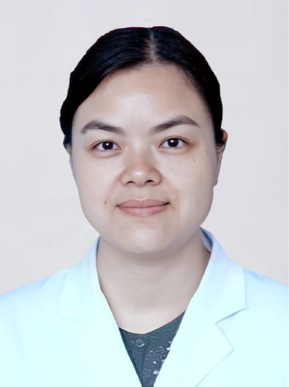 Junxia Yao (姚军霞)合作论文数Union Hospital, Tongji Medical College, Huazhong University of Science and Technology5
 Xiaodong Teng合作论文数5Weigang Ge合作论文数4
 Lu Tang合作论文数Department of Biostatistics, School of Public Health, University of Pittsburgh4Liu Wei合作论文数4Huanhuan Gao合作论文数4Nan Xiang合作论文数4
 N Gopalakrishna Iyer合作论文数Dept Surg Oncol, Natl Canc Ctr Singapore4
 Rudolf Aebersold合作论文数Department of Biology, Eidgenössische Technische Hochschule Zürich4Wen Du (杜雯)合作论文数4Yanli He (何艳丽)合作论文数4
 Sathiyamoorthy Selvarajan合作论文数Department of Anatomy, National University of Singapore Singapore4Oi Lian Kon合作论文数4
 Zhongzhi Luan (栾钟治)合作论文数School of Computer Science and Engineering, Beihang University4Junhong Xiao合作论文数3Danying Liao合作论文数3
 Xiao Liang合作论文数3Juan Li合作论文数3
 Chunhui Yuan合作论文数3Guangzhi Wang合作论文数3
 Shuang Liang合作论文数3Bo Wang合作论文数3Wei Xie合作论文数3Zheng Jin (金政)合作论文数3
 Jingqiong Hu (胡晶琼)合作论文数Union Hospital, Tong Ji Medical College, Huazhong University of Science and Technology3




