Autophagy is a conserved catabolic process that relies on vacuoles or lysosomes. While autophagosome formation is well characterized, the mechanisms that prevent autophagy-related proteins form being enclosed by the autophagosome and degraded in the vacuole remain unclear in yeast. Here, we show that the SWR1 chromatin remodeling complex plays an essential, noncanonical role in this process. Genome-wide screening identified the SWR1 complex as a critical regulator that prevents the vacuolar delivery of multiple autophagy proteins. This process depends on the structural integrity and ATPase activity of the SWR1 complex. Mechanistically, the SWR1 subunit Rvb1 interacts directly with Atg21, and this interaction is important for SWR1 localization to the phagophore assembly site and efficient protein retrieval. Disruption of the Atg21-Rvb1 interaction results in the vacuolar accumulation of autophagy proteins. These findings uncover an unexpected link between a chromatin remodeling complex and the autophagy machinery, highlighting the Atg21-Rvb1 module as a key regulator of autophagy dynamics in yeast.
Astrocytes are essential for neuronal homeostasis, and synaptic modulation and development. While astrocytes exhibit distinct Ca2+ signaling patterns, decoding their physiological roles remains challenging because conventional approaches generate nonphysiological, spatially imprecise Ca2+ surges that obscure endogenous signals. Here, we developed a tunable light-based tool to modulate astrocytic Ca2+ activity, mimicking localized spikes and global waves elicited by graded glutamate through endogenous mGluR-Gq-IP3R signaling. IP3R triple knockout abolished light-evoked Ca2+ elevations, indicating elevated Ca2+ requires ER-IP3Rs. Using this modulation platform, we found that local Ca2+ spikes enhance organelle entry into astrocytic processes, whereas global Ca2+ waves arrest organelle movement. FKBP-FRB motor recruitment assays showed that global Ca2+ elevations acutely suppress motor-driven transport when cargo adaptors are bypassed. Combining our method with astrocyte process outgrowth analysis showed that Ca2+-dependent distal organelle accumulation promotes process elongation. Together, this light-based strategy provides a versatile platform for eliciting endogenous-like astrocytic Ca2+ patterns and reveals how distinct Ca2+ patterns differentially control organelle dynamics and astrocytic structural remodeling.
Cell survival requires tight coordination between growth-promoting metabolism and cellular quality-control pathways, yet how these processes are integrated remains unclear. Here we identify the conserved glycolytic enzyme PGAM1 as a metabolic-autophagy checkpoint that links glycolysis to autophagy initiation independently of its catalytic activity. Using complementary yeast and mammalian systems we show that PGAM1 functions as a molecular scaffold that recruits phosphatidylinositol 3-kinase complex I to the phagophore assembly site, thereby licensing autophagosome biogenesis. This autophagy-regulatory function is genetically essential, evolutionarily conserved and functionally separable from glycolysis. It is regulated by Atg1/ULK1-mediated phosphorylation that enhances Atg14 binding under starvation. Functionally, PGAM1 coordinates anabolic growth and stress-induced survival to maintain cellular homeostasis. In cancer, PGAM1 upregulation enhances both glycolytic flux and autophagy capacity. Disruption of either function markedly impairs tumour growth, establishing PGAM1 as a homeostatic checkpoint that is hijacked in cancer to drive both proliferation and stress tolerance.
Mec1 plays an essential role in both the DNA damage response and glucose starvation-induced autophagy. We recently reported that Mec1 regulates glucose starvation-induced autophagy through its direct binding to Atg13. However, the role of Mec1's kinase activity in autophagy remains unclear. In this study, we demonstrate that the kinase activity of Mec1 is required for glucose starvation-induced autophagy by regulating the phagophore assembly site (PAS) recruitment of Atg9 vesicles. Mechanistic and functional analyses identified Atg9 as a direct phosphorylation substrate of Mec1, with phosphorylation occurring at the S35, T203, and T243 sites. Mutations at these sites reduce the association of Atg9 with Atg17, Atg23, and Atg27, thereby impairing the PAS recruitment of Atg9 vesicles. Notably, we found that the Mec1-Atg13 binding is a prerequisite for the phosphorylation of Atg9 by Mec1. Furthermore, Mec1-mediated phosphorylation of Atg9 is also crucial for the PAS recruitment of Atg9 vesicles in response to DNA damage. We thus propose that Mec1's kinase activity regulates the PAS recruitment of Atg9 vesicles by phosphorylating Atg9 in response to energy stress and DNA damage.
In Saccharomyces cerevisiae, Atg1 is a core autophagy-related (Atg) protein kinase (PK) in regulating macroautophagy/autophagy, by physically interacting with numerous other proteins, or by phosphorylating various substrates. It is unclear how many Atg1-interacting partners and substrates are also involved in regulating autophagy. Here, we conducted transcriptomic, proteomic and phosphoproteomic profiling of Atg1-dependent molecular landscapes during nitrogen starvation-triggered autophagy, and detected 244, 245 and 217 genes to be affected by ATG1 in the autophagic process at mRNA, protein, and phosphorylation levels, respectively. Based on the Atg1 interactome, we developed a novel artificial intelligence (AI) framework, inference of autophagy regulators from multi-omic data (iAMD), and predicted 12 Atg1-interacting partners and 17 substrates to be potentially functional in autophagy. Further experiments validated that Rgd1 and Whi5 are required for bulk autophagy, as well as physical interactions and co-localizations with Atg1 during autophagy. In particular, we demonstrated that 2 phosphorylation sites (p-sites), pS78 and pS149 of Whi5, are phosphorylated by Atg1 to regulate the formation of Atg1 puncta during autophagy initiation. A working model was illustrated to emphasize the importance of the Atg1-centered network in yeast autophagy. In addition, iAMD was extended to accurately predict Atg proteins and autophagy regulators from other PK interactomes, indicating a high transferability of the method. Taken together, we not only revealed new autophagy regulators from the Atg1 interactome, but also provided a useful resource for further analysis of yeast autophagy.
Selective autophagy plays a crucial role in maintaining cellular homeostasis by specifically targeting unwanted cargo labeled with "autophagy cues" signals for autophagic degradation. In this study, we identify Rab GTPases as a class of such autophagy cues signals involved in selective autophagy. Through biochemical and imaging screens, we reveal that human Rab GTPases are common autophagy substrates. Importantly, we confirm the conservation of Rab GTPase autophagic degradation in different model organisms. Rab GTPases translocate to damaged mitochondria, lipid droplets, and invading Salmonella-containing vacuoles (SCVs) to serve as degradation signals. Furthermore, they facilitate mitophagy, lipophagy, and xenophagy, respectively, by recruiting receptors. This interplay between Rab GTPases and receptors may ensure the de novo synthesis of isolation membranes around Rab-GTPase-labeled cargo, thereby mediating selective autophagy. These processes are further influenced by upstream regulators such as LRRK2, GDIs, and RabGGTase. In conclusion, this study unveils a conserved mechanism involving Rab GTPases as autophagy cues signals and proposes a model for the spatiotemporal control of selective autophagy.
Ribophagy is a selective autophagic process that regulates ribosome turnover. Although NUFIP1 has been identified as a mammalian receptor for ribophagy, its homologues do not exist in yeast and nematodes. Here we demonstrate that Rpl12, a ribosomal large subunit protein, functions as a conserved ribophagy receptor in multiple organisms. Disruption of Rpl12-Atg8s binding leads to significant accumulation of ribosomal proteins and rRNA, while Atg1-mediated Rpl12 phosphorylation enhances its association with Atg11, thus triggering ribophagy during starvation. Ribophagy deficiency accelerates cell death induced by starvation and pathogen infection, leading to impaired growth and development and a shortened lifespan in both Caenorhabditiselegans and Drosophila melanogaster. Moreover, ribophagy deficiency results in motor impairments associated with ageing, while the overexpression of RPL12 significantly improves movement defects induced by starvation, ageing and A beta accumulation in fly models. Our findings suggest that Rpl12 functions as a conserved ribophagy receptor vital for ribosome metabolism and cellular homeostasis.
Macroautophagy/autophagy is an evolutionarily conserved intracellular degradation pathway that relies on vacuoles or lysosomes. Over 40 ATG genes have been identified in yeast cells as participants in various types of autophagy, although these genes are non-essential. While some essential genes involved in autophagy have been identified using temperature-sensitive yeast strains, systematic research on essential genes in autophagy remains lacking. To address this, we established an essential protein conditional degradation library using the auxin-inducible degron (AID) system. By introducing the GFP-Atg8 plasmid, we identified 29 essential yeast genes involved in autophagy, 19 of which had not been previously recognized. In summary, the yeast essential protein conditional degradation library we constructed will serve as a valuable resource for systematically investigating the roles of essential genes in autophagy and other biological functions.Abbreviation: AID: auxin-inducible degron; ALP: alkaline phosphatase; ATG: autophagy related; CSG: constitutive slow growth; DAmP: Decreased Abundance by mRNA Perturbation; GFP: green fluorescent protein; MMS: methyl methanesulfonate; ORF: open reading frame; PAS: phagophore assembly site; PCR: polymerase chain reaction; SD-G: glucose starvation medium; SD-N: nitrogen starvation medium; TOR: target of rapamycin kinase; YGRC: yeast genetic resource center; YPD: yeast extract peptone dextrose
Autophagy is essential for maintaining glucose homeostasis. However, the mechanism by which cells sense and respond to glucose starvation to induce autophagy remains incomplete. Here, we show that calcium serves as a fundamental triggering signal that connects environmental sensing to the formation of the autophagy initiation complex during glucose starvation. Mechanistically, glucose starvation instigates the release of vacuolar calcium into the cytoplasm, thus triggering the activation of Rck2 kinase. In turn, Rck2-mediated Atg11 phosphorylation enhances Atg11 interactions with Bmh1/2 bound to the Snf1-Sip1-Snf4 complex, leading to recruitment of vacuolar membrane-localized Snf1 to the PAS and subsequent Atg1 activation, thereby initiating autophagy. We also identified Glc7, a protein phosphatase-1, as a critical regulator of the association between Bmh1/2 and the Snf1 complex. We thus propose that calcium-triggered Atg11-Bmh1/2-Snf1 complex assembly initiates autophagy by controlling Snf1-mediated Atg1 activation in response to glucose starvation.
The autophagy initiation complex is brought about via a highly ordered and stepwise assembly process. Two crucial signaling molecules, mTORC1 and AMPK, orchestrate this assembly by phosphorylating/dephosphorylating autophagy-related proteins. Activation of Atg1 followed by recruitment of both Atg9 vesicles and the PI3K complex I to the PAS (phagophore assembly site) are particularly crucial steps in its formation. Ypt1, a small Rab GTPase in yeast cells, also plays an essential role in the formation of the autophagy initiation complex through multiple regulatory pathways. In this review, our primary focus is to discuss how signaling molecules initiate the assembly of the autophagy initiation complex, and highlight the significant roles of Ypt1 in this process. We end by addressing issues that need future clarification.
Macroautophagy/autophagy, is widely recognized for its crucial role in enabling cell survival and maintaining cellular energy homeostasis during starvation or energy stress. Its regulation is intricately linked to cellular energy status. In this review, covering yeast, mammals, and plants, we aim to provide a comprehensive overview of the understanding of the roles and mechanisms of carbon- or glucose-deprivation related autophagy, showing how cells effectively respond to such challenges for survival. Further investigation is needed to determine the specific degraded substrates by autophagy during glucose or energy deprivation and the diverse roles and mechanisms during varying durations of energy starvation.Abbreviations: ADP: adenosine diphosphate; AMP: adenosine monophosphate; AMPK: AMP-activated protein kinase; ATG: autophagy related; ATP: adenosine triphosphate; ER: endoplasmic reticulum; ESCRT: endosomal sorting complex required for transport; GAPDH: glyceraldehyde-3-phosphate dehydrogenase; GD: glucose deprivation; GFP: green fluorescent protein; GTPases: guanosine triphosphatases; HK2: hexokinase 2; K phaffii: Komagataella phaffii; LD: lipid droplet; MAP1LC3/LC3: microtubule-associated protein1 light chain 3; MAPK: mitogen-activated protein kinase; Mec1: mitosis entry checkpoint 1; MTOR: mechanistic target of rapamycin kinase; NAD (+): nicotinamide adenine dinucleotide; OGD: oxygen and glucose deprivation; PAS: phagophore assembly site; PCD: programmed cell death; PtdIns3K: class III phosphatidylinositol 3-kinase; PtdIns3P: phosphatidylinositol-3-phosphate; ROS: reactive oxygen species; S. cerevisiae: Saccharomyces cerevisiae; SIRT1: sirtuin 1; Snf1: sucrose non-fermenting 1; STK11/LKB1: serine/threonine kinase 11; TFEB: transcription factor EB; TORC1: target of rapamycin complex 1; ULK1: unc-51 like kinase 1; Vps27: vacuolar protein sorting 27; Vps4: vacuolar protein sorting 4.
Ribophagy, the cellular process wherein ribosomes are selectively self-digested through autophagy, plays a pivotal role in maintaining ribosome turnover. Understanding the molecular regulatory mechanisms governing ribophagy is pivotal to uncover its significance. Consequently, the establishment of methods for detecting ribophagy becomes important. In this protocol, we have optimized, enriched, and advanced existing ribophagy detection techniques, including immunoblotting, fluorescence microscopy, and transmission electron microscopy (TEM), to precisely monitor and quantify ribophagic events. Particularly noteworthy is the introduction of TEM technology for yeast ribophagy detection. In summary, the delineated methods are applicable for detecting ribophagy in both yeast and mammals, laying a solid foundation for further exploring the physiological importance of ribophagy and its potential implications in diverse cellular environments.
Autophagy plays a crucial role in maintaining cellular homeostasis in response to various stimuli. Compared to research on nutrient deprivation-induced autophagy, the understanding of the molecular mechanisms and physiological/pathological significance of autophagy triggered by energy deprivation remains limited. A primary focus of our lab is to elucidate how cells sense energy deprivation and initiate autophagy. Using the model organisms Saccharomyces cerevisiae and mammalian cells, we found that cellular reactive oxygen species (ROS), DNA damage sensor Mec1, and mitochondrial aerobic respiration play essential roles in the autophagy induced by energy deprivation. This review aims to provide a concise overview of these research findings.
AbstractCCT2 serves as an aggrephagy receptor that plays a crucial role in the clearance of solid aggregates, yet the underlying molecular mechanisms by which CCT2 regulates solid aggrephagy are not fully understood. Here we report that the binding of Cct2 to Atg8 is governed by two distinct regulatory mechanisms: Atg1-mediated Cct2 phosphorylation and the interaction between Cct2 and Atg11. Atg1 phosphorylates Cct2 at Ser412 and Ser470, and disruption of these phosphorylation sites impairs solid aggrephagy by hindering Cct2-Atg8 binding. Additionally, we observe that Atg11, an adaptor protein involved in selective autophagy, directly associates with Cct2 through its CC4 domain. Deficiency in this interaction significantly weakens the association of Cct2 with Atg8. The requirement of Atg1-mediated Cct2 phosphorylation and of Atg11 for CCT2-LC3C binding and subsequent aggrephagy is conserved in mammalian cells. These findings provide insights into the crucial roles of Atg1-mediated Cct2 phosphorylation and Atg11-Cct2 binding as key mediators governing the interaction between Cct2 and Atg8 during the process of solid aggrephagy.
Macroautophagy/autophagy is essential for maintaining glucose homeostasis, but the mechanisms by which cells sense glucose starvation and initiate autophagy are not yet fully understood. Recently, we reported that the assembly of a Ca2+-triggered Snf1-Bmh1/Bmh2-Atg11 complex initiates autophagy in response to glucose starvation. Our research reveals that during glucose starvation, the efflux of vacuolar Ca2+ increases cytoplasmic Ca2+ levels, which activates the protein kinase Rck2. Rck2-mediated phosphorylation of Atg11 enhances its interaction with Bmh1 and Bmh2. This interaction recruits the Snf1-Sip1-Snf4 complex, which is located on the vacuolar membrane, to the phagophore assembly site (PAS), leading to the activation of Atg1 and the initiation of autophagy. In summary, we have identified a previously unrecognized signaling pathway involved in glucose starvation-induced autophagy, where Ca2+ acts as a fundamental signaling molecule that links energy stress to the formation of the autophagy initiation complex.Abbreviation: AMPK: AMP-activated protein kinase; ATG: autophagy related; co-IP: co-immunoprecipitation; MAPK: mitogen-activated protein kinase; PAS: phagophore assembly site; ULK1: unc-51 like autophagy activating kinase 1.
The regulation of autophagy initiation is a key step in autophagosome biogenesis. However, our understanding of the molecular mechanisms underlying the stepwise assembly of ATG proteins during this process remains incomplete. The Rab GTPase Ypt1/Rab1 is recognized as an essential autophagy regulator. Here, we identify Atg23 and Atg17 as binding partners of Ypt1, with their direct interaction proving crucial for the stepwise assembly of autophagy initiation complexes. Disruption of Ypt1-Atg23 binding results in significantly reduced Atg9 interactions with Atg11, Atg13, and Atg17, thus preventing the recruitment of Atg9 vesicles to the phagophore assembly site (PAS). Likewise, Ypt1-Atg17 binding contributes to the PAS recruitment of Ypt1 and Atg1. Importantly, we found that Ypt1 is phosphorylated by TOR at the Ser174 residue. Converting this residue to alanine blocks Ypt1 phosphorylation by TOR and enhances autophagy. Conversely, the Ypt1S174D phosphorylation mimic impairs both PAS recruitment and activation of Atg1, thus inhibiting subsequent autophagy. Thus, we propose TOR-mediated Ypt1 as a multifunctional assembly factor that controls autophagy initiation via its regulation of the stepwise assembly of ATG proteins.
Atg11 is an adaptor protein required for the induction of selective autophagy via receptor binding. However, our understanding of the molecular mechanisms by which it regulates selective autophagy remains incomplete. Here, we show that Atg11 is phosphorylated by Atg1. Rapamycin treatment or starvation conditions induced slower electrophoretic mobility of Atg11 in an Atg1 kinase activity-dependent manner. Through in vitro kinase assays combined with mutagenesis, we determined that Atg1 phosphorylates S949, S1057, and S1064 residues in CC4 domain of Atg11. Replacing the three residues with alanine suppressed the cleavage of selective autophagy substrates for the cytoplasm-to-vacuole targeting (Cvt) pathway, mitophagy, reticulophagy, and pexophagy. The Atg11 mutant was defective in binding to related selective autophagy receptors. These results demonstrate a previously unknown function of Atg1 in regulation of selective autophagy via Atg11 phosphorylation.
Mec1 is a DNA damage sensor, which performs an essential role in the DNA damage response pathway and glucose starvation-induced autophagy. However, the functions of Mec1 in autophagy remain unclear. In response to glucose starvation, Mec1 forms puncta, which are recruited to mitochondria through the adaptor protein Ggc1. Here, we show that Mec1 puncta also contact the phagophore assembly site (PAS) via direct binding with Atg13. Functional analysis of the Atg13-Mec1 interaction revealed two previously unrecognized protein regions, the Mec1-Binding Region (MBR) on Atg13 and the Atg13-Binding Region (ABR) on Mec1, which mediate their mutual association under glucose starvation conditions. Disruption of the MBR or ABR impairs the recruitment of Mec1 puncta and Atg13 to the PAS, consequently blocking glucose starvation-induced autophagy. Additionally, the MBR and ABR regions are also crucial for DNA damage-induced autophagy. We thus propose that Mec1 regulates glucose starvation-induced autophagy by controlling Atg13 recruitment to the PAS.
Protein aggregation is a hallmark of multiple human pathologies. Autophagy selectively degrades protein aggregates via aggrephagy. How selectivity is achieved has been elusive. Here, we identify the chaperonin subunit CCT2 as an autophagy receptor regulating the clearance of aggregation-prone proteins in the cell and the mouse brain. CCT2 associates with aggregation-prone proteins independent of cargo ubiquitination and interacts with autophagosome marker ATG8s through a non-classical VLIR motif. In addition, CCT2 regulates aggrephagy independently of the ubiquitin-binding receptors (P62, NBR1, and TAX1BP1) or chaperone-mediated autophagy. Unlike P62, NBR1, and TAX1BP1, which facilitate the clearance of protein condensates with liquidity, CCT2 specifically promotes the autophagic degradation of protein aggregates with little liquidity (solid aggregates). Furthermore, aggregation-prone protein accumulation induces the functional switch of CCT2 from a chaperone subunit to an autophagy receptor by promoting CCT2 monomer formation, which exposes the VLIR to ATG8s interaction and, therefore, enables the autophagic function.
 Liqin Zhang (张立钦)合作论文数College of Forestry and Biotechnology, Zhejiang A&F University10
 Choufei Wu (吴酬飞)合作论文数Huzhou University10
 Weijing Yao合作论文数9
 Yuting Chen合作论文数7Wei Liu合作论文数6Yingcong Chen合作论文数6Yigang Wang合作论文数6
 Jingjing Tong (仝晶晶)合作论文数School of Life Sciences, Central China Normal University6
 Qiming Sun (孙启明)合作论文数School of Medicine, Zhejiang University6Shan Feng合作论文数6
 Yuyao Feng合作论文数5
 Yi Zhang (张熠)合作论文数Laboratory of Molecular Cancer Research, College of Pharmaceutical Sciences, Soochow Medical College of Soochow University;Department of Pharmacology, College of Pharmaceutical Sciences, Soochow Medical College of Soochow University5
 Hongguang Xia (夏宏光)合作论文数Molecular Medical Center, School of Medicine, Zhejiang University5
 Li Yu (俞立)合作论文数School of Life Sciences, Tsinghua University5
 Mingzhu Fan合作论文数Great Lakes Bioenergy Res Ctr, Michigan State Univ4Xiaoyong Wu合作论文数4
 Pengwei Zhao合作论文数4Yixing Li合作论文数4
 Du Feng (冯杜)合作论文数State Key Laboratory of Respiratory Disease, Guangzhou Medical University;School of Basic Medical Sciences, Guangzhou Medical University4
 Di Peng (彭迪)合作论文数College of Life Science and Technology, Huazhong University of Science and Technology3
 Zhi Hong (洪智)合作论文数Zhejiang University-University of Edinburgh Institute3
 Liefeng Wang (王烈峰)合作论文数Gannan Medical University3Jing Liu合作论文数3
 Zhiping Xie (谢志平)合作论文数School of Life Science and Biotechnology, Shanghai Jiao Tong University3
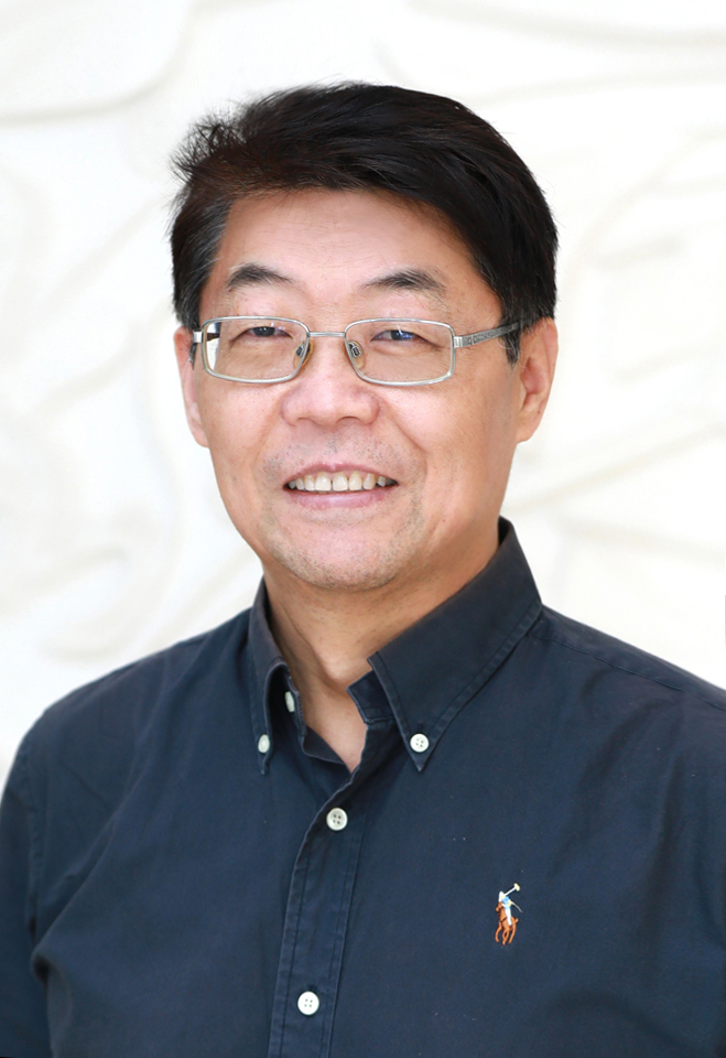 Haiteng Deng (邓海腾)合作论文数School of Life Sciences, Tsinghua University3
 Yu Xue (薛宇)合作论文数Department of Bioinformatics & Systems Biology, College of Life Science and Technology, Huazhong University of Science and Technology;Key Laboratory of Molecular Biophysics of Ministry of Education, Huazhong University of Science and Technology3Miaojuan Ye合作论文数2Huiming Zhong合作论文数2Chenjun Lu合作论文数2Qiang Jiang合作论文数2
 Yi Ting Zhou合作论文数2Qingwei Zhao合作论文数2Shaojin Zhang合作论文数Tsinghua Univ, State Key Lab Biomembrane & Membrane Biotechnol, Tsinghua Univ Peking Univ Joint Ctr Life Sci, Sch Life Sci, Beijing 100084, Peoples R China2Xufeng Cen合作论文数Department of Biochemistry & Research Center of Clinical Pharmacy of The First Affiliated Hospital, Zhejiang University School of Medicine,Hangzhou 310058,China2Yu Xie合作论文数2Di Miao合作论文数2
 Najafov Ayaz合作论文数Dept Cell Biol, Harvard Med Sch2
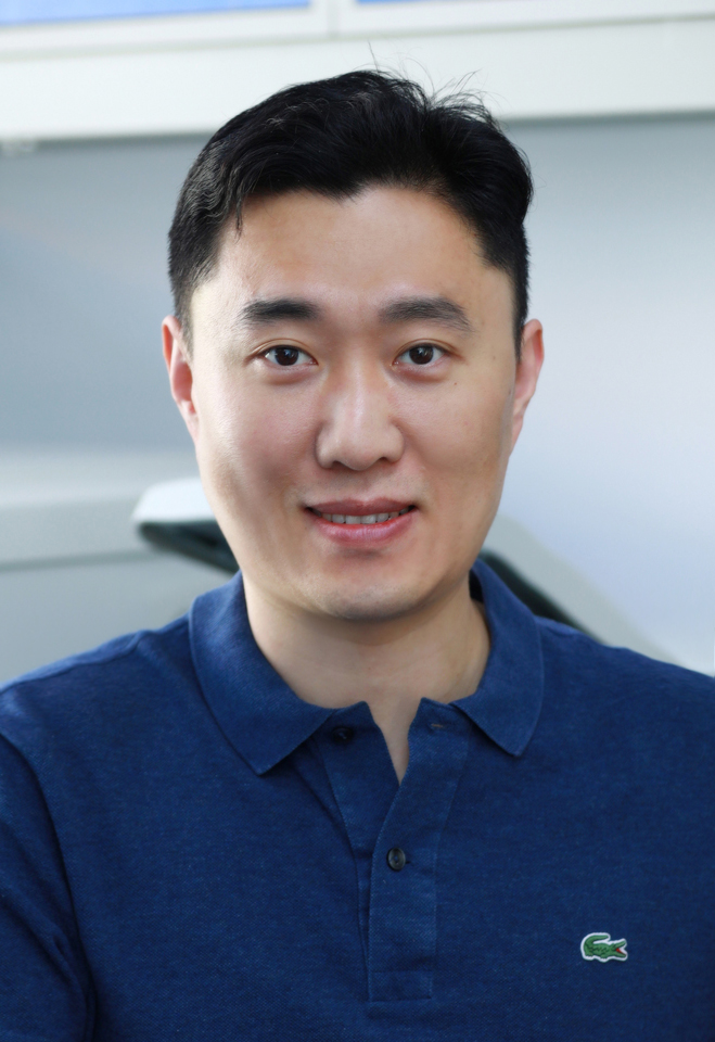 Liang Ge (葛亮)合作论文数School of Life Sciences, Tsinghua University2Kunrong Mei (梅坤荣)合作论文数2Yan Le (乐雁)合作论文数2
 Wei Zou (邹炜)合作论文数Institute of Translational Medicine, Zhejiang University2Xue Bai (白雪)合作论文数2Zhaoxiang He合作论文数1Yanying Chen (陈颖妍)合作论文数1Puzhong Lu合作论文数1Xi Sheng Rao合作论文数1Tingxue Xie合作论文数1Jiaxin Hu合作论文数1




