This international multisociety consensus statement, developed by 21 thoracic radiologists and reviewed by four pulmonology experts, aims to standardize chest CT indications, acquisition, and reporting of post–COVID-19 residual CT lung abnormalities.
Connective tissue disease, also referred to as collagen vascular disease, encompasses a group of systemic inflammatory disorders characterized by autoimmune-mediated end-organ damage. Connective tissue diseases may result in a variety of interstitial lung disease patterns on computed tomography including usual interstitial pneumonia, nonspecific interstitial pneumonia (NSIP), organizing pneumonia and lymphoid interstitial pneumonia, with NSIP being the most common. As interstitial lung disease may be the only manifestation of connective tissue disease or present years before positive serologies, radiologists must be familiar with these patterns to provide diagnostic value.
Coronavirus disease 2019 (COVID-19) pneumonia has had catastrophic effects worldwide. Radiology, in particular computed tomography (CT) imaging, has proven to be valuable in the diagnosis, prognostication, and longitudinal assessment of those diagnosed with COVID-19 pneumonia. This article will review acute and chronic pulmonary radiologic manifestations of COVID-19 pneumonia with an emphasis on CT and also highlighting histopathology, relevant clinical details, and some notable challenges when interpreting the literature.
CT angiography (CTA) of the aortoiliofemoral (AIF) arteries in the abdomen, pelvis, and lower extremities has become an invaluable tool in assessment of patients with peripheral arterial disease (PAD) and lower extremity trauma. AIF CTA provides rapid and comprehensive assessment of arterial inflow and outflow, guiding management of patients with chronic claudication and those with more acute manifestations, including atherothrombotic occlusion, embolic disease, or thrombosis of prior interventions such as bypass graft or stent placement. Careful attention to technique is critical in performing diagnostic AIF CTA, as pitfalls related to imaging too early or too late relative to the arrival of contrast material in the legs can lead to misdiagnosis or diagnostic uncertainty. The protocol can be tailored to the clinical scenario; for instance, the chest can be additionally imaged when aortic dissection affecting lower extremity perfusion, trauma, and embolic disease are clinical considerations. Additionally, when one is imaging patients with prior endovascular aortic repair, an additional delayed acquisition to assess for endoleak can be performed. Newer techniques such as dual-energy and photon-counting CT can improve endovascular enhancement, facilitate material decompensation, and produce virtual noniodine images. The authors detail normal vascular anatomy and key collateral pathways; AIF CTA protocols; and imaging of PAD, vascular trauma, and infectious or inflammatory disorders affecting the lower extremities. This knowledge, combined with a comprehensive approach to interpreting AIF CTA studies, will prepare the radiologist to offer optimal diagnostic value to guide clinical care. ©RSNA, 2025 Supplemental material is available for this article.
Coronavirus disease 2019 (COVID-19) pneumonia has had catastrophic effects worldwide. Radiology, in particular computed tomography (CT) imaging, has proven to be valuable in the diagnosis, prognostication, and longitudinal assessment of those diagnosed with COVID-19 pneumonia. This article will review acute and chronic pulmonary radiologic manifestations of COVID-19 pneumonia with an emphasis on CT and also highlighting histopathology, relevant clinical details, and some notable challenges when interpreting the literature.
Flow artifacts are commonly encountered at contrast-enhanced CT and can be difficult to discern from true pathologic conditions. Therefore, radiologists must be comfortable distinguishing flow artifacts from true pathologic conditions. This is of particular importance when evaluating the pulmonary arteries and aorta, as a flow artifact may be mistaken for a pulmonary embolism or dissection flap. Understanding the mechanics of flow artifacts and how these artifacts are created can help radiologists in several ways. First, this knowledge can help radiologists appreciate how the imaging characteristics of flow artifacts differ from true pathologic conditions. This information can also help radiologists better recognize the clinical conditions that predispose patients to flow artifacts, such as pneumonia, chronic lung damage, and altered cardiac output. By understanding when flow artifacts may be confounding the interpretation of an examination, radiologists can then know when to pursue other troubleshooting methods to assist with the diagnosis. In these circumstances, the radiologist can consider several troubleshooting methods, including adjusting the imaging protocols, recommending when additional imaging may be helpful, and suggesting which imaging study would be the most beneficial. Finally, flow artifacts can also be used as a diagnostic tool when evaluating the vascular anatomy, examples of which include the characterization of shunts, venous collaterals, intimomedial flaps, and alternative patterns of blood flow, as seen in extracorporeal membrane oxygenation circuits.
Pleural effusions are categorized as transudative or exudative, with transudative effusions usually reflecting the sequala of a systemic etiology and exudative effusions usually resulting from a process localized to the pleura. Common causes of transudative pleural effusions include congestive heart failure, cirrhosis, and renal failure, whereas exudative effusions are typically due to infection, malignancy, or autoimmune disorders. This document summarizes appropriateness guidelines for imaging in four common clinical scenarios in patients with known or suspected pleural effusion or pleural disease. The American College of Radiology Appropriateness Criteria are evidence-based guidelines for specific clinical conditions that are reviewed annually by a multidisciplinary expert panel. The guideline development and revision process support the systematic analysis of the medical literature from peer reviewed journals. Established methodology principles such as Grading of Recommendations Assessment, Development, and Evaluation or GRADE are adapted to evaluate the evidence. The RAND/UCLA Appropriateness Method User Manual provides the methodology to determine the appropriateness of imaging and treatment procedures for specific clinical scenarios. In those instanceswhere peer reviewed literature is lacking or equivocal, experts may be the primary evidentiary source available to formulate a recommendation.
This study evaluates the prognostic value of CT findings, including volumetric measurements, in predicting outcomes for patients with Fournier gangrene (FG), focusing on mortality, ICU admission, hospital stay length, and healthcare costs. A retrospective study was conducted on 38 FG patients who underwent CT scans before surgical debridement. We analyzed demographic data, CT volumetric measurements, and clinical outcomes using logistic and linear regression models. No single CT measurement significantly predicted mortality or ICU admission. The best model for mortality prediction included age, air volume, NSTI score, and male sex, with an AUC of 0.911. Intubation likelihood was modeled with an AUC of 0.913 using age, NSTI score, and visceral to subcutaneous fat ratio. The ICU admission model achieved an AUC of 0.677. Hospital stay was predicted by air volume (β = 0.0002656, p = 0.0505) with an adjusted R-squared of 0.1287. Air volume significantly predicted hospital costs (β = 2.859, p = 0.00558), resulting in an adjusted R-squared of 0.2165. Volumetric CT findings provide valuable prognostic insights for FG patients, suggesting a basis for informed clinical decisions and resource allocation. Further validation in larger, multi-center studies is recommended to develop robust predictive models for FG outcomes.
Biphenotypic sinonasal sarcoma (BSNS) is a rare low-grade malignancy occurring in the sinonasal tract that is characterized by dual neural and myogenic differentiation. Rearrangements involving the PAX3 gene, usually with MAML3, are a hallmark of this tumor type and their identification are useful for diagnosis. Rarely, a MAML3 rearrangement without associated PAX3 rearrangement has been described. Other gene fusions have not been previously reported. Herein, we report a 22 year-old woman with a BSNS harboring a novel gene fusion involving the PAX7 gene (specifically PAX7::PPARGC1A), which is a paralogue of PAX3. The histologic features of the tumor were typical with two exceptions: a lack of entrapment of surface respiratory mucosa and no hemangiopericytoma-like vasculature. Immunophenotypically, the tumor was notably negative for smooth muscle actin, which is usually positive in BSNS. However, the classic S100 protein-positive, SOX10-negative staining pattern was present. In addition, the tumor was positive for desmin and MyoD1 but negative for myogenin, a pattern that is common among BSNS with variant fusions. Awareness of the possibility of PAX7 gene fusions in BSNS is important as it may aid in the diagnosis of PAX3 fusion negative tumors.
Diffuse alveolar damage (DAD), which represents the pathologic changes seen after acute lung injury, is caused by damage to all three layers of the alveolar wall and can ultimately result in alveolar collapse with loss of the normal pulmonary architecture. DAD has an acute phase that predominantly manifests as airspace disease at CT owing to filling of the alveoli with cells, plasma fluids, and hyaline membranes. DAD then evolves into a heterogeneous organizing phase, with mixed airspace and interstitial disease characterized by volume loss, architectural distortion, fibrosis, and parenchymal loss. Patients with DAD have a severe clinical course and typically require prolonged mechanical ventilation, which may result in ventilator-induced lung injury. In those patients who survive DAD, the lungs will remodel over time, but most will have residual findings at chest CT. Organizing pneumonia (OP) is a descriptive term for a histologic pattern characterized by intra-alveolar fibroblast plugs. The significance and pathogenesis of OP are controversial. Some authors regard it as part of a spectrum of acute lung injury, while others consider it a marker of acute or subacute lung injury. At CT, OP manifests with various forms of airspace disease that are most commonly bilateral and relatively homogeneous in appearance at individual time points. Patients with OP most often have a mild clinical course, although some may have residual findings at CT. In patients with DAD and OP, imaging findings can be combined with clinical information to suggest the diagnosis in many cases, with biopsy reserved for difficult cases with atypical findings or clinical manifestations. To best participate in the multidisciplinary approach to patients with lung injury, radiologists must not only recognize these entities but also describe them with consistent and meaningful terminology, examples of which are emphasized in the article. © RSNA, 2023 See the invited commentary by Kligerman et al in this issue. Quiz questions for this article are available in the supplemental material.
Lung cancer remains the leading cause of cancer-related mortality for men and women in the United States. Screening for lung cancer with annual low-dose CT is saving lives, and the continued implementation of lung screening can save many more. In 2015, the CMS began covering annual lung screening for those who qualified based on the original United States Preventive Services Task Force (USPSTF) lung screening criteria, which included patients 55 to 77 year of age with a 30 pack-year history of smoking, who were either currently using tobacco or who had smoked within the previous 15 years. In 2021, the USPSTF issued new screening guidelines, decreasing the age of eligibility to 80 years of age and pack-years to 20. Lung screening remains controversial for those who do not meet the updated USPSTF criteria, but who have additional risk factors for the development of lung cancer. The American College of Radiology Appropriateness Criteria are evidence-based guidelines for specific clinical conditions that are reviewed annually by a multidisciplinary expert panel. The guideline development and revision process support the systematic analysis of the medical literature from peer reviewed journals. Established methodology principles such as Grading of Recommendations Assessment, Development, and Evaluation or GRADE are adapted to evaluate the evidence. The RAND/UCLA Appropriateness Method User Manual provides the methodology to determine the appropriateness of imaging and treatment procedures for specific clinical scenarios. In those instances where peer reviewed literature is lacking or equivocal, experts may be the primary evidentiary source available to formulate a recommendation.
Purpose The effect of obesity in penetrating trauma outcomes is poorly understood. The purpose of this study was to determine if there is a protective effect of subcutaneous or visceral fat from stab and gunshot wounds.Methods 443 patients admitted after penetrating traumatic injury of the torso were retrospectively identified from our institution’s trauma registry. CT scans performed at presentation were used to determine cross-sectional area of visceral and subcutaneous fat at the level of the umbilicus via manual segmentation. Obesity-associated parameters including body mass index, visceral and subcutaneous fat were compared with injury severity score, length of hospital/intesive care unit (ICU) stay, and number of operating room (OR) visits. Parameters were compared between patients who sustained stab wounds versus gunshot injuries.Results Comparing all patients with gunshot injuries with those with stab injuries, gunshots resulted in increased hospital and ICU length of stay, and injury severity score (ISS). For patients with gunshot wounds, all obesity-related parameters correlated with increased length of stay and total ICU stay; subcutaneous fat and visceral fat were correlated with increased OR visits, but there was no significant correlation between obesity-related parameters and ISS. In contrast, with stab wounds there were no statistically significant associations between obesity parameters and any of the outcome measures.Conclusion For penetrating trauma in the torso, obesity is correlated with worse outcomes with gunshot injuries but not in stab injuries.Level of evidence Level III, prognostic and epidemiological.
Diffusion-weighted imaging (DWI) is a fundamental sequence not only in neuroimaging but also in oncologic imaging and has emerging applications for MRI evaluation of the chest. DWI can be used in clinical practice to enhance lesion conspicuity, tissue characterization, and treatment response. While the spatial resolution of DWI is in the order of millimeters, changes in diffusion can be measured on the micrometer scale. As such, DWI sequences can provide important functional information to MRI evaluation of the chest but require careful optimization of acquisition parameters, notably selection of b values, application of parallel imaging, fat saturation, and motion correction techniques. Along with assessment of morphologic and other functional features, evaluation of DWI signal attenuation and apparent diffusion coefficient maps can aid in tissue characterization. DWI is a noninvasive noncontrast acquisition with an inherent quantitative nature and excellent reproducibility. The outstanding contrast-to-noise ratio provided by DWI can be used to improve detection of pulmonary, mediastinal, and pleural lesions, to identify the benign nature of complex cysts, to characterize the solid portions of cystic lesions, and to classify chest lesions as benign or malignant. DWI has several advantages over fluorine 18 (18F)-fluorodeoxyglucose PET/CT in the assessment, TNM staging, and treatment monitoring of lung cancer and other thoracic neoplasms with conventional or more recently developed therapies. © RSNA, 2023 Quiz questions for this article are available in the supplemental material. Supplemental material and the slide presentation from the RSNA Annual Meeting are available for this article.
Incidental pulmonary nodules are common. Although the majority are benign, most are indeterminate for malignancy when first encountered making their management challenging. CT remains the primary imaging modality to first characterize and follow-up incidental lung nodules. This document reviews available literature on various imaging modalities and summarizes management of indeterminate pulmonary nodules detected incidentally. The American College of Radiology Appropriateness Criteria are evidence-based guidelines for specific clinical conditions that are reviewed annually by a multidisciplinary expert panel. The guideline development and revision process support the systematic analysis of the medical literature from peer reviewed journals. Established methodology principles such as Grading of Recommendations Assessment, Development, and Evaluation or GRADE are adapted to evaluate the evidence. The RAND/UCLA Appropriateness Method User Manual provides the methodology to determine the appropriateness of imaging and treatment procedures for specific clinical scenarios. In those instances where peer reviewed literature is lacking or equivocal, experts may be the primary evidentiary source available to formulate a recommendation.
 Sanjeev Bhalla合作论文数Mallinckrodt Institute of Radiology, Washington University School of Medicine56
 Vincent M Mellnick合作论文数38
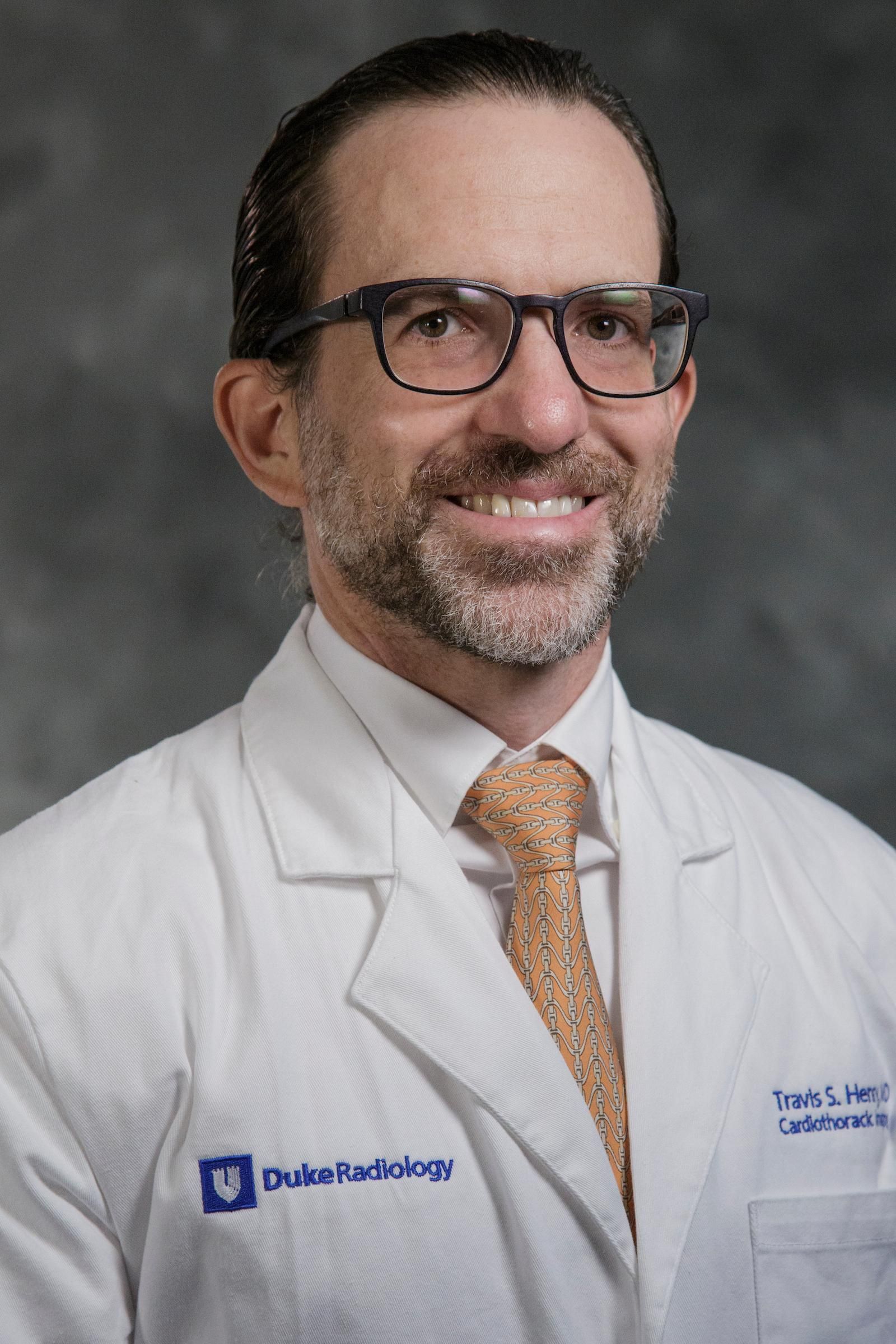 Travis Scott Henry合作论文数Division of Cardiothoracic Imaging, Department Of Radiology, School of Medicine, Duke University24
 Mark M. Hammer合作论文数23
 Kathryn Fowler合作论文数Department of Radiology, University of California San Diego;Jacobs Medical Center, University of California San Diego;Hillcrest Medical Center, University of California San Diego18Raptis Demetrios A合作论文数14
 David H Ballard合作论文数School of Medicine, Louisiana State University Health-Shreveport11Kaitlin M Marquis合作论文数9
 Edwin F. Donnelly合作论文数Department of Radiology, Ohio State University Wexner Medical Center9Christine O. Menias合作论文数Dept Radiol, Mayo Clin9Christopher T. Kuzniewski合作论文数8Jeffrey P Kanne合作论文数8Jordi Broncano合作论文数University Health Network, University of Toronto;Faculty of Medicine, University of Toronto;Concord Hospital Clinical School, The University of Sydney;Qscan Imaging Group, Clayfield, Australia (L.I.V.);Department of Radiology, Gold Coast University Hospital;HT Médica, Hospital San Juan de Dios;University Health Network, University of Toronto;Ottawa Hospital Research Institute Clinical Epidemiology Program, University of Ottawa;University Health Network, University of Toronto;Sinai Health System, University of Toronto;University Health Network, University of Toronto;University of Toronto, University of Toronto8Morris Michael F合作论文数8
 Michael D Hope合作论文数UCSF, San Francisco, CA, USA8Kacie L Steinbrecher合作论文数7Aine Marie Kelly合作论文数7Vamsi R. Narra合作论文数7Maria D. Martin合作论文数6Caroline Robb合作论文数6Arlene Sirajuddin合作论文数6
 Richard Tsai合作论文数6Wu Carol C合作论文数6
 Shetty Anup S合作论文数Mallinckrodt Institute of Radiology, Washington University School of Medicine6
 Daniel R Ludwig合作论文数5Archana T Laroia合作论文数5
 Elizabeth Lee合作论文数Department of Radiology, University of Michigan5
 Seth Kligerman合作论文数From the Departments of Diagnostic Radiology and Nuclear Medicine (Chest Imaging) (S.J.K., J.R.G.) and Internal Medicine (Pulmonary/Critical Care) (J.R.G.),, University of Maryland School of Medicine;Joint Task Force National Capital Region Medical, The Joint Pathology Center;and Department of Chest Imaging, American Institute for Radiologic Pathology;American Institute for Radiologic Pathology, University of Maryland School of Medicine5
 Richard L. Wahl合作论文数Precision Radiotheranostics Translation Center, Mallinckrodt Institute of Radiology, Washington University School of Medicine in St. Louis5
 Antonio Luna合作论文数5Sebastian Mcwilliams合作论文数National University of Ireland, Cork University Hospital5Daniel B. Green合作论文数4Parisa Mazaheri合作论文数4
 Douglas J E Schuerer合作论文数4Shah Amar B合作论文数4
 Pamela K. Woodard合作论文数Barnes-Jewish Hospital;St. Louis Children's Hospital;Mallinckrodt Institute of Radiology, Washington University School of Medicine in St. Louis;Department of Radiology, Washington University School of Medicine in St. Louis4
 Jonathan Mcconathy合作论文数4
 Alpesh N. Amin合作论文数Department of Medicine, School of Medicine, University of California, Irvine;Institute for Precision Health, University of California, Irvine;Henry Samueli School of Engineering, University of California, Irvine;School of Nursing, University of California, Irvine;UCI Health4
 Andrew J Bierhals合作论文数4Kyungran Shim合作论文数3Obeid N Ilahi合作论文数3Muhammad Naeem合作论文数3Mark J Hoegger合作论文数3
 Joseph W Owen合作论文数University of Kentucky;state of Kentucky;University of Kentucky3Kim L Sandler合作论文数Vanderbilt Univ, Med Ctr, Dept Radiol, Nashville, TN 37232 USA3
 Rebecca Chernock合作论文数Department of Pathology & Immunology, School of Medicine, Washington University in St. Louis;Barnes-Jewish Hospital;Barnes-Jewish West County Hospital3Cummings Kristopher W合作论文数3
 Perry W. Grigsby合作论文数Institute of Clinical and Translational Sciences, Department of Radiation Oncology, School of Medicine, Washington University in St. Louis3
 Fabien Maldonado合作论文数University Of Missouri Kansas City3




