This clinical policy from the American College of Emergency Physicians is a revision of the 2015 clinical policy: critical issues in the evaluation and management of adult patients with suspected acute nontraumatic thoracic aortic dissection. A writing subcommittee conducted a systematic review of the literature to derive evidence-based recommendations to answer the following clinical question: "In adult patients presenting to the emergency department with suspected acute nontraumatic thoracic aortic dissection, can a clinical decision rule plus additional laboratory testing or imaging (plain film or point-of-care ultrasound) identify a group of patients at very low risk for the diagnosis of thoracic aortic dissection?" Evidence was graded, and recommendations were made based on the strength of the available data.
This clinical policy from the American College of Emergency Physicians addresses key issues in the evaluation and management of adult emergency department patients presenting with asymptomatic elevated blood pressure. A writing committee conducted a systematic review of the literature to derive evidence-based recommendations to answer the following clinical question: In adult emergency department patients being discharged with asymptomatic elevated blood pressure, is initiation of outpatient antihypertensive medications from the emergency department safe and effective? Evidence was graded and recommendations were made based on the strength of the available data.
INTRODUCTION:Antipsychotic medications are often associated with QT interval prolongation, which can lead to ventricular dysrhythmias, including torsade de pointes. However, unstable or significant dysrhythmic events are rare. Evidence-based recommendations on the assessment and management of poisoned patients at risk for QT interval prolongation are lacking. Current practice often involves costly and lengthy cardiac monitoring, leading to delayed disposition of the patient from emergency departments or prolonged monitoring times in other hospital units. To address this issue, the QT Interval Prolongation in Clinical Toxicology Workgroup was formed by the Clinical Toxicology Recommendations Collaborative to develop evidence-based recommendations for the management and treatment of QT interval prolongation in acute overdose patients. This article reports our findings on patients with antipsychotic poisoning. METHODS:A systematic review of the literature regarding QT interval prolongation in all acute overdoses was undertaken, and the evidence was summarized for antipsychotic medication overdose. Voting statements were drafted using a predetermined format for monitoring the QT interval and use of continuous cardiac monitoring. A two-round modified Delphi method was used to reach a consensus. The strength of consensus was measured using the disagreement index as defined by the RAND/UCLA Appropriateness Method. RESULTS:From the 327 articles that were included in the systematic review of QT interval prolongation in all acute overdoses, a total of 55 articles were relevant to antipsychotic medication-induced QT prolongation in acute overdose. Medications with more than three articles were reviewed individually for recommendations, whilst medications with less than three were grouped together for discussion as miscellaneous. Individual medication recommendations were made for amisulpride (15 articles), thioridazine (11 articles), ziprasidone (eight articles) and quetiapine (13 articles), whilst consensus statements based on limited data were made for acute ingestions of clozapine, haloperidol, iloperidone, pimozide, pipamperone, olanzapine and risperidone (14 articles in total). The electrocardiogram is suggested as a risk assessment tool for ingestion of all antipsychotic medication. We recommend continuous cardiac monitoring for patients ingesting more than amisulpride 2 g, thioridazine 1 g, and ziprasidone 3 g. We suggest continuous cardiac monitoring for the above-mentioned medications for lower dose ingestions as well as for ingestions of haloperidol, iloperidone, pimozide and pipamperone. We do not suggest continuous cardiac monitoring for acute clozapine and risperidone poisoning, and do not recommend continuous cardiac monitoring for the QT interval in acute olanzapine or quetiapine poisoning. The need for ongoing cardiac monitoring should be guided by an individual risk assessment considering the medication and dose ingested, the time since ingestion, as well as other factors such as heart rate or co-ingestions. DISCUSSION:The quality of evidence for the risk of QT interval prolongation and torsade de pointes is heterogeneous among different antipsychotics and inherently constrains the recommendations. Available data suggest that amisulpride, thioridazine and ziprasidone are associated with QT interval prolongation and torsade de pointes, while the risk is likely overstated for quetiapine, olanzapine and risperidone. Ongoing research is needed to improve management strategies for acute antipsychotic overdose-induced QT interval prolongation and dysrhythmias. CONCLUSIONS:The QT Interval Prolongation in Clinical Toxicology Workgroup recommends the use of screening electrocardiograms in all patients with acute antipsychotic medication overdose and cardiac monitoring in patients with at-risk overdoses from thioridazine, amisulpride, and ziprasidone. The QT Interval Prolongation in Clinical Toxicology Workgroup suggests the same approach for patients with overdoses of haloperidol, iloperidone, pipamperone and pimozide. The risk of torsade de pointes is likely overstated for acute antipsychotic medication overdose as a general class group, and concern should rather focus on a few specific medications.
Pleural effusions are categorized as transudative or exudative, with transudative effusions usually reflecting the sequala of a systemic etiology and exudative effusions usually resulting from a process localized to the pleura. Common causes of transudative pleural effusions include congestive heart failure, cirrhosis, and renal failure, whereas exudative effusions are typically due to infection, malignancy, or autoimmune disorders. This document summarizes appropriateness guidelines for imaging in four common clinical scenarios in patients with known or suspected pleural effusion or pleural disease. The American College of Radiology Appropriateness Criteria are evidence-based guidelines for specific clinical conditions that are reviewed annually by a multidisciplinary expert panel. The guideline development and revision process support the systematic analysis of the medical literature from peer reviewed journals. Established methodology principles such as Grading of Recommendations Assessment, Development, and Evaluation or GRADE are adapted to evaluate the evidence. The RAND/UCLA Appropriateness Method User Manual provides the methodology to determine the appropriateness of imaging and treatment procedures for specific clinical scenarios. In those instanceswhere peer reviewed literature is lacking or equivocal, experts may be the primary evidentiary source available to formulate a recommendation.
This clinical policy from the American College of Emergency Physicians (ACEP) is the revision of a clinical policy approved in 2015 addressing a critical question regarding the use of thrombolytics for the management of acute ischemic stroke. A writing committee conducted a systematic review of the literature to derive evidence-based recommendations to answer the following clinical question: In adult stroke patients who are a candidate for mechanical thrombectomy, is the use of intravenous thrombolysis prior to mechanical thrombectomy (Bridge therapy) beneficial and safe versus mechanical thrombectomy alone? Evidence was graded, and recommendations were made based on the strength of the available data.
IntroductionHydrogen peroxide ingestions cause significant morbidity and mortality due to oxygen gas emboli and are treatable with hyperbaric oxygen therapy. Recommendations for observation are based on small case series.ObjectivesThe aim of this systematic review is to define the time of onset of embolic phenomena after hydrogen peroxide exposure and to describe the proportion of patients who received hyperbaric oxygen therapy.MethodsCases from a systematic literature search were combined with those from a prior study that used data derived from the American Association of Poison Control Centers National Poison Data System. Air-gas emboli were defined as embolic phenomena (stroke, myocardial infarction, obstructive shock) potentially reversed with hyperbaric oxygen therapy. Simple counts, mean, and interquartile range were used for description and comparisons.ResultsA total of 766 records were identified in the literature search. Three-hundred and eighty-three duplicate records were identified and removed. Of the 383 remaining records, 156 met inclusion criteria; 88 were excluded based on predetermined criteria yielding 68 records with 85 unique cases. Forty-one cases were extracted from the 2017 National Poison Data System study resulting in a total of 126 cases for analysis. Case descriptions: We analyzed these 126 cases and documented 213 discrete clinical events, excluding deaths. There were 108 high-concentration exposures, 10 low-concentration exposures, and 8 were unknown. Thirty-five cases were intentional ingestions but not for self-harm, and 84 were unintentional or accidental. Only 4 cases were for self-harm, and there were 23 pediatric cases. There were 99 air-gas emboli reported in 78 patients. Time to onset: The time to onset of air-gas embolic was documented in 70/78. Time to symptom onset ranged from immediate to 72 h after hydrogen peroxide exposure. Over 90% of embolic symptoms occurred within 10 h of ingestion. Hyperbaric oxygen therapy: A total of 54/126 cases received hyperbaric oxygen therapy. Of those 54 cases, 31 had primary portal venous gas while the remaining 23 had air-gas emboli. Of the 23 air-gas emboli cases treated with hyperbaric oxygen therapy, 13 made full recoveries while 10 had residual symptoms or died. Mean time from air-gas emboli symptom onset to hyperbaric oxygen therapy in the full recovery group was 9 h compared to 18.2 h in the partial recovery/death group. Portal venous gas: There were 63 total reported cases of portal venous gas. Forty-nine of these cases were primary portal venous gas, 13 were secondary findings in patients with air-gas emboli and one case was secondary to non-air-gas emboli symptoms. Twenty-seven of 49 patients with portal venous gas (55%) as the primary finding had gastrointestinal bleeding. Thirty of the 63 cases received hyperbaric oxygen therapy for portal venous gas without any documented air-gas emboli. Deaths: Seventeen deaths occurred in the combined cohort. Of these, 13 were associated with high-concentration exposures. All deaths with reported time to symptom onset had symptoms within 1 h of exposure.ConclusionThis review of hydrogen peroxide exposure cases suggests that clinically significant embolic phenomena occur within 10 h of exposure, although delayed air-gas emboli do happen and should considered when deciding duration of observation. It remains equivocal whether hyperbaric oxygen therapy is beneficial in cases of primary portal venous gas without systemic involvement.
Won, Kimberly; Jacknin, Gabrielle; Kiser, Tyree; Mueller, Scott; Fish, Douglas; MacLaren, Robert; Hatten, Benjamin Author Information
Small-bowel obstruction is a common cause of abdominal pain and accounts for a significant proportion of hospital admissions. Radiologic imaging plays the key role in the diagnosis and management of small-bowel obstruction as neither patient presentation, the clinical examination, nor laboratory testing are sufficiently sensitive or specific enough to diagnose or guide management. This document focuses on the imaging evaluation of the two most commonly encountered clinical scenarios related to small-bowel obstruction: the acute presentation and the more indolent, low-grade, or intermittent presentation. This document hopes to clarify the appropriate utilization of the many imaging procedures that are available and commonly employed in these clinical settings. The American College of Radiology Appropriateness Criteria are evidence-based guidelines for specific clinical conditions that are reviewed annually by a multidisciplinary expert panel. The guideline development and revision include an extensive analysis of current medical literature from peer reviewed journals and the application of well-established methodologies (RAND/UCLA Appropriateness Method and Grading of Recommendations Assessment, Development, and Evaluation or GRADE) to rate the appropriateness of imaging and treatment procedures for specific clinical scenarios. In those instances where evidence is lacking or equivocal, expert opinion may supplement the available evidence to recommend imaging or treatment.
Introduction: Salicylate toxicity is a common cause of morbidity and hospitalization. Animal and human studies suggest that salicylates cause a dose-dependent inhibition of the activation of factors 2, 7, 9, and 10. However, limited reports of coagulopathy or major bleeding from salicylate toxicity exist. Methods: This is a retrospective study examining subjects from January 1, 2001 to December 31, 2011 in whom at least one serum salicylate concentration was measured above 30 mg/dL. Cases were patients with elevated salicylate concentration and coagulopathy (INR > 1.5). Major bleeding cases were those with elevated salicylate concentration who developed hemorrhagic death; or bleeding from an intracranial, intraspinal, intraocular, retroperitoneal, pericardial, intramuscular site; or hemoglobin decrease of >2 g/dL, or transfusion of at least 2 units of packed RBCs during hospitalization. Results: Twelve percent of all cases of elevated salicylate concentration developed coagulopathy, 6% developed major bleeding, and 3% died. In a multivariate model, duration of elevated salicylate concentration and renal impairment were associated with coagulopathy and no variable was associated with major bleeding. Patients were more likely to develop major bleeding if they had coagulopathy, but not all cases of major bleeding had coagulopathy. Discussion: Coagulopathy and major bleeding during salicylate toxicity has been underrecognized. Renal impairment and duration of salicylate elevation contribute to the risk of coagulopathy, but no factors predict major bleeding. Patients with coagulopathy have a high risk of bleeding but some bleeding occurs without coagulopathy, suggesting that other factors, such as platelet dysfunction, may play a role. Conclusion: Coagulopathy and major bleeding develop in a clinically relevant percentage of cases of salicylate toxicity.
This clinical policy from the American College of Emergency Physicians addresses key issues in opioid management in adult patients presenting to the emergency department. A writing subcommittee conducted a systematic review of the literature to derive evidence-based recommendations to answer the following clinical questions: (1) In adult patients experiencing opioid withdrawal, is emergency department-administered buprenorphine as effective for the management of opioid withdrawal compared with alternative management strategies? (2) In adult patients experiencing an acute painful condition, do the benefits of prescribing a short course of opioids on discharge from the emergency department outweigh the potential harms? (3) In adult patients with an acute exacerbation of noncancer chronic pain, do the benefits of prescribing a short course of opioids on discharge from the emergency department outweigh the potential harms? (4) In adult patients with an acute episode of pain being discharged from the emergency department, do the harms of a short concomitant course of opioids and muscle relaxants/sedative-hypnotics outweigh the benefits? Evidence was graded and recommendations were made based on the strength of the available data.
 Robert G Hendrickson合作论文数17
 Charles J. Gerardo合作论文数Division of Emergency Medicine, Duke University Medical Center11Sharon E. Mace合作论文数11
 Steven A Godwin合作论文数11
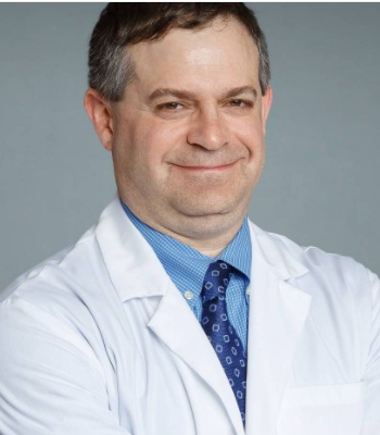 Stephen P. Wall合作论文数Department of Emergency Medicine, Grossman School of Medicine, New York University10
 Heemun Kwok合作论文数Department of Emergency Medicine, University of Washington10Silvers Scott M合作论文数10
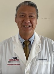 Richard Shih合作论文数Department of Emergency Medicine, Morristown Memorial Hospital10Susan B. Promes合作论文数San Francisco Medical Center (SBP),, University of California10Jonathan H Valente合作论文数9
 Molly Thiessen合作论文数Dept Emergency Med, Univ Colorado9
 Zane Horowitz合作论文数9
 Lauren M Westafer合作论文数Med Sch Baystate, Univ Massachusetts8Michael D. Smith合作论文数8
 Chris R. Carpenter合作论文数Department of Emergency Medicine, Mayo Clinic8
 John T Finnell合作论文数Department of Emergency Medicine, Indiana University School of Medicine, Indianapolis, IN7
 Jason Haukoos合作论文数Department of Emergency Medicine, School of Medicine, University of Colorado Anschutz Medical Campus;Department of Epidemiology, Colorado School of Public Health, University of Colorado Anschutz Medical Campus;Denver Health Medical Center;University of Colorado Hospital7
 Deborah Diercks合作论文数Department of Emergency Medicine, University of Texas Southwestern Medical Center7
 Benjamin W. Friedman合作论文数Department of Emergency Medicine, Albert Einstein College of Medicine, Montefiore Einstein7
 Stacy A Trent合作论文数5French L Keith合作论文数5
 Amal Mattu合作论文数Department of Emergency Medicine, University of Maryland School of Medicine5Christian Tomaszewski合作论文数4Stephen J. Wolf合作论文数Clinical Policies Subcommittee, From the American College of Emergency Physicians4
 Bruce Lo合作论文数4Shana Kusin合作论文数3
 Andrea Slivinski合作论文数3
 Kaushal Shah合作论文数Department of Emergency Medicine, Weill Cornell Medicine, Cornell University3
 Michael Brown合作论文数Department of Kinesiology, University of Maryland3Amy H. Kaji合作论文数3Stephen V. Cantrill合作论文数3
 Darren M Roberts合作论文数2Seth R. Gemme合作论文数2Antonio Bueso合作论文数2
 Brian Wright合作论文数2Megan Fee合作论文数2Sasha Kaiser合作论文数2Marc A. Camacho合作论文数2Jeffrey Brent合作论文数Department of Medicine, University of Colorado School of Medicine2Nathanael J. McKeown合作论文数2Christopher Yates合作论文数2
 Richard Byyny合作论文数Department of Emergency Medicine, Denver Health Medical Center;University of Colorado School of Medicine;Denver Health Medical Center, University of Colorado School of Medicine2
 Laura Carucci合作论文数2
 Jason A. Pietryga合作论文数The Warren Alpert Medical School, Brown University2
 Ingrid Berling合作论文数2
 Gillian A Beauchamp合作论文数2
 Sophie Gosselin合作论文数McGill University Health Centre, McGill University2
 Kathryn Fowler合作论文数Department of Radiology, University of California San Diego;Jacobs Medical Center, University of California San Diego;Hillcrest Medical Center, University of California San Diego2King Andrew M合作论文数2




