Purpose: This study aimed to investigate foot kinematics during gait in individuals with idiopathic clubfoot initially treated with the Ponseti method, focusing on clubfoot-specific deformities, to improve the understanding of posttreatment functional impairments.Methods: In this prospective cohort study, 23 patients with treated idiopathic clubfoot (34 feet) were compared with 15 age-matched healthy controls (30 feet). Gait analysis was performed using the Heidelberg Foot Model. To characterize residual clubfoot deformities, selected kinematic parameters included medial arch and subtalar angles, as well as intersegmental angles between hindfoot-shank and forefoot-hindfoot, based on more functional joint centers. Within the patient group, potential influence of additional surgical interventions was explored.Results: Compared with controls, treated idiopathic clubfoot exhibited significant reductions in hindfoot sagittal and transverse range of motion, forefoot sagittal range of motion, and medial arch mobility. Equinus deformity was not present posttreatment, as dorsiflexion metrics during stance did not differ significantly. However, plantarflexion range of motion during the transition to swing was reduced. In addition, treated idiopathic clubfoot showed persistent subtalar inversion and hindfoot varus, increased mid-stance adductus in both hindfoot and forefoot, and pronounced arch stiffness during transition to swing. Within the patient group, ankle and subtalar capsulotomy was associated with increased medial arch cavus.Conclusion: Patients with treated idiopathic clubfoot exhibit a functionally stiffer foot with altered alignment during gait, contributing to impaired limb advancement. The results expand the understanding of residual clubfoot deformities and highlight the importance of detailed functional assessment posttreatment, providing a foundation for future research.Level of evidence: Level II, prognostic study.
The relationship between femoral deformity and gait deviation is complex. Femoral anteversion can be assessed using the trochanter prominence angle test or by imaging techniques. Hip rotation during gait can be determined using conventional 3D gait analysis methods including palpation of femoral epicondyles or by using functional calibration. This study re-evaluates the indications for femoral osteotomies in this context. Hip rotation was analysed using predictive and functional methods in 80 patients who were referred for gait analysis due to rotational gait issues. Femoral anteversion was determined both manually and via MRI. In severe cases of femoral malalignment, the trochanter prominence angle test systematically underestimates the deformity by up to 15° compared to MRI results. Hip rotation, as measured by functional methods, also underestimates the outcome obtained by conventional methods, by up to 5°. Regardless of the method used, significant variability in hip rotation is observed during gait when the femoral deformation is moderate (anteversion between 0° and 30°). More severe deformities are not fully compensated for during gait. In cases of severe femoral malalignment, the functional change after osteotomy does not match the amount of derotation. Furthermore, both the trochanter prominence angle test and hip rotation during gait, as monitored via functional methods, underestimate the problem in the transverse plane.
Multisegment foot models have become increasingly important in biomechanical research and clinical gait analysis but often face limitations in defining joint positions. Often, they rely on simplified methods, such as using the midpoint between two markers to represent a joint, which lacks functional verification. In contrast, phenomenological angles, such as the medial arch angle, bypass joint center calculations, and offer sensitive, radiologically aligned indicators of foot mechanics. The Heidelberg functional foot model (HFFM) integrates functionally verified joint positions in combination with clinically relevant phenomenological measures, thereby enhancing clinical interpretability in gait analysis. The marker placement of the HFFM is based on the Heidelberg foot measurement method (HFMM). A four-segment model (shank, hindfoot, forefoot, and hallux) is defined. Anatomical coordinate systems are established via regression formulas derived from functional joint parameter determination. Kinematic angles are compared with radiological measures. Additionally, six clinically relevant angles of the HFMM are integrated into the HFFM. The method is applied to cavovarus (CV, 19 feet), equinovarus (EV, 31 feet), and typically developed feet (TD, 88 feet). EV feet show more pronounced hindfoot varus and forefoot adduction than CV and TD feet. Within the parameters adopted from the HFMM, EV feet exhibit increased subtalar inversion and a stronger medial arch than CV. Significant correlations are identified between hindfoot/shank flexion, forefoot/hindfoot flexion and medial arch, and radiological angles. The HFFM is sensitive for analyzing equinvarus and cavovarus deformities without applying static offsets due to the functional approach. It enables calculating kinetics to better understand the biomechanics of foot deformities.
Instrumented gait analysis provides objective data for clinical assessment, with surface electromyography (EMG) serving as a key tool in identifying abnormal muscle activation. However, reliable reference data considering both age and gender remain limited. Age- and gender-related differences in lower-limb EMG during gait in typically developing individuals were examined in this study using statistical parametric mapping (SPM). We also determined the minimum sample size required for robust clinical reference data. Our findings revealed significant differences in muscle activation patterns across age and gender. Children exhibited increased rectus femoris activation in initial swing and greater hamstring activation in the midstance, whereas adults demonstrated greater semimembranosus activity at initial contact, increased soleus activation at push-off, and greater rectus femoris activity in late swing. Gender-based differences included greater tibialis anterior activation in females during the terminal stance and increased vastus lateralis activity during swing, whereas males showed greater vastus lateralis and biceps femoris activation during terminal swing. Additionally, significant age–gender interaction effects were observed in the biceps femoris and semimembranosus, with gender-related differences becoming more pronounced in adulthood. Power analysis indicates that at least 47 participants, with a minimum of 12 per subgroup (male children, female children, male adults, and female adults), are required to detect age–gender interactions reliably. We strongly recommend incorporating both age and gender in clinical norm bands to enhance the accuracy of gait assessments and improve clinical and research comparisons.
People with neurological disorders and foot drop may suffer from cognitive-motor interference during walking. Functional Electrical Stimulation (FES) targets foot drop during gait but its effects on cognition remain underexplored. Fifteen individuals (4 males, 11 females; mean age 35.5 ± 12.5 years) with various neurological disorders, who had been using FES for at least three months, were recruited from our outpatient clinic. Subjects were assessed during walking with and without FES (FES CONDITION) and under single- and dual-task walking (TASK). The dual-task consisted of counting backwards from a number near 100 in steps of seven while walking. Cognition rate served as outcome parameter, with high values indicating subjects could efficiently maintain both cognitive task performance and walking speed. A linear mixed model analysis was conducted with FES CONDITION and TASK as fixed and SUBJECTS as random effects. The cognition rate during dual-task walking was significantly better with FES vs. without. FES showed minor effects on dorsiflexion in swing but larger effects on overall gait, as reflected in walking speed, step length and step width. While dual-task led to inferior results in gait, FES counteracted this effect and improved cognition rate. These findings suggest that FES not only addresses gait pattern and stability but also frees cognitive resources for walking. This shift in focus may enhance environmental awareness, social interaction or multitasking and thereby improving overall independence and quality of life, which is particularly relevant for patients with neurological disorders and an increased risk of falls.
Patients with musculoskeletal tumors who undergo amputation show reduced physical function. To give these patients better expectations of their future physical capabilities and to enhance the evaluation and development of postoperative rehabilitation, we investigated high-level activity and access to sport using objective measurements and questionnaires. We then compared the results to those of amputees who had undergone amputation due to trauma. In a prospective, monocentric study, we evaluated and correlated the results of the conventional mobility tests such as “timed-up-and-go” test (TUG), “2-Minute-Walk-Test” (2MWT), as well as the “10-Meter-Walk-Test” (10MWT) with a test for high-level activity. In this contribution, the Comprehensive High-Level Activity Mobility Predictor (CHAMP), originally developed for male servicemembers, was tested for feasibility in our cohort. We examined eleven patients who underwent amputation due to primary malignant bone or soft-tissue sarcomas and compared the results with ten patients who experienced traumatic amputation, along with seventeen patients in the healthy control group. Patients with lower leg amputations due to malignant musculoskeletal tumors exhibited superior outcomes in general mobility and high-level activity mobility compared to those with traumatic amputations. Using a questionnaire, we were able to demonstrate that patients suffering after amputation because of musculoskeletal tumor exhibited higher motivation and a greater sense of health and well-being compared to participants who had undergone traumatic amputation. The CHAMP can be utilized as a complementary tool in the rehabilitation of amputees to objectively assess high-level mobility and to guide targeted training and therapeutic interventions.
INTRODUCTION:To determine whether differences exist in the measured range of motion (ROM) by the goniometer, equinometer, and 3-dimensional (3D) gait analysis in different settings (under general anesthesia (GA) or in the clinic) among patients with cerebral palsy. METHODS:A prospective comparative cohort study was conducted at our orthopaedics center, where 15 patients with diplegic cerebral palsy (30 limbs) were evaluated, all of whom had Gross Motor Function Classification System (GMFCS) level I and II. ROM was measured by (1) goniometer under GA (benchmark), (2) goniometer analysis during clinical examination, (3) equinometer under GA, (4) equinometer in the clinic, and (5) 3D gait analysis. ROM was measured during both knee flexion and extension. Subgroup analysis based on the GMFCS level was performed. RESULTS:Nine patients were male with GMFCS level I and a mean age of 14.3 (SD=7.2) years. Statistically significant differences were noted between all studied measurement techniques in terms of ROM, dorsiflexion, and plantarflexion during both knee flexion and extension. The GMFCS level was an effect modifier of ROM measurements. DISCUSSION:The ROM parameters during both knee flexion and extension differed from one measurement device/technique to another. The GMFCS level played a notable effect-modifying role on the ROM parameters.
BACKGROUND:Foot drop is a common condition for patients with upper motor neuron syndrome such as cerebral palsy (CP). This study aimed to investigate the effects of functional electrical stimulation (FES) on gait function, quality of life, and FES satisfaction in adults with CP and foot drop. To analyze effects over time, an observational, longitudinal study was performed. METHODS:In this observational prospective cohort study, 8 adults with CP (median age of 22 years; range: 18.5-33.25) received a FES system for 12 weeks. 3D gait analysis was performed at beginning (T1) and end of the study period (T2). To detect effects of FES, ankle kinematics, kinetics, and spatiotemporal parameters were monitored for the conditions FES-OFF and -ON at each examination. RESULTS:Over time, walking speed increased significantly from T1ON (1.08 m/s) to T2OFF (1.21 m/s) with further improvement at T2ON. The ON-OFF difference of maximum dorsiflexion during swing tended to increase at T2 (2.49 degrees) compared with T1 (1.73 degrees) but with a more plantarflexed ankle position at T2 (range: -7.1 to 1.31 degrees) than T1 (range: -1.53 to 4.51 degrees) resulting in a significant decrease between T1ON and T2OFF. With FES, push-off energy tended to decrease for both examinations and lead to lower values over time. CONCLUSION:Positive therapeutic effects are represented in walking speed over time regardless of FES being OFF or ON even with a reduction in push-off energy. Despite no significant increase in maximum dorsiflexion in swing, over time, participants showed the tendency of an improvement in foot lift with FES-ON.
Die orthopädietechnische Versorgung von Patienten mit Extremitätenverlängerung und -rekonstruktion spielt eine entscheidende Rolle für die Verbesserung der Funktion und Mobilität. Die Behandlungsstrategie basiert auf einer präzisen Klassifizierung des Defekts und berücksichtigt Faktoren wie Instabilität der Gelenke, Muskelpathologie und Beinlängendefizite. Die Wahl der orthoprothetischen Maßnahmen erfolgt in Abhängigkeit von der Fehlbildung, ihrer Klassifikation und der individuellen Ansprüche des Patienten. Durch eine interdisziplinäre Entscheidungsfindung und individuelle Anpassungen der Orthoprothesen kann eine optimale Funktion und ein ästhetisch ansprechendes Ergebnis erreicht werden. Die Verwendung von Orthoprothesen mit individuell gefertigten Bauteilen wie Traktionsvorrichtungen in Kombination mit elastischen Linern kann den langfristigen Behandlungserfolg überhaupt erst ermöglichen und chirurgische Eingriffe unterstützen. Eine gute Kenntnis des behandelnden Arztes ist entscheidend für die richtige Wahl der orthopädietechnischen Versorgung nach Extremitätenverlängerung und -rekonstruktion.
ABSTRACT Introduction Lower-limb orthoses are usually part of conservative therapy in patients with pathological gait due to neurological disorders. A modular design, defined as a detachable combination of different orthotic designs, offers the possibility to adapt orthosis functionality to individual needs. Currently, however, knowledge about the actual impact of modularity on wearing times is limited. The aim of the present study was therefore to examine the wearing time of modular and nonmodular orthoses to determine whether there are differences between the different orthotic concepts of the lower limb on weekdays and weekends and whether modularity affects wearing time. Material and Methods In this prospective clinical study, 53 patients with neurological gait disorder were included. All wore modular or nonmodular orthoses in one of these three groups: dynamic ankle-foot orthoses (DAFOs), ankle-foot orthoses (AFOs), and knee-ankle-foot orthoses (KAFOs). Wearing time was recorded by temperature sensors for 3 months. Data analysis included both descriptive and further parametric statistical testing (t-test for dependent and independent samples) as well as Pearson correlations. A univariate analysis of variance was used when comparing multiple groups. Results Participants wore DAFOs and AFOs, but not KAFOs, significantly longer on weekdays. Wearing time correlated significantly between weekdays and weekends for all groups. There is no significant difference in the mean daily wearing time between the groups. Modularity influenced the wearing time only for the DAFO/AFO combination. Conclusion Orthotic design and anatomic height affect the average daily wearing time. A change in wearing behavior between weekdays and weekends and influences by modularity can be found solely in DAFOs and AFOs.
Background: Orthopedic technical care plays a crucial role in improving function and mobility for patients undergoing limb lengthening and reconstruction. The treatment strategy is based on a precise classification of the defect, taking into account factors such as joint instability, muscle pathology and limb length discrepancy.Choice of aids: The selection of orthopedic aids depends on the specific deformity, its classification and the individual needs of the patient. Through interdisciplinary decision-making and customized adaptations of prostheses and orthoses, optimal function and aesthetically pleasing outcomes can be achieved. The utilization of custom-made technical components, such as traction devices combined with elastic liners, is essential for long-term treatment success and can support surgical interventions. The treating physician's expertise is crucial in selecting the appropriate orthopedic technical device following limb lengthening and reconstruction.
Background: Foot deformities in children are common, and the majority can be treated conservatively. Nevertheless, there are deformities that require surgical treatment. These include rigid clubfeet, severe forms of pes planovalgus, pes cavus and several more. We retrospectively analysed the pseudarthrosis rate of surgical treatment of foot deformities with transcutaneous K-wire osteosynthesis in neurologically healthy children and adolescents. The aim of the study was to show that the results with K-wires are comparable to those with other osteosynthesis methods in the literature. Methods: A total of 46 paediatric patients aged 6 to 17 years treated between January 2010 and December 2015 met the inclusion criteria. Depending on the diagnosis, different surgical interventions were necessary. In clubfoot and pes planovalgus, representing n = 81, 70% of the whole collective triple arthrodesis with fusion of the talonavicular, calcaneocuboid and subtalar joints or Evans osteotomy was usually performed. Radiographs were taken at least 6 months post-surgery, and bony consolidation of the subtalar, talonavicular (TN), and calcaneocuboidal (CC) joints and the metatarsal I (MT I) osteotomy were assessed. If there was no evidence of fusion at this time, it was considered non-union. Results: In total, 117 arthrodesis procedures with K-wires were performed. Overall, 110 of the arthrodesis (94%) healed, and only 7 joints (6%) showed non-union (subtalar 0%, TN 7.7%, CC 6.5% and MT I 6.7%). All non-unions occurred in subjects with clubfoot deformities. No significant risk factors were observed. Conclusion: This study replicated the good consolidation rates reported in the literature with screws, plates, intramedullary nails or staples in arthrodesis of the adolescent foot in neurologically healthy subjects and confirmed the efficacy of K-wires. The main advantages of transcutaneous K-wire treatment are easy metal removal, lower osteosynthesis material costs and less concomitant damage. Further studies, especially randomised controlled trials, are needed to further investigate this topic.
BACKGROUND:This study aimed to evaluate gait outcomes and strength following the surgical correction of equinus in cerebral palsy (CP) based on different surgical procedures. We included the Baumann and Strayer procedures, as well as the Achilles tendon lengthening (ATL). METHODS:A retrospective analysis was performed in patients with infantile, bilateral CP who received instrumental 3D gait analysis before and after surgical correction (18.66 months postoperatively). Patients were divided into 3 groups: Strayer surgery, Baumann surgery, and ATL. Gait performance and muscle strengths were compared between studied surgeries. RESULTS:A total population of 204 patients (15.43 years) with 341 operated lower limbs (LLs) was analyzed. Dorsiflexion in swing and stance phases significantly improved in all groups postoperatively. The Strayer and the ATL group showed higher postoperative dorsiflexion than the Baumann group. However, no loss of strength was observed with the Baumann method. Maximum power improved in this group postoperatively. An 8.2% loss in calf muscle strength was recorded in the Strayer group. CONCLUSION:Operative pes equinus treatment successfully improved the gait of children and adults with CP postoperatively. There were differences in postoperative results between studied operative techniques regarding range of motion and power. LEVEL OF EVIDENCE:Level III, retrospective cohort study.
Objective The aim of the investigation was to assess the contribution of pain to functional, social, and health-related quality-of-life outcomes in adults with cerebral palsy. Design This monocentric, prospective study included long-term data for ambulant adults with cerebral palsy and at levels I–III in the Gross Motor Function Classification System who had received orthopedic multilevel surgery in adulthood and in whom preoperative and postoperative three-dimensional gait analysis was conducted, including the Gait Profile Score. A questionnaire including the Visual Analog Scale, Oswestry Disability Index, and European Quality of Life 5 Dimensions 3 Level was analyzed. Results Twenty-four adults at a mean age of 38.0 yrs were included. Sixteen patients (66.7%) indicated that they experienced pain and 10 patients (41.7%) had pain for more than 6 mos. Overall, 13 patients (54%) experienced both pain and anxiety. There was no correlation of the Gait Profile Score with pain, Gross Motor Function Classification System, and Oswestry Disability Index. The Oswestry Disability Index showed a positive correlation with the Gross Motor Function Classification System ( r = 0.39, P = 0.05) and pain ( r = 0.57, P = 0.004). Conclusions Pain and anxiety represent relevant comorbidities in adults with cerebral palsy. The occurrence of pain correlates with the Oswestry Disability Index and is higher than in children with cerebral palsy.
Developmental dysplasia of the hip (DDH) is one of the most common disorders of hips in children. The deformity can remain asymptomatic into adolescence and adulthood; however, it is considered to be a form of prearthritis and is the main cause of premature osteoarthritis of the hip. The deformity affects the acetabulum but can also be accompanied by changes in the shape of the proximal femur. If conservative treatment for mild DDH is insufficient, or in cases of moderate to severe DDH, operative treatment should be carried out, for example by corrective osteotomy of the pelvis and/or the proximal femur and hip arthroscopy may be considered adjunctively in order to resolve the prearthritis and prevent premature osteoarthritis of the hip. This manuscript elucidates the deformity, the diagnostic measures required to make the diagnosis and the treatment options available for prevention of arthritis.
Background/Aim: The pattern of lower-limb amputation, indications, complications, and revision in pediatric cases differs globally. Therefore, we conducted this study to describe the patterns of lower-limb amputation at our institution. Methods: During a set period between 2010 and 2020, adolescent patients undergoing lower-limb amputation within the orthopedic department of Heidelberg University Hospital were retrospectively collected and analyzed. The retrieved dataset included two parts: data on lower-limb amputations and data on subsequent complications and revision surgeries at the same time. Besides patients' general information (age, gender), the dataset included data regarding amputation patterns (number, indications, and level of amputation, complications, and revision surgeries and their indications). Results: Twenty-two patients undergoing lower-limb amputation were examined, of which the majority were males (63.6%) with a mean age of 12 (5.1) years. Tumor was the most common indication for amputation (72.7%), and transfemoral amputation was the most frequent level (68.2%). Complications occurred in 10 patients, mostly due to stump impalement or bony overgrowth. Of all recorded patients requiring revision, nine were regarding bone and one case regarding soft tissue. Conclusions: Lower-limb amputation in adolescents is a rare encounter and it is commonly indicated due to bone tumors. The thigh is the most common level of amputation. Postoperative complications are frequent, mainly secondary to bony overgrowth, and often require revision surgery.
 Sebastian Wolf合作论文数Universitäts Klinikum Heidelberg23
 Block Julia合作论文数16
 Thomas Dreher合作论文数Department of Orthopaedic Surgery, Heidelberg, Germany16Merkur Alimusaj合作论文数11Bernd Wiedenhofer合作论文数11Daniel Heitzmann合作论文数10Geisbüsch Andreas合作论文数9Axel Horsch合作论文数9
 Rudiger Rupp合作论文数Orthopaedic University Hospital8Fürstenberg Carl Hans合作论文数7Gerner Hans Jürgen合作论文数7s gantz合作论文数6
 Martin Schwarze合作论文数6Götze Marco合作论文数6
 Michael Akbar合作论文数6Hans Jürgen Gerner合作论文数6Tobias Renkawitz合作论文数6H-J Gerner合作论文数5
 Maher Ghandour合作论文数5
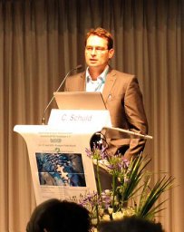 Christian Schuld合作论文数Spinal Cord Injury Ctr, Heidelberg Univ Hosp5
 Matthias Klotz合作论文数5C. H. Fürstenberg合作论文数5Norbert Weidner合作论文数5
 Babak Moradi合作论文数Orthopaedic Clinic, University Heidelberg5
 Julian Maximilian Deisenhofer合作论文数4
 Firooz Salami合作论文数4Marcus Schiltenwolf合作论文数4
 Sebastien Hagmann合作论文数4T Kaib合作论文数3Carl Hans Fuerstenberg合作论文数3Thomas Grieser合作论文数3Bernd Wiedenhoefer合作论文数3L. Döderlein合作论文数3Niklasch Mirjam合作论文数3Martin H Pouw合作论文数3
 Lars Helbig合作论文数3Cornelia Hensel合作论文数2
 Paul Mick合作论文数2Simone Gantz合作论文数2
 Frank Braatz合作论文数Universitätsmedizin Göttingen2
 Mirjam Thielen合作论文数BG Trauma Ctr Ludwigshafen, Heidelberg Univ2b greitemann合作论文数2Mesrian Alireza合作论文数2Rosshirt Nils合作论文数2
 Karimi Mohammad Taghi合作论文数Muscoloskeletal Research Centre, Isfahan University of Medical sciences2
 Esrafilian Amir合作论文数Department of Mechanics, Islamic Azad University2Schönit Eva合作论文数2




