BACKGROUND:Surgical treatment of shoulder instability with glenoid bone loss using a distal tibial allograft has achieved success, with the bone graft placed parallel, flush, or recessed to the articular surface, but questions remain about optimal graft positioning. HYPOTHESIS:Given evidence that glenoid concavity and congruence influence stability, we hypothesized that angled bone blocks better restore native concavity and biomechanics compared to flat graft placement. STUDY DESIGN:Controlled laboratory study. METHODS:A total of 9 cadaveric shoulders were dissected, separating rotator cuff muscles from the capsule. Native glenoid dimensions were measured with a digitizer. A 20% anterior bone defect was created and then reconstructed with a distal tibial allograft (bone graft). There were 4 conditions tested: intact, defect, flat bone block, and angled bone block (30°). Using a testing machine, the humeral head was translated anteriorly 10 mm, and distraction and contact forces were recorded. RESULTS:Mean peak distraction forces were as follows: 48.28 ± 8.06 N (intact), 33.99 ± 10.54 N (defect; 70.4% of intact), 41.83 ± 9.07 N (flat; 89.6% of intact), and 45.64 ± 9.03 N (angled; 98.5% of intact). Humeral head contact with the graft occurred in 100.0% of angled trials versus 70.0% of flat trials. Mean glenohumeral contact forces were as follows: 71.81 ± 16.42 N (intact), 31.39 ± 16.60 N (defect), 46.91 ± 4.12 N (flat), and 62.17 ± 9.26 N (angled). One-way analysis of variance showed a significant effect of construct type on both contact and distraction forces (P < .001). Pairwise comparisons showed significant differences in distraction forces between intact and defect conditions and between defect and angled bone block conditions. Comparisons also demonstrated significant differences in contact forces between intact and defect, intact and flat bone block, defect and flat bone block, defect and angled bone block, and flat bone block and angled bone block conditions. No significant difference was observed between the intact shoulder and angled bone block repair. CONCLUSION:Angled bone blocks more closely restored native glenoid concavity and were biomechanically superior to flat bone blocks in re-establishing stability and centering the humeral head after glenoid bone loss. CLINICAL RELEVANCE:This study provides a quantitative comparison between different bony glenoid augmentation methods.
Purpose: To biomechanically assess the impact of different patellar defect sizes on the ultimate load to failure (ULTF) and fracture risk immediately after bone–patellar tendon–bone harvest. Methods: Twelve fresh-frozen mid femur to mid tibia–fibula cadaveric specimens (8 female specimens) with an average age of 75 years (range, 64-88 years) were randomly assigned to a defect length of 20 or 25 mm. A triangular wedge bone block was harvested from the patella, and the defect was measured. The extensor mechanism was loaded until failure with the knee in 30° of flexion. ULTF and failure modes were recorded. Statistical analyses were performed between the 2 groups to identify significance (P < .05). Results: Analyses were performed on 10 specimens because 2 were excluded. The average ULTF was not significantly different between the 20- and 25-mm groups (2,313 ± 863 N and 2,443 ± 839 N, respectively; P = .815) despite a significantly different percentage of total patellar defect length between the 2 groups (39.5% ± 3.9% and 49.1% ± 4.1%, respectively; P = .005). In 7 specimens, the first point of bony failure was at the patella, whereas in the remaining 3 specimens, it was distal to the patella. Conclusions: The results of this biomechanical study show that up to a 25-mm-long patellar bone block harvest can be performed safely with a low risk of patellar fracture. Clinical Relevance: There is concern for complications including an increased risk of patellar fracture at the harvest site after bone–patellar tendon–bone autograft harvest. The risk of this complication based on patellar harvest size is not well understood. This study provides biomechanical insights into assessing fracture risk of patellar harvest between 20 and 25 mm in length.
Objectives: Pre- and Peri-Operative nerve blocks are traditionally administered by the anesthesia team as an adjunctive for operative pain management. Providing safe and effective peri-operative pain control helps to limit narcotic use by patient, postoperative nausea and improves post-operative patient flow through the PACU. Recently, there have been significant advancements in perioperative nerve blocks, local anesthesia, and multi-modal pain regimens to improve patient comfort and reduce reliance on opiate pain medications. Infiltration of 10 mL mixture of 30mL of 0.5% Marcaine, 20mL of 0.9% Normal Saline, 1mL of Morphine 10mg/1mL, and 1mL of Toradol 30mg/1mL between the popliteal artery and capsule of the knee or IPACK block has been described as a motor sparring nerve block which can provide effective analgesia following knee surgery and serve as an adjunct to commonly used adductor canal or femoral nerve blocks. We have recently reported on a posterior knee capsular block technique administered under direct arthroscopic visualization, intra-operatively by the orthopedic surgeon providing coverage identical to a traditional ultrasound guided Infiltration Between Popliteal Artery and Capsule of the Knee (IPACK Block). We have devised a simple, and reproducible technique for intraoperative, arthroscopic administration of local anesthetic in the space directly posterior to the knee capsule. This technique has been implemented by sports medicine orthopedic surgeons at our institution over the past couple of years. While the practice anecdotally seems to have improved post operative pain control in our patients, the safety and efficacy have not been formally studied. The purpose of this study is to investigate the effect of this practice on the post operative pain control as well as identify any adverse events over the period that this block was added to our standard practice. Methods: Electronic medical records of patients undergoing primary anterior cruciate ligament (ACL) reconstruction by three fellowship-trained sports medicine surgeons at our institution between March 2019 and February 2022 were retrospectively reviewed. Patients were included if they underwent primary arthroscopic assisted ACL reconstruction with or without concomitant meniscus procedure, chondroplasty, or microfracture. Patients who underwent revision ACL reconstruction, multi-ligament reconstruction, a concomitant realignment osteotomy, or a concomitant osteochondral allograft or meniscus allograft transplantation were excluded. Patients were divided into two groups according to the receival of arthroscopic IPACK block. All patients received a nerve block, but no patients received an IPACK done in the preoperative setting with anesthesia, any anesthesia IPACK block performed was in the PACU as a rescue block. The primary outcome was the need for a postoperative rescue nerve block. The secondary outcome was the incidence of complications related to the surgeon-directed IPACK block Results: Three hundred and twenty-five patients were included. Among them, 117 received the IPACK block and 208 did not receive it. There was no statistically significant difference in patient’s age and gender between the groups. There was a trend towards more rescue blocks in patients who did not receive the IPACK block compared to those who receive it (10.5% vs 6.8% respectively, p=0.264), and this trend reached the statistical significance when we looked at a subgroup of patients who received concomitant meniscus repair (17.4% vs 4.7% respectively, p=0.017). Logistic regression analysis showed that IPACK block decreased the risk of receiving a rescue block by approximately 50%. No complications related to IPACK block were reported. Conclusions: Implementing the arthroscopic assisted IPACK block reduced the need for postoperative rescue block in patients undergoing primary ACL reconstruction, and more significantly in those who had concomitant meniscus repair. It is a safe procedure with no reported complications or adverse events in our cohort. Implementation of the block adds very little time or cost to the surgical procedure. It has the potential to improve postoperative pain control, decrease reliance on postoperative opiate medications, facilitate early rehabilitation, and contribute to overall patient satisfaction after surgery.
Objectives: Patients with anterior knee pain caused by articular cartilage lesions specific to the patellofemoral joint are difficult to treat because of the joint biomechanics and shear forces experienced by this joint. There are very few studies looking at biologic restoration procedures within this joint and no study looking at only patella-based lesions. Current surgical treatments include chondroplasty, microfracture, autologous chondrocyte implantation, osteochondral autograft (OAT) and allograft (OCA). OAT and OCA procedures may help restore or replace articular cartilage while other treatment options may limit the natural ability of the knee to resist compression. The purpose of this study was to evaluate the clinical outcomes of a large series of patients undergoing a primary osteochondral allograft (OCA) or autograft (OAT) procedures to treat painful osteochondral lesions of the patella. Methods: This study is a retrospective clinical outcomes study of patients who underwent an OCA or OAT procedure performed by two fellowship trained surgeons at a single institution, between January 1 st , 2009 and May 31 st , 2019. All patients included in this study had a minimum 1-year follow-up. OAT procedures were performed for patients with a lesion <10 mm per standard of care practice guidelines via autografts harvested from a non-weight-bearing portion of the distal femur. OCA procedures were performed for patients with lesions >10 mm using fresh donor allograft using standard press-fit techniques. Patients were evaluated retrospectively through a chart review to collect demographic data, indications for surgery, preoperative outcome scores, intraoperative notes and recorded postoperative outcome scores. Surgical data including laterality and concomitant surgeries was also obtained via chart review. A postoperative questionnaire was utilized either in clinic, via email or telephone to collect Kujala and Knee Injury and Osteoarthritis Outcome Score (KOOS). Demographic data was collected including age, BMI, and sex. Results: We identified 57 patients who underwent either an OCA or OAT of which 51 had 1 year or more of follow up. The final study population consisted of 28 patients which included 22 OCA and 6 OAT procedures (16 females, 12 males) with an average age of 29.8 ± 6.6 years (16 - 43). The average lesion size was 23.0 mm (range 10 - 35mm). Average KOOS and Kujala scores of the patient cohort was 60.7 ± 22.4 (range 18.5 - 95.1) and 56.6 ± 24.2 (range 22 – 96) respectively. Age and lesion size were found to negatively correlate to KOOS and Kujala scores (p < 0.05) and the greatest predictors of clinical outcomes for both KOOS and Kujala. The effect of age was not found to be statistically significant when accounting for the size of the lesion (p = 0.073). Likewise, size was not found to be statistically significant when age was accounted for (p = 0.073). The combined effect of age and size on KOOS and Kujala was found to be statistically significant (p < 0.05). Conclusions: We were able to explicitly identify worsening clinical outcomes with increasing patient age and graft size. The results of this study can be used while counseling patients on the efficacy and success rate of an OAT and OCA procedure. Based on our results, younger patients with smaller lesions will exhibit better clinical outcomes when compared to older patients with larger lesions.
Purpose:To evaluate whether cumulative impact load and serum biomarkers are related to lower-extremity injury and to determine any impact load and cartilage biomarker relationships in collegiate female basketball athletes. Methods:This was a prospective longitudinal study evaluating lower-extremity impact load, serum cartilage biomarkers, and injury incidence over the course of a single collegiate women's basketball season. Data were collected from August 2022 to April 2023; no other follow-up after the cessation of the season was conducted in this cohort. Inclusion criteria for the study included collegiate women's basketball athletes, ages 18 to 25 years, who were noninjured at the start of the study time frame (August 2024). Cartilage synthesis (procollagen II carboxy propeptide and aggrecan chondroitin sulfate 846 epitope) and degradation (collagen type II cleavage) biomarkers were evaluated at 6 season timepoints. Impact load metrics (cumulative bone stimulus, impact intensity) were collected during practices using inertial measurement units secured to the distal medial tibiae. Injury was defined as restriction of participation for 1 or more days beyond day of initial injury. Cumulative impact load metrics were calculated over the week before any documented injury and blood draws for analysis. Point biserial and Pearson product moment correlations were used to determine the relationship between impact load metrics, serum biomarkers, and injury. Results:Eleven collegiate women's basketball athletes (height: 1.86 meters, mass: 82.0 kg, age: 20.54 years) participated. Greater medium-range (6-20 g) cumulative impact intensities during week 5 and 6 for both limbs (r = 0.674, P = .023) and high-range (20-200 g) during week 8 for both limbs (0.672, P = .024) were associated with injury. Greater cumulative bone stimulus was associated with increased procollagen II carboxy propeptide levels before conference playoffs for right (r = 0.694, P = .026) and left (r = 0.747, P = .013) limbs. Greater chondroitin sulfate 846 epitope levels at off-season-1 (r = 0.729, P = .017), and at the beginning of the competitive season (r = 0.645, P = .044) were associated with season-long injury incidence. Conclusions:In this study, we found that moderate-to-high intensity impacts (6-200 g) early in the season were associated with subsequent injury among female collegiate basketball players. Increased cartilage synthesis at various time points was correlated with increased cumulative bone stimulus metrics and season-long injury incidence in this population. Level of Evidence:Level IV, prognostic case series.
Meniscus allograft transplantation (MAT) has been shown to be a feasible surgical option for younger patients, below 50 years of age who have meniscal insufficiency and have failed conservative treatment measures. In this technical note, we describe a procedure of harvesting and injecting bone marrow aspirate concentrate in a meniscus allograft during a MAT procedure, which may allow for longer lasting transplants and improve patient outcomes. In this technical note, bone marrow aspirate concentrate is harvested arthroscopically from the intercondylar notch at the surgical site, which prevents additional donor site morbidity, as seen with harvesting from other locations, such as the iliac crest. This also reduces operating time, since harvesting from the iliac crest requires different patient positioning and usually additional anesthesia. The authors of this surgical technique believe that biological augmentation during MATs will assist surgeons in maximizing graft survivorship and, ultimately, lead to better patient outcomes.
Advances in regional anesthesia techniques for knee surgery have led to drastic improvements in postoperative pain control and have reduced reliance on perioperative opioid analgesics. The infiltration between the popliteal artery and capsule of the knee (IPACK) block has been a useful tool for providing posterior knee analgesia as an adjuvant to traditional femoral or adductor canal blocks in knee surgery. We present a simple and reproducible technique for the arthroscopic administration of this block.
Clavicle fractures are a common injury resulting from a high-energy force, such as a fall onto the shoulder, motor vehicle accident, or sporting activity. Although some clavicle fractures may be treated nonoperatively, operative treatment results in higher union rates and faster return to activity. Here we discuss the operative treatment options for plating of clavicle fractures; specifically, a single plate placed either superiorly or anteriorly or two plates placed orthogonally. Because both techniques provide adequate stability, fracture and patient characteristics should guide the surgical decision making regarding single versus dual plating of clavicle fractures.
Clavicle fractures are a common injury resulting from a high-energy force, such as a fall onto the shoulder, motor vehicle accident, or sporting activity. Although some clavicle fractures may be treated nonoperatively, operative treatment results in higher union rates and faster return to activity. Here we discuss the operative treatment options for plating of clavicle fractures; specifically, a single plate placed either superiorly or anteriorly or two plates placed orthogonally. Because both techniques provide adequate stability, fracture and patient characteristics should guide the surgical decision making regarding single versus dual plating of clavicle fractures.
Revision anterior cruciate ligament reconstruction (rACLR) procedures are often technically and intellectually challenging. However, with careful preoperative evaluation and planning, the likelihood of success can be maximized. Understanding the various etiologies of and contributors to primary ACLR failure can guide the surgical plan in terms of whether concomitant procedures are needed. Although successful outcomes have been reported with both one-stage and two-stage rACLRs, appropriate patient selection is important. Overall, clinical outcomes including patient-reported outcomes, graft failure rates, and return to sport are worse after rACLR compared with primary ACLR. It is important to review the preoperative evaluation, surgical considerations, and outcomes of rACLR.
Background: Patients with recurrent patellar dislocations with trochlear dysplasia are commonly treated surgically with a tibial tubercle osteotomy (TTO). Recovery and rehabilitation processes are often nonoperative out of concern for fixation failure or fracture. A more accelerated rehabilitation protocol allowing for early weightbearing and quadriceps strengthening may help to improve patient outcomes as long as complications are not increased. Purpose: To evaluate the safety and effectiveness of an accelerated weightbearing and early strengthening postoperative rehabilitation program for patients who undergo TTO. Study Design: Case series; Level of evidence, 4. Methods: Included were patients who underwent unilateral/staged bilateral TTO performed by a single surgeon between August 2013 and February 2018 with ≥6 months of follow-up. The surgical indication was primarily for patients with recurrent patellar instability. In all cases, a diagnostic arthroscopy was performed to evaluate the cartilage surfaces and document patellar tracking. The TTO was performed using a freehand technique and two 3.5-mm fully threaded screws for fixation. Patients underwent an accelerated postoperative rehabilitation program that allowed for weightbearing and lower extremity strengthening starting at 4 weeks. Objective and subjective outcome measures included any postoperative complications, knee range of motion, and patient-reported outcome scores (Kujala Anterior Knee Pain Scale [AKPS] and Knee injury and Osteoarthritis Outcome Score composite [(KOOS5]). Results: A total of 51 knees in 50 patients (38 female, 12 male) with a mean age of 31.24 ± 12.57 years were included in the final analysis. Compared with preoperative values, postoperative maximum knee flexion was significantly improved (117.67° ± 32.65° vs 131.12° ± 9.02°, respectively; P = .022). Postoperative complications included 6 patients with arthrofibrosis requiring manipulation under anesthesia, 4 with removal of symptomatic hardware, 1 tibial fracture (due to a fall), and 1 conversion to patellofemoral arthroplasty. The mean postoperative AKPS and KOOS5 scores were 72.98 ± 21.51 and 75.05 ± 16.02, respectively. Conclusion: Accelerated postoperative rehabilitation in TTO patients was an effective means of treatment with good subjective and objective outcomes and complication rates lower than traditional rehabilitation protocols.
Case: A 21-year-old collegiate baseball pitcher presented with transient lateral arm pain and wrist extension weakness after pitching more than 1 inning. Physical examination was unremarkable at rest. Ultrasound-guided injection of the radial nerve at the level of the lateral intramuscular septum improved his symptoms. After decompression of the radial nerve, the patient noted resolution of his symptoms while pitching. Conclusion: Atraumatic radial neuropathy is a rare but documented phenomenon. As far as we know, this is the first reported case of a transient exertional radial neuropathy in an athlete at the level of the lateral intermuscular septum.
Background: Suspensory fixation of anterior cruciate ligament (ACL) reconstruction (ACLR) grafts has emerged as a popular device for femoral graft fixation. However, improper deployment of the suspensory fixation can compromise proper graft tensioning, leading to failure and revision. Also, soft tissue interposition between the button and bone has been associated with graft migration and pain, occasionally requiring revision surgery. Many surgeons rely on manual testing and application of distal tension to the graft to confirm proper button deployment on the lateral cortex of the femur for ACL graft fixation. Purpose: To determine the reliability of the manual resistance maneuver when applying distal tension to deploy the suspensory device along the lateral cortex of the femur. Study Design: Case series; Level of evidence, 4. Methods: All patients undergoing ACLR with a suture button suspensory device for femoral fixation were eligible for enrollment in the study. The surgeries were performed by 3 board-certified, sports medicine fellowship-trained orthopaedic surgeons at a single outpatient surgical center between May 2018 and June 2019. All grafts were passed in a retrograde manner into the femoral tunnel, and a vigorous manual tensioning maneuver in a distal direction was placed on the graft to deploy and secure along the lateral cortex of the femur. Intraoperative mini c-arm fluoroscopy was obtained to demonstrate proper suture button positioning. If interposing tissue or an improperly flipped button was identified, rectifying steps were undertaken and recorded. Results: A total of 51 patients with a mean age of 33.3 years were included in the study. Of these patients, 74.5% had normal suture button positioning identified via intraoperative fluoroscopic imaging, while 15.7% had interposed soft tissue and 9.8% had an improperly flipped button. In all cases, the surgeon was able to rectify the malpositioning intraoperatively. Conclusion: Despite the manual sensation of proper suspensory button positioning, intraoperative fluoroscopy identified suture button deployment errors in ACLR 25% of the time. Correcting the malpositioning is not technically demanding. These findings advocate for routine intraoperative surveillance to confirm appropriate suture button seating during ACLR.
BACKGROUND:Superior capsular reconstruction (SCR) treatment of massive, symptomatic, irreparable rotator cuff tears (RCTs) has become a more recently used procedure. However, there is a lack of consensus surrounding optimal graft choice for the SCR technique, and current dermal grafts have increased cost and are technically challenging because of a need for multiple implants. The purpose of this study was to biomechanically investigate a biological lower-cost alternative as a support for the superior capsule reconstruction concept: an isolated semitendinosus tendon (STT) allograft and a combination graft with the long head of the biceps tendon (LHBT) in an established massive posterosuperior RCT cadaver model. METHODS:Ten fresh-frozen cadaveric shoulders (53.3 ± 12.4 years: range: 26-65) were tested on an established dynamic shoulder simulator using dynamic muscle loading. Cumulative deltoid forces, maximum abduction angle, and superior humeral head translation were compared across 4 testing conditions: (1) intact state, (2) massively retracted (Patte III), irreparable posterosuperior RCT, (3) SCR repair using an STT allograft, and (4) SCR repair using a combined STT-LHBT repair. RESULTS:Intact shoulders required a mean deltoid force of 154.2 ± 20.41 N to achieve maximum glenohumeral abduction (55.3° ± 2.3°). Compared with native shoulders, the maximum abduction angle decreased following a massively retracted posterosuperior RCT by 52% (28.3° ± 8.4°; P < .001), whereas the cumulative deltoid forces increased by 48% (205.3 ± 40.9 N; P = .001). The STT repair and the STT-LHBT repair improved shoulder function compared with the tear state, with a mean maximum abduction angle of 30.6° ± 9.0° and 31.8° ± 7.7° and a mean deltoid force of 205.3 ± 40.9 N and 201.0 ± 34.0 N, respectively, but this was not statistically significant (P > .05). The STT-LHBT repair significantly improved the range of motion with respect to the tear state (P = .04). CONCLUSIONS:In a dynamic shoulder simulator model, both the STT and the STT-LHBT repair techniques improved glenohumeral joint kinematics in an amount similar to previously reported "traditional" SCR techniques for treatment of an irreparable posterosuperior RCT.
Background: Suspensory fixation of anterior cruciate ligament (ACL) reconstruction (ACLR) grafts has emerged as a popular device for femoral graft fixation. However, improper deployment of the suspensory fixation can compromise proper graft tensioning, leading to failure and revision. Also, soft tissue interposition between the button and bone has been associated with graft migration and pain, occasionally requiring revision surgery. Many surgeons rely on manual testing and application of distal tension to the graft to confirm proper button deployment on the lateral cortex of the femur for ACL graft fixation. Purpose: To determine the reliability of the manual resistance maneuver when applying distal tension to deploy the suspensory device along the lateral cortex of the femur. Study Design: Case series; Level of evidence, 4. Methods: All patients undergoing ACLR with a suture button suspensory device for femoral fixation were eligible for enrollment in the study. The surgeries were performed by 3 board-certified, sports medicine fellowship–trained orthopaedic surgeons at a single outpatient surgical center between May 2018 and June 2019. All grafts were passed in a retrograde manner into the femoral tunnel, and a vigorous manual tensioning maneuver in a distal direction was placed on the graft to deploy and secure along the lateral cortex of the femur. Intraoperative mini c-arm fluoroscopy was obtained to demonstrate proper suture button positioning. If interposing tissue or an improperly flipped button was identified, rectifying steps were undertaken and recorded. Results: A total of 51 patients with a mean age of 33.3 years were included in the study. Of these patients, 74.5% had normal suture button positioning identified via intraoperative fluoroscopic imaging, while 15.7% had interposed soft tissue and 9.8% had an improperly flipped button. In all cases, the surgeon was able to rectify the malpositioning intraoperatively. Conclusion: Despite the manual sensation of proper suspensory button positioning, intraoperative fluoroscopy identified suture button deployment errors in ACLR 25% of the time. Correcting the malpositioning is not technically demanding. These findings advocate for routine intraoperative surveillance to confirm appropriate suture button seating during ACLR.
Femoral nerve block (FNB) is a routinely used regional analgesic technique for anterior cruciate ligament (ACL) reconstruction. One method to balance the analgesic effect and functional impairment of FNBs may be to control the concentration of local anesthetics utilized for the block. Retrospective chart review was performed on 390 consecutive patients who underwent ACL reconstruction between June 2014 and May 2017. Patients were divided into those who received a standard (0.5%-bupivacaine) or low (0.1–0.125%-bupivacaine) concentration single-shot FNB performed with ultrasound guidance. Maximum postoperative VAS, Post-Anaesthesia Care Unit (PACU) time prior to discharge, need for additional ‘rescue’ block, and intravenous postoperative narcotic requirements were recorded. A total of 268 patients (28.4 ± 11.9 years) were included for final analysis, with 72 patients in the low-concentration FNB group and 196 patients receiving the standard concentration. There were no differences in the maximum postoperative VAS between the low (6.4 ± 2.5) and standard (5.7 ± 2.9) concentration groups (P = 0.08). Similarly, the time from PACU arrival to discharge was not different between groups (P = 0.64). A sciatic rescue block was needed in 22% of patients with standard-dose FNB compared to 30% of patients receiving the low-concentration FNB (P = 0.20). Patients with a hamstring autograft harvest were more likely to undergo a postoperative sciatic rescue block compared to a bone-patellar tendon autograft (P = 0.005), regardless of preoperative block concentration. Quadriceps activation was preserved with low-concentration blocks. Using 1/5th to 1/4th the standard local anesthetic concentration for preoperative femoral nerve block in ACL reconstruction did not significantly differ in peri-operative outcomes, PACU time, need for rescue blockade, or additional immediate opioid requirements. III.
Surgical treatment of patellofemoral instability and associated cartilaginous lesions can be technically challenging. Visualization of patellar tracking and underlying osteochondral lesions is paramount to operative success. To treat these conditions effectively, a comprehensive arthroscopic assessment of the patellofemoral joint as well as dynamic visualization of patella tracking must be achieved. Visualization of the patellofemoral joint—in particular, the articular cartilage of the patella and trochlea morphology—can be difficult when using traditional anteromedial or anterolateral portals and a 30° arthroscope lens. The technique described here uses an accessory superolateral portal and a 70° arthroscope to achieve significantly improved visualization of the patellofemoral articulation, in particular the chondral surfaces. This vantage point aids the surgeon in effectively evaluating patellar tracking, trochlea morphology, and importantly, osteochondral lesion location to help guide treatment algorithms in the patellofemoral joint.
Osteochondral lesions of the talus vary both in terms of patient symptoms and treatment options. Patient presentation is variable, commons symptoms include minor previous trauma with persistent pain, recurrent ankle swelling, and mechanical symptoms. Treatment often depends on the stability of the cartilage fragment and typically includes a course of conservative treatment. Surgical options include simple arthroscopy with loose body removal with or without microfracture, open reduction and internal fixation of the lesion, autologous chondrocyte implantation with structural allograft, or an osteochondral autograft transplant (OAT), typically using the knee as a donor site. Although a range of successful surgical treatments has been published, OATs demonstrates reliable functional outcomes and is a good option for large osteochondral lesions (>1.5 cm2), after a failed microfracture or in situations with cystic lesions. In this article, we aim to provide a comprehensive description of OAT of the talus. Level of Evidence: Diagnostic Level V. See Instructions for Authors for a complete description of levels of evidence.
Meniscal injuries in athletes present a challenging problem. Surgeons must balance the needs of the healing meniscus with the desire of the athlete to return to play as quickly as possible. Evidence-based rehabilitation protocols are important for ensuring a successful meniscal repair and preventing athletes from returning to play prematurely. Ultimately, however, the return to play determination requires a shared decision-making approach between the physician, the athlete, and the providers involved in the athlete's rehabilitation process. This decision considers not only the athlete's ability to meet return-to-play criteria but also their season-specific and career goals.
 Robert A. Arciero合作论文数Department of Orthopedic Surgery, UConn Health14
 Mark Coté合作论文数Department of Digital Humanities, Faculty of Arts & Humanities, King’s College London;Computing Education Research Centre, King’s College London9
 Mazzocca Augustus D合作论文数9
 Elifho Obopilwe合作论文数9
 John Pryor Fulkerson合作论文数Department of Orthopaedics & Rehabilitation, Yale School of Medicine6Anthony A. Schepsis合作论文数6Andreas Voss合作论文数5
 Ryan Bell合作论文数Joan and Sanford I. Weill Department of Medicine, Weill Cornell Medicine, Cornell University5Jessica DiVenere合作论文数4
 Julie P. Burland合作论文数University of Connecticut4Kumar Neil S合作论文数4
 Thomas Einhorn合作论文数Department of Orthopedic Surgery, Chobanian & Avedisian School of Medicine, Boston University;Orthopaedic Research Laboratory, Boston University;Boston Medical Center4
 Connor G Ziegler合作论文数Department of Human Genetics Biozentrum, University of Würzburg4
 Tiahna Spencer合作论文数3Courtney Morgan合作论文数3
 Yang Justin S合作论文数Department of Orthopedics, Washington University3Louis C. Gerstenfeld合作论文数3Sanjeev Kakar合作论文数3James K Ware合作论文数2Dale N Reed合作论文数2Lisa Tamburini合作论文数2
 Andrew Esteban Jimenez合作论文数American Hip Institute Research Foundation2
 Jeb Struder合作论文数Korey Stringer Inst, Univ Connecticut2Michael R. Szymanski合作论文数2
 Daniel P. Berthold合作论文数2
 Lukas N. Munch合作论文数2
 Margaret C Morrissey合作论文数Korey Stringer Institute, University of Connecticut2
 Thomas Michael DeBerardino合作论文数Dept Orthopaed Surg, Univ Connecticut2
 Coyner Katherine合作论文数Department of Orthopedic Surgery, University of Texas Southwestern Medical Center2Murakami Akira M合作论文数2
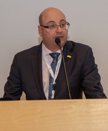 Christian Lattermann合作论文数Ferguson Laboratory, University of Pittsburgh School of Medicine2Craig M. Rodner合作论文数2
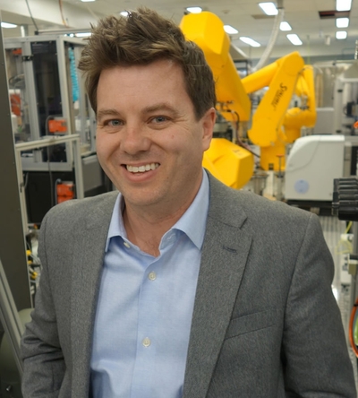 Matthew D. Hall合作论文数Early Translation Branch, Division of Preclinical Innovation, National Center for Advancing Translational Sciences, National Institutes of Health2Andrew L. Haas合作论文数2
 Knut Beitzel合作论文数2Mary K. Mulcahey合作论文数2Joshua Hornstein合作论文数2
 Ryan Charette合作论文数1Prashanth Vishwanath合作论文数1Kimberly A Jacobsen合作论文数1Benjamin J. Levy合作论文数1Laura A. Vogel合作论文数1
 Stephen E. Marcaccio合作论文数1
 Danielle Kriscenski合作论文数1Jeffrey D Trojan合作论文数1Jordan Gross合作论文数1Victoria Stoj合作论文数1Scott Zimmer合作论文数1




