The maintenance of extracellular fluid (ECF) osmolality and sodium concentration ([Na+]o) near optimal "set point" values sustains physiological functions and prevents pathological states such as hypo- and hypernatremia. The peptide hormones vasopressin (antidiuretic hormone) and oxytocin (a natriuretic hormone in rats) play key roles in this process. These hormones are synthesized by hypothalamic magnocellular neurosecretory cells (MNCs) that project to the neurohypophysis and are released into the systemic circulation in response to rises in ECF osmolality or [Na+]o. These homeostatic responses are highly sensitive. For example, vasopressin release is elicited by an increase in ECF osmolality as small as ≥1%. The osmotic and sodium-dependent control of vasopressin and oxytocin release at the neurohypophysis is directly regulated by the electrical activity of MNCs. This regulation involves an array of mechanisms that include synaptic inputs from the brain and periphery, the effects of chemicals released by glial cells, and intrinsic sensory properties of MNCs. These overlapping mechanisms may offer an important degree of redundancy for the homeostatic control of vasopressin and oxytocin release and contribute to the high sensitivity of these responses. Recent work has shown that the intrinsic sodium sensitivity and osmosensitivity of MNCs play an important role in the control of these neurons in vivo. This review provides an update of our current understanding of the molecular and cellular mechanisms that contribute to the cell-autonomous sensory properties of MNCs.
Central autonomic and endocrine dysfunctions following traumatic brain injury (TBI) are believed to involve the hypothalamus; however, underlying mechanisms are unknown. Although chronic deficits might be caused by irreversible tissue damage, various neuroendocrine and autonomic symptoms are only observed transiently, suggesting they might result from a temporary alteration in the activity of hypothalamic neurons. We therefore examined if a mouse model of mild TBI could induce reversible autonomic phenotypes and cause acute changes in c-Fos expression within corresponding regions of the hypothalamus. Adult C57Bl/6 male mice were lightly anesthetized with isoflurane and subjected to TBI by lateral head impact using a Gothenburg impactor. Mice treated the same way, but without the head impact served as controls (shams). We monitored body weight and core body temperature by infrared thermography and performed immunohistochemistry against c-Fos in various regions of the hypothalamus. We determined that a projectile velocity of 9 m/s significantly delayed recovery from the anesthesia without inducing skull fractures and signs of discomfort disappeared within 3 h, as assessed by grimace scale. Compared with shams, TBI mice displayed a rapid decrease in core body temperature which resolved within 48 h. Daily body weight gain was also significantly lower in TBI mice on the day following injury but recovered thereafter. c-Fos analysis revealed a significantly higher density of c-Fos-positive cells in the paraventricular nucleus and a significantly lower density in the median preoptic nucleus and medial preoptic area. We conclude that mild TBI induced by a single lateral head impact in mice at 9 m/s produces acute and reversible symptoms associated with hypothalamic dysfunction accompanied by significant changes in c-Fos expression within relevant areas of the hypothalamus. These findings support the hypothesis that a temporary alteration of neuronal activity may underlie the expression of reversible central autonomic and neuroendocrine symptoms.
Increases in core body temperature cause secretion of vasopressin (vasopressin, antidiuretic hormone) to promote water reabsorption and blunt water losses incurred through homeostatic evaporative cooling. Subtypes of transient receptor potential vanilloid (Trpv) channels have been shown to contribute to the intrinsic regulation of vasopressin‐releasing magnocellular neurosecretory cells (MNCs) in the supraoptic nucleus (SON) and paraventricular nucleus (PVN). However, MNCs in vivo can also be excited by local heating of the adjacent preoptic area, indicating they receive thermosensory information from other areas. Here, we investigated whether neurons in the organum vasculosum lamina terminalis (OVLT) contribute to this process using in vitro electrophysiological approaches in male rats. We found that the majority of OVLT neurons are thermosensitive in the physiological range (36–39°C) and that this property is retained under conditions blocking synaptic transmission. A subset of these neurons could be antidromically activated by electrical stimulation in the SON. Whole cell recordings from SON MNCs revealed that heating significantly increases the rate of spontaneous excitatory postsynaptic currents (sEPCSs), and that this response is abolished by lesions targeting the OVLT, but not by bilateral lesions placed in the adjacent preoptic area. Finally, local heating of the OVLT caused a significant excitation of MNCs in the absence of temperature changes in the SON, and this effect was blocked by inhibitors of ionotropic glutamate receptors. These findings indicate that the OVLT serves as an important thermosensory nucleus and contributes to the activation of MNCs during physiological heating.
Our understanding of neuropeptide function within neural networks would be improved by methods allowing dynamic detection of peptide release in living tissue. We examined the usefulness of sniffer cells as biosensors to detect endogenous vasopressin (VP) release in rat hypothalamic slices and from isolated neurohypophyses. Human embryonic kidney cells were transfected to express the human V1a VP receptor (V1aR) and the genetically encoded calcium indicator GCaMP6m. The V1aR couples to Gq11, thus VP binding to this receptor causes an increase in intracellular [Ca2+] that can be detected by a rise in GCaMP6 fluorescence. Dose-response analysis showed that VP sniffer cells report ambient VP levels >10 pM (EC50 = 2.6 nM), and this effect could be inhibited by the V1aR antagonist SR 49059. When placed over a coverslip coated with sniffer cells, electrical stimulation of the neurohypophysis provoked a reversible, reproducible, and dose-dependent increase in VP release using as few as 60 pulses delivered at 3 Hz. Suspended sniffer cells gently plated over a slice adhered to the preparation and allowed visualization of VP release in discrete regions. Electrical stimulation of VP neurons in the suprachiasmatic nucleus caused significant local release as well as VP secretion in distant target sites. Finally, action potentials evoked in a single magnocellular neurosecretory cell in the supraoptic nucleus provoked significant VP release from the somatodendritic compartment of the neuron. These results indicate that sniffer cells can be used for the study of VP secretion from various compartments of neurons in living tissue. NEW & NOTEWORTHY The specific functional roles of neuropeptides in neuronal networks are poorly understood due to the absence of methods allowing their real-time detection in living tissue. Here, we show that cultured "sniffer cells" can be engineered to detect endogenous release of vasopressin as an increase in fluorescence.
Castillo, Juan P., Sánchez-Rodríguez, Jorge E., Hyde, H. Clark, Zaelzer, Cristian A., Aguayo, Daniel, Sepúlveda, Romina V., Luk, Louis Yu Pan, Kent, Stephen B. H., Gonzalez-Nilo, Fernando D., Bezanilla, Francisco and Latorre, Ramón 2016. 1 subunit-induced structural rearrangements of β the Ca2+and voltage-activated (BK) channel. Proceedings of the National Academy of Sciences of the United States of America 113 (23) , E3231-E3239. 10.1073/pnas.1606381113 file
Large-conductance Ca(2+)- and voltage-activated K(+) (BK) channels are involved in a large variety of physiological processes. Regulatory β-subunits are one of the mechanisms responsible for creating BK channel diversity fundamental to the adequate function of many tissues. However, little is known about the structure of its voltage sensor domain. Here, we present the external architectural details of BK channels using lanthanide-based resonance energy transfer (LRET). We used a genetically encoded lanthanide-binding tag (LBT) to bind terbium as a LRET donor and a fluorophore-labeled iberiotoxin as the LRET acceptor for measurements of distances within the BK channel structure in a living cell. By introducing LBTs in the extracellular region of the α- or β1-subunit, we determined (i) a basic extracellular map of the BK channel, (ii) β1-subunit-induced rearrangements of the voltage sensor in α-subunits, and (iii) the relative position of the β1-subunit within the α/β1-subunit complex.
Thirst and antidiuretic hormone secretion occur during hyperthermia or hypertonicity to preserve body hydration. These vital responses are triggered when hypothalamic osmoregulatory neurons become depolarized by ion channels encoded by an unknown product of the transient receptor potential vanilloid-1 gene (Trpv1). Here, we show that rodent osmoregulatory neurons express a transcript of Trpv1 that mediates the selective translation of a TRPV1 variant that lacks a significant portion of the channel's amino terminus (ΔN-TRPV1). The mRNA transcript encoding this variant (Trpv1dn) is widely expressed in the brains of osmoregulating vertebrates, including the human hypothalamus. Transfection of Trpv1dn into heterologous cells induced the expression of ion channels that could be activated by either hypertonicity or by heating in the physiological range. Moreover, expression of Trpv1dn rescued the osmosensory and thermosensory responses of single hypothalamic neurons obtained from Trpv1 knockout mice. ΔN-TRPV1 is therefore a co-detector of core body temperature and fluid tonicity.
Magnocellular neurosecretory cells (MNCs) in the rat supraoptic nucleus display clustered firing during hyperosmolality or dehydration. This response is beneficial because this type of activity potentiates vasopressin secretion from axon terminals in the neurohypophysis and thus promotes homoeostatic water reabsorption from the kidney. However, the mechanisms which lead to the generation of clustering activity in MNCs remain unknown. Previous work has shown that clustered firing can be induced in these neurons through the pharmacological activation of NMDA receptors (NMDARs) and that silent pauses observed during this activity are mediated by apamin-sensitive calcium activated potassium (SK) channels. However, it remains unknown if clustered firing can be induced in situ by endogenous glutamate release from axon terminals. Here we show that electrical stimulation of glutamatergic osmosensory afferents in the organum vasculosum lamina terminalis (OVLT) can promote clustering in MNCs via NMDARs and apamin-sensitive channels. We also show that the rate of spike clustering induced by NMDA varies as a bell-shaped function of voltage, and that partial inhibition of SK channels can increase cluster duration and reduce the rate of clustering. Finally, we show that MNCs express neurotensin type 2 receptors, and that activation of these receptors can simultaneously depolarize MNCs and suppress clustered firing induced by bath application of NMDA or by repetitive stimulation of glutamate afferents. These studies reveal that spike clustering can be induced in MNCs by glutamate release from afferent nerve terminals and that that this type of activity can be fine-tuned by neuromodulators such as neurotensin.
Mammalian osmosensory neurons exposed to hypertonicity display a shrinking-induced increase in non-selective cation conductance. That causes membrane depolarization and excitation triggering homeostatic responses such as thirst and antidiuretic hormone release. The molecular identity of the osmoreceptor channel is not known, but appears to involve a capsaicin-insensitive N-terminal variant of the transient receptor potential vanilloid type 1 (TRPV1) channel (Ciura and Bourque, 2006; Sharif-Naeini et al., 2006).
The high conductance voltage- and Ca(2+)-activated K(+) channel is one of the most broadly expressed channels in mammals. This channel is named BK for 'big K' because of its single-channel conductance that can be as large as 250 pS in 100 mm symmetrical K(+). BK channels increase their activity by membrane depolarization or an increase in cytosolic Ca(2+). One of the key features that defines the behaviour of BK channels is that neither Ca(2+) nor voltage is strictly necessary for channel activation. This and several other observations led to the idea that both Ca(2+) and voltage increase the open probability by an allosteric mechanism. In this type of mechanism, the processes of voltage sensor displacement, Ca(2+) binding and pore opening are independent equilibria that interact allosterically with each other. These allosteric interactions in BK channels reside in the structural characteristics of the BK channel in the sense that voltage and Ca(2+) sensors and the pore need to be contained in different structures or 'modules'. Through electrophysiological, mutagenesis, biochemical and fluorescence studies these modules have been identified and, more important, some of the interactions between them have been unveiled. In this review, we have covered the main advances achieved during the last few years in the elucidation of the structure of the BK channel and how this is related with its function as an allosteric protein.
We describe two approaches for the construction of a 3D molecular model for BK. First, we used a LRET technique in which an encoded Lanthanide Binding Tag (LBT) that binds Tb3+ plays the role of the donors and SulphoRodhamine Methanothiosulfonate (TMSR) attached to Charibdotoxin (ChTX) ) plays the role of the acceptor. The data obtained allowed us to determine the distances from the center of symmetry of the channel to the external aspect of S0, S1, S2, S3-S4 linker in the α subunit. The distances from the channel center of symmetry to TM1, TM2 and three positions in the β1 loop were obtained using the same methodology, by inserting LBT's in the β1 subunit. Second, molecular simulations of ChTX-TMSR complex bound to the BK pore were used to obtain the conformational space of TMSR bound to ChTX. This simulation was used to evaluate the population of the different conformations that TMSR can adopt. The conformational sampling of TMSR was used to recalculate all the distances and estimate the position of the donors in 3D coordinates. The new distance values were used as distance restrains to build a final model of BK. The initial homology model of BK was built using as reference structure the Kv chimeric crystal of Kv1.2-Kv2.1 mammalian channel. Our model refined with LRET experiments shows a structure of α subunit alone with the extra-transmembrane segment S0 located in a pocket in the voltage sensor domain, and a concave shape for BK extracellular face. The β1 external loop, on the other hand, lies very near to the BK pore and forms a structure similar to an alpha greek letter. Supported by FONDECYT Nº1070049, AnilloCientifico ACT/24 and NIHGM030376
The large-conductance Ca2+ and voltage-activated K+ (BKCa) channel possesses an Alfa pore-forming subunit and 4 different tissue specific regulatory Beta subunits. The Alfa subunit of BKCa channels contains the S4-based voltage sensor of Kv channels, but is an exception inside the S4 superfamily of ion channels because it is made up of seven (instead of 6) transmembrane segments (S0-S6) with the N terminus facing the extracellular side. Our knowledge is limited of how this extra transmembrane segment affects the architecture of BKCa channels. Equally mysterious is the actual structure of the Alfa-Beta subunit complex. Here we use the genetically encoded Lanthanide Binding Tag (LBT) that binds Tb3+ as LRET donor and the Charibdotoxin (CTX) labeled with Tetramethylrhodamine (TMR) for in vivo spectroscopic studies of intramolecular distances and interactions between Alfa and Beta1 subunit of BKCa channel. We have measured the distance between the extracellular end of S0, S1, S2, and S3 in the Alfa subunit to the TMR in the CTX blocking the pore with and without Beta1 subunit. We have also measured the distance from TM1, TM2 and loop of Beta1 subunit to the TMR in the toxin. We found that segment S0 is further away than the other segments with respect to the center of the pore in the absence of Beta1 subunit, locating it in the periphery of the molecule. However, it becomes closer to the center when Alfa is co expressed with Beta1 subunit. Beta1 co-expression also changes the position of S2. We found that some transmembrane domains of Alfa are further away than Beta1's transmembranes domains, indicating that the Beta subunit is embedded in the protein. Support: NIHGM30376 and FONDECYT 1070049.
Although a unifying characteristic common to all transient receptor potential (TRP) channel functions remains elusive, they could be described as tetramers formed by subunits with six transmembrane domains and containing cation-selective pores, which in several cases show high calcium permeability. TRP channels constitute a large superfamily of ion channels, and can be grouped into seven subfamilies based on their amino acid sequence homology: the canonical or classic TRPs, the vanilloid receptor TRPs, the melastatin or long TRPs, ankyrin (whose only member is the transmembrane protein 1 [TRPA1]), TRPN after the nonmechanoreceptor potential C (nonpC), and the more distant cousins, the polycystins and mucolipins. Because of their role as cellular sensors, polymodal activation and gating properties, many TRP channels are activated by a variety of different stimuli and function as signal integrators. Thus, how TRP channels function and how function relates to given structural determinants contained in the channel-forming protein has attracted the attention of biophysicists as well as molecular and cell biologists. The main purpose of this review is to summarize our present knowledge on the structure of channels of the TRP ion channel family. In the absence of crystal structure information for a complete TRP channel, we will describe important protein domains present in TRP channels, structure-function mutagenesis studies, the few crystal structures available for some TRP channel modules, and the recent determination of some TRP channel structures using electron microscopy.
Ion channels activate by sensing stimuli such as membrane voltage, ligand binding or temperature and transduce this information into conformational changes that open the channel pore. Thus, a key question in understanding ion channel function is how do the protein domains involved in sensing stimuli (sensors) and opening the pore (gates) communicate. In this regard, transient receptor potential (TRP) channels that confer thermosensation [A. Dhaka, V. Viswanath, A. Patapoutian, TRP ion channels and temperature sensation, Annu. Rev. Neurosci. 29 (2006) 135-161; I.S. Ramsey, M. Delling, D.E. Clapham, An introduction to TRP channels, Annu. Rev. Physiol. 68 (2006) 619-647] (thermoTRP; Q(10)>10) are unique to the extent that they integrate a variety of physical and chemical stimuli. In some cases such as, for example, the vanilloid receptor TRPV1 [M.J. Caterina, M.A. Schumacher, M. Tominaga, T.A. Rosen, J.D. Levine, D. Julius, The capsaicin receptor: a heat-activated ion channel in the pain pathway, Nature 389 (1997) 816-824] and TRPA1 [G.M. Story, A.M. Peier, A.J. Reeve, S.R. Eid, J. Mosbacher, T.R. Hricik, T.J. Earley, A.C. Hergarden, D.A. Andersson, S.W. Hwang, P. McIntyre, T. Jegla, S. Bevan, A. Patapoutian, ANKTM1, a TRP-like channel expressed in nociceptive neurons, is activated by cold temperatures, Cell 112 (2003) 819-829; S. Jordt, D. Julius, Molecular basis for species-specific sensitivity to "hot" chilli peppers, Cell 108 (2002) 421-430] the integration of these stimuli elicit pain [M. Tominaga, M.J. Caterina, A.B. Malmberg, T.A. Rosen, H. Gilbert, K. Skinner, B.E. Raumann, A.I. Basbaum, D. Julius, The cloned capsaicin receptor integrates multiple pain-producing stimuli, Neuron 21 (1998) 531-543; M. Bandell, A. Dubin, M. Petrus, A. Orth, J. Mathur, S. Hwang, A. Patapoutian, High-throughput random mutagenesis screen reveals TRPM8 residues specifically required for activation by menthol, Nat. Neurosci. 9 (2006) 466-468; S. Zurborg, B. Yurgionas, JA. Jira, O. Caspani, P.A. Heppenstall, Direct activation of the ion channel TRPA1 by Ca(2+), Nat. Neurosci. 10 (2007) 277-279]. These stimuli include voltage, pH, agonist binding, and temperature. Understanding how each of these distinct physiological signals regulate channel opening will be informative about the mechanical linkages that can act either independently or in concert to influence channel activation. In this paper we show that thermoTRP channel-forming proteins are modular in the sense that certain structure or structures (modules) confer temperature-dependent regulation, whereas others confer voltage-dependent regulation. We also discuss the thermodynamic basis of heat and cold activation in an effort to elucidate what confer to these channels the capability to be gated by temperature directly.
 Ramón Latorre合作论文数Center Interdisciplinary for Neuroscience of Valparaiso, Universidad de Valparaíso;The University of Chicago;Department of Biology, Faculty of Sciences, University of Chile7
 Charles Bourque合作论文数Centre for Research in Neuroscience, McGill University;Department of Neurology and Neurosurgery, Faculty of Medicine and Health Sciences, McGill University;Department of Physiology, School of Biomedical Sciences, McGill University7
 H. Clark Hyde合作论文数4
 Francisco Bezanilla合作论文数Department of Biochemistry and Molecular Biology, University of Chicago4
 Fernando Danilo Gonzalez Nilo合作论文数Centro de Bioinformatica y Biologia Integrativa, Facultad de Ciencias de la Vida, Universidad Andres Bello3
 Juan P. Castillo合作论文数Calif Inst Quantitat Biosciences, Univ Calif2
 Francisco J Morera合作论文数Integrat Biol Grp, Univ Austral Chile2
 Louis Y. P. Luk合作论文数School of Chemistry and Cardiff Catalysis Institute, Cardiff University;School of Chemistry and Cardiff Catalysis Institute, Cardiff University2
 Ciura Sorana合作论文数Research Center of the Institute for Brain and Spinal Cord CRICM, Pierre and Marie Curie University2
 Walter Sandtner合作论文数University of Chicago2
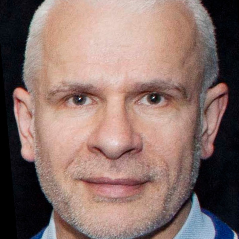 Wolfgang Liedtke合作论文数Regeneron2Pierce Hua合作论文数2Daniel Aguayo合作论文数Computer Science and Artificial Intelligence Laboratory M.I.T.2
 Sebastian Brauchi合作论文数Escuela de Ingeniería Bioquímica;Instituto de Química;Universidad Católica de Valparaíso;Instituto de Química, Universidad Católica de Valparaíso2
 Jessica R Sudbury合作论文数1Sukhee Lee合作论文数1Maria Prager-Khoutorsky合作论文数1Zahra Simone Thirouin合作论文数Hlth Ctr, McGill Univ1Claire Gizowski合作论文数McGill Univ, Ctr Hlth, Ctr Res Neurosci, 1650 Cedar Ave, Montreal, PQ H3G 1A4, Canada1
 Masha Prager Khoutorsky合作论文数Department of Physiology, Faculty of Medicine and Health Sciences, McGill University1
 Keith Murai合作论文数Centre for Research in Neuroscience, McGill University1Sánchez-Rodríguez Jorge E合作论文数Instituto de Física, Universidad Autónoma de San Luis Potosí1
 Romina V. Sepulveda合作论文数Universidad Andrés Bello, Universidad Andrés Bello1Christopher Salmon合作论文数1
 Stephen Kent合作论文数Department of Biochemistry and Molecular Biology, The University of Chicago1Okuda Tika合作论文数1Gagnon Ariane合作论文数1
 Eric Trudel合作论文数1
 Choe Katrina Y合作论文数Montreal General Hospital, Research Institute of the McGill University Health Centre1
 Michael R. Walsh合作论文数Cold Regions Research and Engineering Laboratory, US Army Engineer Research and Development Center1
 Horacio Poblete合作论文数Center for Bioinformatics and Molecular Simulations, Universidad de Talca1David Aguayo合作论文数1
 Gerardo Orta合作论文数1Ariela Vergara-Jaque合作论文数Ctr Bioinformat & Simulac Mol1Daniel L. Voisin合作论文数1




