Rationale: Diffuse idiopathic pulmonary neuroendocrine cell hyperplasia (DIPNECH) is a pulmonary condition characterised by neuroendocrine cell proliferation along respiratory bronchioles resulting in tumourlet formation which may progress to pulmonary carcinoid. DIPNECH is a rare and under-recognised condition that predominantly affects females, with an estimated worldwide incidence of 5-6 people per million population. Given it's rarity and the poor understanding around the pathogenesis and treatment of this condition, we undertook the largest international case series to date of DIPNECH patients. We aim to further characterise this patient cohort to better understand the clinical features, identify common pulmonary function test (PFT) patterns and radiographic findings and describe current treatment practices. Methods: We collated data from 17 centres across 8 different countries comprising a total of 258 patients. Data collected included patient demographics, smoking history, symptoms, co-morbidities, pathological diagnosis, imaging findings, PFT pattern and therapies. Results: In total 258 patients were identified. The median age was 65 (SD+/- 10.6). 241 (93%) were female. 39% (n= 94/243) were ex-smokers. 79% (n=186/234) of patients reported pulmonary symptoms with cough being the most common symptom reported in 81% (n=151/186) of patients. Many patients were polysymptomatic, with cough and dyspnoea being the most common combination of symptoms described. (50.54%; n=94/186). Complete spirometry pattern was documented for 180 patients (69.78%) with an obstructive pattern being the most commonly observed (n=76/180; 42%). 27.5% (n=41/139) had clinically significant FEV1 reversibility post bronchodilator administration. Pathology specimens were available in 219 cases. Most common pathological features were neuroendocrine hyperplasia which was observed in 75% (n=165/219) and tumourlets in 74% (n=162/219) of cases. Carcinoid, either typical or atypical, was observed in 64% (n=141/219) of samples. 252 patients had computed tomography (CT) imaging performed. Pulmonary nodules were the most common feature identified in 99.60% of cases (248/250). Mosaic attenuation (69.30%; n=50/173) and airway dilatation (28.90%; n=50/173) were also frequently reported. There was no significant correlation between symptoms (cough, dyspnoea, wheeze) and airway dilatation (p=0.496), mosaic attenuation (p=0.496) and PFT pattern (p=0.837). There was a significant association between presence of symptoms and mosaic attenuation on CT (p=0.003). Conclusions: DIPNECH, while a rare disorder, is understudied. This international case series is the largest performed to date and highlights the complexity in management of these patients.
Background: Hypersensitivity pneumonitis (HP) represents a chronic lung disease with an unpredictable clinical course. There is a pressing need for clinically applicable prognostic biomarkers in patients with HP. Methods: This was an observational, retrospective study. We investigated the prognostic potential of complete blood count parameters in treatment-naïve patients diagnosed with HP between 15 December 2010 and 1 October 2023. Receiver operating characteristic (ROC) curve analysis identified the optimal cut-off thresholds for each parameter in terms of mortality prediction. Results: We included 129 patients diagnosed with HP [median age: 68.0 years (95% CI: 65.0 to 69.0), fibrotic HP: n = 85, 65.9%]. Patients with HP and an eosinophil count >160 cells/μL [ROC curve, area under curve (AUC): 0.61] exhibited increased mortality risk compared to patients with HP and an eosinophil count ≤ 160 cells/μL [Kaplan–Meier, HR: 2.95 (95% CI: 1.36 to 6.42), p = 0.006]. Patients with HP and a monocyte count > 350 cells/μL (ROC curve, AUC: 0.52) had worse survival compared to patients with HP and a monocyte count lower than this threshold [Kaplan–Meier, HR: 2.48 (95% CI: 1.03 to 5.09), p = 0.04]. Patients with HP and an eosinophil–lymphocyte ratio (ELR) > 0.09 (ROC curve, AUC: 0.64) had a higher risk of mortality compared to patients with HP and ELR ≤ 0.09 [Kaplan–Meier, HR: 2.75 (95% CI: 1.3 to 5.78), p = 0.008]. Conclusions: This study demonstrated that eosinophil count, monocyte count, and ELR could be prognostic biomarkers in patients with HP. Further studies aiming to validate the prognostic potential of complete blood count parameters in patients with HP are greatly anticipated.
Background:Suspected interstitial lung disease (ILD) patients may be referred to an ILD-specialist centre or a non-ILD-specialist centre for diagnosis and treatment. Early referral and management of patients at ILD-specialist centres has been shown to improve survival and reduce hospitalisations. The COVID-19 pandemic has affected the ILD patient diagnostic pathway and prompted centres to adapt. This study investigates and contrasts ILD patient pathways in ILD-specialist and non-ILD-specialist centres, focusing on referrals, caseloads, diagnostic tools, multi-disciplinary team (MDT) meeting practices and resource accessibility. Methods:Conducted as a cross-sectional study, a global self-selecting survey ran from September 2022 to January 2023. Participants included ILD specialists and healthcare professionals (HCPs) from ILD-specialist centres and non-ILD-specialist centres. Results:Of 363 unique respondents from 64 countries, 259 were from ILD-specialist centres and 104 from non-ILD-specialist centres. ILD centres had better resource availability, exhibiting higher utilisation of diagnostic tests (median: 12 tests) than non-ILD centres (nine tests) and better access to specialist professions attending MDT meetings (median: six professions at meeting) in specialist centres than non-ILD centres (three professions at meeting). Transitioning to virtual MDT meetings allowed HCPs from other locations to join meetings in nearly 90% of all centres, increasing regular participation in 60% of specialist centres and 72% of non-ILD centres. For treatment of patients, specialist centres had better access to antifibrotic drugs (91%) compared to non-ILD centres (60%). Conclusions:Diagnostic pathways for ILD patients diverged between specialist centres and non-ILD centres. Disparities in resource and specialist availability existed between centres.
Background: Depression and anxiety represent significant comorbidities in idiopathic pulmonary fibrosis (IPF) patients, affecting their quality of life. The COVID-19 pandemic has had an uneven impact on global mental health. The Hospital Anxiety and Depression Scale (HADS) constitutes a validated tool to identify anxiety disorders and depression. The aim of this multicentre study was to evaluate the effect of COVID-19 vaccination on depression and anxiety in IPF patients. Methods: Consecutive IPF patients (median 73.5 years) who are regularly followed-up with were included in the study. Demographics, functional, and clinical were recorded. The HADS score was calculated before and one month after vaccination against COVID-19 in all participants. A Wilcoxon signed ranks test was conducted. Results: A total of 180 IPF patients (median 73.5 years) were included in the study. Among them, 145 patients (81%) received antifibrotic treatment. A significant reduction in HADS, both in anxiety and depression scales, was observed one month after vaccination against SARS-COV-2), independent of age, smoking, lung function impairment, and prior history of depression (p < 0.01). Conclusions: A higher Hospital Anxiety and Depression Scale score was detected before vaccination against COVID-19. It seems that vaccination also offered a beneficial effect on depression and anxiety in IPF patients, independent of age, smoking, lung function impairment, and prior history of depression.
Case PresentationA 74-year-old man presented to our department with progressive dyspnea on exertion over the last year. The patient did not report any other symptoms. He had previously smoked with a 60 pack-year history. He worked in an office and did not report any environmental, occupational, or domestic exposures. His history included asymptomatic Waldenström’s macroglobulinemia that was diagnosed 18 months before respiratory symptoms. He was not receiving any treatment and was monitored regularly by the hematology department.
BackgroundLung involvement in the context of idiopathic inflammatory myopathies has significant impact on outcome; early and accurate diagnosis is important but can be difficult to achieve. In particular, patients without clinically evident muscle involvement pose a significant diagnostic challenge.MethodsA computer-assisted search was conducted to identify patients with amyopathic interstitial lung disease associated with the presence of myositis-specific autoantibodies. Medical records and chest imaging studies were reviewed to identify clinical and radiologic features at presentation.ResultsOf the 35 patients with amyopathic interstitial lung disease associated with myositis-specific autoantibodies, the median age was 65 years (range 43–78) and 20 were women (57%). Of the patients, 34% had previously visited the rheumatology department. Presenting symptoms consisted of dyspnea (94%), cough (43%), and arthritis (23%). Raynaud phenomenon, “mechanic hands,” Gottron papules, and inspiratory crackles were present in 23, 31, 9, and 74% of patients, respectively. After a detailed history, none of the patients reported muscle weakness, while four (11%) exhibited increased CK levels; of these four, two had a concomitant increase in aldolase levels. Median FVC was 79% predicted (range: 49–135) and median DLco was 50% predicted (range: 17–103). HRCT pattern was suggestive of an alternative to UIP pattern in 31/33 (94%) patients; the most common imaging patterns were NSIP (49%) and NSIP/OP (39%).ConclusionIn patients with NSIP and NSIP/OP pattern, the presence of amyopathic interstitial lung disease associated with myositis-specific autoantibodies should be considered even in the absence of clinical evident myositis.
BACKGROUND:Human telomerase reverse transcriptase (hTERT) is the catalytic subunit of telomerase enzyme, which adds nucleotides to telomeres and counteracts their length shortening. The development of a telomere maintenance mechanism represents a hallmark of cancer. On the other hand, idiopathic pulmonary fibrosis (IPF) is associated with mutations in telomerase genes and shorter telomeres. IPF is frequently complicated with lung cancer.AIM:To investigate the expression of hTERT in lung cancer with co-existing IPF and to compare with lung cancer without fibrosis.METHODS:Diagnostic lung cancerous biopsies were retrieved from 18 patients with lung cancer and concomitant IPF, as well as 18 age and gender matched controls with lung cancer without pulmonary fibrosis. The expression of hTERT was studied with immunohistochemistry. ImajeJ software was used to quantitate subcellular stain intensity. Immunohistochemical investigation of two senescence-associated markers, p16 and p21, was also performed in all 36 cases.RESULTS:Both groups highly expressed hTERT, without significant difference (100% vs 95%, p = 0.521). Evaluation of p16 and p21 immunostaining revealed negative to minimal immunoreactivity in both groups. hTERT localization exhibited higher median nuclear intensity in the group of lung cancer with IPF (0.62 vs 0.45, p = 0.016), while cytoplasmic intensity did not differ significantly (0.17 vs 0.15, p = 0.463). Higher median nuclear intensity was also correlated with small cell lung cancer subtype in the whole study sample (0.69 vs 0.45, p = 0.09).CONCLUSION:hTERT is highly expressed in lung cancer with concomitant IPF, but with differential localization compared to lung cancer without IPF, implying differences in pathogenicity and requiring further investigation.
1. Rajan SK, Cottin V, Dhar R, et al. Progressive pulmonary fibrosis: an expert group consensus statement. Eur Respir J. 2023;61(3):2103187. doi:10.1183/13993003.03187-2021 CrossRef Google Scholar
OBJECTIVE:Monocyte count and red cell distribution width (RDW) have shown prognostic potential in patients with fibrotic lung diseases. Their kinetics and prognostic usefulness of peripheral blood indices in patients with interstitial lung diseases (ILDs) undergoing surgical lung biopsy for diagnostic reasons have not been studied. PATIENTS AND METHODS:We retrospectively included consecutive patients with ILD who underwent surgical lung biopsy for diagnostic purposes Between 07/11/2019 and 11/10/2022. RESULTS:Fifty-five (n=55) patients were included in the study. Median age was 65.0 years (95% CI: 63.0 to 66.0). Postoperative peripheral blood monocyte count on Day 1 was significantly higher compared to preoperative, perioperative, and postoperative values on Day 90 (repeated measures ANOVA, p<0.0001). Patients in the high postoperative monocyte count group had significantly increased length of postoperative hospital stay [Mann-Whitney test, p=0.007] and significantly lower Forced Vital Capacity (FVC)% predicted 3 months after surgery [Mann-Whitney test, p=0.029] compared to patients in the low postoperative monocyte count group. Postoperative RDW on Day 90 was significantly higher compared to preoperative, perioperative and postoperative-Day 1 RDW (repeated measures ANOVA, p=0.008, p=0.006, p<0.0001, respectively). Patients in the high postoperative RDW group did not have increased hospital stay (Mann-Whitney test, p=0.49) or decreased FVC% predicted at 3 months compared to patients in the low postoperative RDW group (Mann-Whitney test, p=0.91). CONCLUSIONS:Peripheral blood monocyte count could be a prognostic biomarker for patients with ILDs undergoing diagnostic surgical lung biopsies. RDW does not seem to represent an acute phase biomarker but seems to increase over time following disease progression. Larger studies are urgently required.
Background: In the pathogenesis of sarcoidosis, the presentation of unknown antigen(s) by MHC (HLA in humans) molecules initiates altered immune and inflammatory responses. This study, the first in the Greek population, investigates if variants in HLA genes and in related single nucleotide polymorphisms (SNP) associate with sarcoidosis and/or its clinical phenotypes.Methods: 103 patients diagnosed by ATS/ERS/WASOG guidelines were enrolled. Genotyping of 7 HLA loci (HLA-A,-B,-C,-DQA1,-DQB1,-DRB1,-DPB1) was performed by next-generation sequencing and 7 SNPs were determined by MassARRAY. HLA allele frequencies in the patients were compared with those in healthy Greeks from the allelic frequency databases; SNP allele distributions in the patients were compared with those in 100 healthy Greeks.Results Sarcoidosis associated with alleles in the HLA class II loci. The alleles HLA-DQB1*05:03, and HLA‑DQB1*06:03 conferred the risk of sarcoidosis. By contrast, HLA-DRB1*11:04, and particularly HLA-DQA1*05:01 acted as protective alleles. HLA-A*02:01 was the prominent allele associated with the chest-X ray (CXR) stage I, while HLA-DRB1*07:01 and HLA-A*01:01 were linked to more advanced CXR stages (II-IV). SNP rs4713607 protected from extrapulmonary manifestation.Conclusions: We re-affirm in this Greek populations some associations between HLA and sarcoidosis which have previously been reported in other ethnicities. Importantly, we report here for the first time two alleles strongly associated with sarcoidosis as risk (HLA-DQB1*06:03) or protective factors (HLA‑DRB1*11:04). Also, we describe other specific associations between sarcoidosis phenotypes and HLA and/or SNP alleles. Further work, particularly genotyping of the expanded patient cohort(s) is required to confirm the clinical relevance of the observed associations.
Purpose: A(1)Antitrypsin deficiency (AATD) pathogenic mutations are expanding beyond the PI*Z and PI*S to a multitude of rare variants. Aim: to investigate genotype and clinical profile of Greeks with AATD. Methods: Symptomatic adult-patients with early-emphysema defined by fixed airway obstruction and computerized-tomography scan and lower than normal serum AAT levels were enrolled from reference centers all over Greece. Samples were analyzed in the AAT Laboratory, University of Marburg-Germany. Results: Included are 45 adults, 38 homozygous or compound heterozygous for pathogenic variants and 7 heterozygous. Homozygous were 57.9% male, 65.8% ever-smokers, median (IQR) age 49.0(42.5-58.5) years, AAT-levels 0.20(0.08-0.26) g/L, FEV1(%predicted) 41.5(28.8-64.5). PI*Z, PI*Q0, and rare deficient allele's frequency was 51.3%, 32.9%,15.8%, respectively. PI*ZZ genotype was 36.8%, PI*Q0Q0 21.1%, PI*MdeficientMdeficient 7.9%, PI*ZQ0 18.4%, PI*Q0Mdeficient 5.3% and PI*Zrare-deficient 10.5%. Genotyping by Luminex detected: p.(Pro393Leu) associated with M-Heerlen (M1Ala/M1Val); p.(Leu65Pro) with M-Procida; p.(Lys241Ter) with Q0(Bellingham); p.(Leu377Phefs*24) with Q0(Mattawa) (M1Val) and Q0(Ourem) (M3); p.(Phe76del) with M-Malton (M2), M-Palermo (M1Val), M-Nichinan (V) and Q0(LaPalma) (S); p.(Asp280Val) with P-Lowell (M1Val)(;) P-Duarte (M4)(,) Y-Barcelona (p.Pro39His). Gene-sequencing (46.7%) detected Q0(GraniteFalls), Q0(Saint-Etienne), Q0(Amersfoort(M1Ala),) M-W & uuml;rzburg, N-Hartfordcity and one novel-variant (c.1A>G) named Q0(Attikon).Heterozygous included PI*MQ0(Amersfoort(M1Ala),) PI*MMProcida, PI*Mp.(Asp280Val), PI*MOFeyzin. AAT-levels were significantly different between genotypes (p = 0.002). Conclusion: Genotyping AATD in Greece, a multiplicity of rare variants and a diversity of rare combinations, including unique ones were observed in two thirds of patients, expanding knowledge regarding European geographical trend in rare variants. Gene sequencing was necessary for genetic diagnosis. In the future the detection of rare genotypes may add to personalize preventive and therapeutic measures. (c) 2023 Sociedade Portuguesa de Pneumologia. Published by Elsevier Esparta, S.L.U. This is an open access article under the CC BY-NC-ND license (http://creativecommons.org/licenses/by-nc-nd/4.0/).
AIM:To investigate the perspective of physicians treating chronic airway diseases on the importance of device and substance characteristics influencing the compliance of patients with chronic obstructive airways diseases.OBJECTIVE:We surveyed physicians... perspective on the impact of device and substance characteristics on patients... compliance.METHODS:This study was carried out by running a structured questionnaire, to a total of 144 physicians, conducting personal interviews and evaluating answers on a scale from 1 for most to 6 for least important influencing parameter.RESULTS:Overall, the most important parameters influencing patients... compliance according to physicians... perspective were rapid onset of action, type of inhalation device and duration of action. Adverse events were considered as the least important parameter. When COPD and asthma were examined separately, the most important parameters influencing compliance were rapid onset of action, ease of use and duration of action. Rapid onset of action was significantly more important in asthma than COPD.CONCLUSION:Onset and duration of action and ease of use were classified higher as important parameters to increase patients... compliance, according to physicians...PERSPECTIVE:
BACKGROUND:Interstitial lung diseases (ILDs) are extremely challenging in terms of diagnosis. Extreme bronchoalveolar lavage (BAL) lymphocytosis is thought to strongly point towards the diagnosis of hypersensitivity pneumonitis (HP). OBJECTIVES:Explore the range of different ILD that can present with BAL lymphocytosis, including cases of pronounced lymphocytosis and its diagnostic utility. METHODS:Patients with ILD that were subjected to BAL were identified retrospectively from a cohort of consecutive patients. RESULTS:BAL lymphocytosis ≥20% was recorded in 106 patients (27%), while pronounced BAL lymphocytosis ≥40% was recorded in 49 patients (12.5%). The most common diagnoses in patients with BAL lymphocytosis ≥20% and ≥40%, were HP (32.1%), connective tissue disease (CTD)-ILD (26.4%), sarcoidosis (16%), and HP (38.8%), CTD-ILD (24.5%), sarcoidosis (14.3%), respectively. CONCLUSIONS:Neither the presence nor the degree of BAL lymphocytosis can point to a specific diagnosis.
Objectives: Data on COVID-19 in patients with interstitial lung disease are scarce and whether SARS-CoV-2 may trigger interstitial lung disease progression remains unknown. We aimed to analyze outcomes of COVID-19 in patients with systemic sclerosis–associated interstitial lung disease, including possible thoracic radiographic progression. Patients and Methods: All 43 patients with systemic sclerosis–associated interstitial lung disease followed in our center (mean ± SD, 55.2 ± 11.6 years, 36 female) with confirmed SARS-CoV2 infection up to 1 September 2022 were analyzed. Individual interstitial lung disease extent on high resolution CT (HRCT) performed before (up to 3 months) and after COVID-19 (2–5 months) was compared. Results: At SARS-CoV-2 infection, 9/43 patients were unvaccinated, whereas 5, 26, and 3 had received 2, 3, or 4 doses of an mRNA vaccine, respectively. Thirty-one patients were either on monotherapy with immunosuppressives (mycophenolate, n = 7; cyclophosphamide, n = 2; methotrexate, n = 10; tocilizumab, n = 7; rituximab, n = 1; etanercept, n = 1), or their combinations ( n = 3). Eight patients (20%), of whom four unvaccinated, required hospitalization for pneumonia and three (7%) died of acute respiratory failure ( n = 2, both unvaccinated) or cardiac arrest. Lack of vaccination was the only independent predictor for hospitalization (OR = 7.98, 95% CI: 1.25–51.09) and marginally for death (OR = 32.7, 95% CI: 0.97–1110.98), regardless of the presence of diffuse systemic sclerosis, interstitial lung disease extent greater than 20% or immunosuppressive treatment. In 22 patients with available HRCT pairs (vaccinated = 20), the interstitial lung disease extent before COVID-19 (20.4%± 17.8%) remained unchanged (22.4% ± 18.5%) in all but one patient. Conclusion: SARS-CoV-2 vaccination is of outmost importance for every systemic sclerosis patient with interstitial lung disease. COVID-19 does not seem to promote progression of systemic sclerosis–associated interstitial lung disease in vaccinated patients, but further studies are warranted.
IntroductionMyositis associated interstitial lung disease (ILD) seems to be an under-recognized entity. MethodsIn this multicenter, retrospective study, we recorded between 9/12/2019 and 30/9/2021 consecutive patients who presented in five different ILD centers from two European countries (Greece, France) and received a multidisciplinary diagnosis of myositis associated-ILD. The primary outcome was all-cause mortality over 1 year in specific subgroups of patients. Secondary outcomes included comparison of disease characteristics between patients diagnosed with the amyopathic subtype and patients with evidence of myopathy at diagnosis. ResultsWe identified 75 patients with myositis associated-ILD. Median age (95% CI) at the time of diagnosis was 64.0 (61.0-65.0) years. Antinuclear antibody testing was positive in 40% of the cohort (n = 30/75). Myopathy onset occurred first in 40.0% of cases (n = 30), ILD without evidence of myopathy occurred in 29 patients (38.7%), while 16 patients (21.3%) were diagnosed concomitantly with ILD and myopathy. The commonest radiographic pattern was cellular non-specific interstitial pneumonia (NSIP) and was observed in 29 patients (38.7%). The radiographic pattern of organizing pneumonia was significantly more common in patients diagnosed with the amyopathic subtype compared to patients that presented with myopathy [24.1% (n = 7/29) vs. 6.5% (n = 3/46), p = 0.03]. One year survival was 86.7% in the overall population. Kaplan-Meier analysis demonstrated significantly higher all-cause 1-year mortality in patients with the amyopathic subtype compared to patients with evidence of myopathy [H R 4.24 (95% CI: 1.16-15.54), p = 0.03]. Patients diagnosed following hospitalization due to acute respiratory failure experienced increased risk of 1-year all-cause mortality compared to patients diagnosed in outpatient setting [HR 6.70 (95% CI: 1.19-37.81), p = 0.03]. Finally, patients with positive anti-MDA5 presented with higher 1-year all-cause mortality compared to anti-MDA5 negative patients [HR 28.37 (95% CI: 5.13-157.01), p = 0.0001]. ConclusionSpecific ILD radiographic patterns such as NSIP and organizing pneumonia may herald underlying inflammatory myopathies. Hospitalized patients presenting with bilateral organizing pneumonia refractory to antibiotics should be meticulously evaluated for myositis associated-ILD even if there is no overt muscular involvement. Incorporation of ILD radiological patterns in the diagnostic criteria of inflammatory myopathies may lead to timely therapeutic interventions and positively impact patients' survival.
 Argyris Tzouvelekis合作论文数123
 Tzilas Vasilios合作论文数Medical School, Democritus University of Thrace98
 Paschalis Steiropoulos合作论文数Medical School, Democritus University of Thrace91
 Marios Froudarakis合作论文数Medical School, Democritus University of Thrace71
 Theodoros Karampitsakos合作论文数Dept Resp Med, Univ Hosp Patras64
 Katerina Antoniou合作论文数University General Hospital of Heraklion "PAGNI";Laboratory of Cellular and Molecular Pulmonology, University of Crete59Nm Siafakas合作论文数56
 Evangelos Bouros合作论文数56
 Nikolaos (Nikos) G. Tzanakis合作论文数Department of Mathematics, University of Crete47Stavros Anevlavis合作论文数Medical School, Democritus University of Thrace45
 Effrosyni D. Manali合作论文数Athens Medical School, National and Kapodistrian University of Athens;Department of 2nd Pulmonary Medicine, Attikon University Hospital34
 Evangelia Nena合作论文数Laboratory of Social Medicine and Sociology, Department of Medicine, School of Health Science, Democritus University of Thrace33Ntolios Paschalis合作论文数32
 Spyros Papiris合作论文数2nd Pulmonary Medicine Department, School of Medicine, National and Kapodistrian University of Athens;"ATTIKON" University Hospital26
 Vassilis Aidinis合作论文数Institute of Immunology, Biomedical Sciences Research Center "Alexander Fleming"25P Panagou合作论文数24Spyridon Papiris合作论文数“Attikon” General Hospital, National and Kapodistrian University of Athens Medical School23
 Serafeim Chrysikos合作论文数5th Department of Pneumonology, Hospital for Thoracic Diseases "SOTIRIA,",22
 Vlassis Polychronopoulos合作论文数3rd Department of Pneumonology, “Sismanoglion” General District Hospital22Panagiotis Boglou合作论文数21Nikolaos Papanas合作论文数20Lykourgos Kolilekas合作论文数18
 Andreas Koulelidis合作论文数18
 Anastasia Oikonomou合作论文数Department of Medical Imaging, Temerty Faculty of Medicine, University of Toronto17
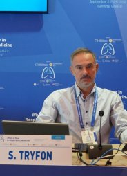 Stavros Tryfon合作论文数1st Department of Pneumonology, GH "G Papanikolaou",17
 Katerina Dimakou合作论文数17A. Tzouvelekis合作论文数16Ourania Papaioannou合作论文数16
 Athol Wells合作论文数National Heart & Lung Institute, Faculty of Medicine, Imperial College London;Royal Brompton Hospital16Z Daniil合作论文数Department of Respiratory Medicine, University of Thessaly16Michael G Alexandrakis合作论文数15Dimitrios Mikroulis合作论文数15
 George Zacharis合作论文数15
 Efstratios Maltezos合作论文数Democritus Univ Thrace, Alexandroupolis Univ Gen Hosp14
 George Kolios合作论文数Democritus Univ Thrace, Fac Med, Lab Pharmacol, Alexandroupolis, Greece14Maria Xanthoudaki合作论文数14Bruno Crestani合作论文数14Matthaios Katsaras合作论文数13
 Nikolaos Siafakas合作论文数Department of Thoracic Medicine, University of Crete13
 Ioannis Pneumatikos合作论文数Alexandroupolis University Hospital, Democritus University of Thrace13Likurgos Kolilekas合作论文数13Konstantinos Kagouridis合作论文数13
 Georgia Trakada合作论文数Medical School, National University of Athens13Ioannis Tomos合作论文数12
 Ilias Papanikolaou合作论文数12Despina Papakosta合作论文数12George Kouliatsis合作论文数12




