mechanism and therapy of angina pectoris. Am. J. Cardiol. 33 (1974), 737—743 [2] Burger, E., G. A. Harten, N. V. Philips: A heartwork index (HWJ) for the evaluation of working capacity. International congress of occupational heart, Helsinki, 1957,113—117 [3] Burger, G. E.: Heart rate and the concept of curculatory load. Ergonomics 12 (1969), 857 [4] Clausen, J. P., J. Trap-Jensen: Heart rate and arterial blood pressure during exercise in patients with angina pectors. Circulation 53 (1976), 436—442 [5] Holmberg, S. W., W. Serzysko, E. Varnauskas: Coronary circulation during heavy exercise in control subjects and patients with coronary heart disease. Acta Med. Scand. 190 (1971), 465—480 [6] Jorgensen, Ch. R., K. Wang, Y. Wang: Effect of propanolol on myocardial oxygen consumption and its hemodynamics correlates during upright exercise. Circulation, 48 (1973), 1173—1192 [7] Katz, N. L., H. Feinberg: The relation of cardiac effect to myocardial oxygen consumption and coronary flow. Circ. Res. 6 (1958), 656 [8] Kitamura, K., Ch. R. Jorgensen, F. L. Gobel, H. L. Taylor, I. Wang: Hemodynamic correlates of myocardial oxygen consumption during upright exercise. J. Appl. Physiol. 32 (1972), 516—522 [9] Kohn, R.: Determinants of maximal exercise. in: Zohmann, R., R. E. Phillips: Progress in cardiac rehabilitation. Thieme, Stuttgart, 1974 [10] Meyer-Erkelenz, J. D., J. Lipinsky, P. Schaefer: Unblutige Abschätzung der unterschiedlichen Herzarbeit unter Ergometer-Belastung bei Coronarkranken und -gesunden. Med. Welt 27 (1976), 1121 bis 112 [11] Meyer-Erkelenz, J. D., P. Schaefer, J. Lipin&ki, S. Eifert: Puls Druck-Produkte als neue Belastungsstandardisation. Ergebnisse bei Gesunden und Coronarkranken. Verhandlungen Deutscher Gesellschaft für Innere Medizin, 82 (1977), 1183 bis 1188 [12] Nelson, R. R., F. L. Gobel, Ch. R. Jorgensen: Hemodynamic perdictors of myocardial oxygen consumption during static and dynamic exercise. Circulation, 50 (1974), 1179—1189 [13] Robinson, B. F.: Relation of heart rate and systolic pressure to onset of pain in angina pectoris. Circulation, 35 (1967), 1073—1083 [14] Semple, T., H. Blackburn, E. Cay, H. Denolin, D. Dorossiev, Z. Fejfar, S. M. Fox, K. König, R. Messing, Z. Pisa, J. Stamler, E. Varnauskas: Herzinfarktverhütung Rehabilitation. BoehringerMannheim, 1973 [15] Vandermooten, P., R. Messin, E. Degre, H. Denolin: Interpretation of blood pressure in ergometry. Malettic cardiovasculari 10 (1969), 207—221
Electron energy loss spectroscopic analysis of squid giant axons in a phosphorus energy window yielded bright signals, which were shown to originate from highly phosphorylated neurofilaments. The frequency and distribution of these signals were analysed at defined intervals in cross-sections of the giant axon, starting from its origin in the stellate ganglion and extending distally along the stellar nerve. The analysis revealed a proximodistal gradient of increasing neurofilament phosphorylation. Within the stellate ganglion and for some distance beyond, the increase in frequency of signals correlated with the widening of the neurofilament meshwork and the radial growth of the axon. This agrees with the hypothesis that neurofilament phosphorylation regulates axon calibre by affecting interfilament spacing. In distal axon domains where the axon diameter diminished, contrary to expectations, the spacing of signals increased and the signals were significantly larger. Hyperphosphorylation apparently compensated for a diminishing supply of neurofilament protein. Contrary to predictions, the presynaptic terminal of the giant synapse contained a distinct and highly phosphorylated neurofilament meshwork. We conclude that the growth of the axon diameter is a function of neurofilament phosphorylation, interfilament spacing and neurofilament density. A mature and highly phosphorylated neurofilament cytoskeleton completely filled the presynaptic terminal of the giant synapse.
Background Neutral protamine Hagedorn (NPH) insulin is one of the most commonly used insulins in insulin pens. NPH in pen cartridges is in a two-phase solution with either a solvent or a short-acting insulin, and needs adequate mixing for complete resuspension. We assessed whether NPH insulin is accurately resuspended by patients and the association of suspension errors with diabetes control.Methods 109 patients (39 with type 1 diabetes) who had received conventional diabetic education had the NPH content of their cartridges measured by an optical system; a control cartridge was designated as 100%. A questionnaire was used to assess clinical details and insulin suspension habits. After the information about residual insulin error was known, all 109 patients were instructed to resuspend their insulin by rolling and tipping the pen 20 times. 52 patients were randomly selected to have cartridges reanalysed 3 months or 6 months later and to complete another questionnaire.Findings Only 10 (9%) of 109 patients tipped and rolled their pen more than ten times. NPH insulin content ranged from 5% to 214% and varied by more than 20% in 71 (65%) of 109 cartridges. There was no relation between inadequate suspension and the frequency of hypoglycaemic episodes (r=0.2, p=0.08). For all patients, there was a correlation between the absolute error of NPH suspension and cycles of rolling and tipping the pen (r=-0.23, p<0.05). After education on resuspending the pen's contents, data were available from 44 of 52 patients; suspension errors decreased in 35 (80%), were unchanged in three (7%), and increased in six (13%). The 35 patients with improved NPH insulin suspension had fewer mean hypoglycaemic episodes per month compared with the previous period (0.4 [SD 0.1] vs 1.0 [0.3], p<0.05). Mean HbA(1c) values in patients with improved suspension quality did not differ from baseline (8.4% [0.3] vs 8.9% [0.4], p=0.07). Mixing of NPH insulin by a mechanical device showed that at least 20 cycles were necessary before complete resuspension was obtained.Interpretation Inadequate NPH insulin suspension is common. We recommended that patients tip pens that contain NPH insulin at least 20 times, since inadequate mixing may impair diabetes control.
Electron energy-loss spectroscopic imaging (ESI) yields high-resolution, element-sensitive images. However, ESI suffers from difficulties in distinguishing element-specific and background contributions. New methods have therefore been introduced which use grey-level measurements in micrographic images for a more accurate detection of element distributions. A videodensitometric method allowed the detection of low phosphorus levels in axoplasmic neurofilaments of squid giant axons. Here we further verify these results by investigating the relationship of videodensitometry and electron energy-loss spectroscopy (EELS), particularly considering the peculiarities of these methods in terms of automatic background correction and representation of the results. Six biological specimens and two nonbiological specimens were examined both by EELS and by videodensitometry. In all cases comparable results were obtained. The overlapping PL2,3 and SL2,3 ionization edges could clearly be recognized individually by both methods, and controls showed that mass density variations within the specimens did not impair elemental analysis. Additional evidence supporting the detection of phosphorylation sites in squid neurofilaments was obtained in both EELS and videodensitometric measurements of neurofilament-enriched pellets and of aggregated axoplasmic particles. Thus, video-densitometry appears to be a useful tool for an improved exploration of the full imaging capabilities of energy filtering electron microscopy.
We demonstrate that clusters of phosphorus atoms can be detected in energy loss spectroscopic images (ESI) of cytoskeletal proteins of squid axons. In series of images taken at four energy windows below and three windows above the phosphorus P-L2,3 ionization edge, signal-to-background intensity differences were analyzed by videodensitometry. A distinct increase of relative intensities was recorded above the phosphorus edge in neurofilaments of the peripheral giant axon and in those of the presynaptic terminal. A high level of neurofilament phosphorylation in the peripheral axon supports previous biochemical and immunochemical results, but our finding of phosphorylated neurofilaments in the presynaptic axon conflicts with these studies. Our method may be advantageous for analysis with high elemental and spatial resolution of the phosphorylation state of cytoskeletal protein molecules in situ.
The present study examines intracranial pressure (ICP), cerebral perfusion pressure (CPP), and cerebral circulation immediately after experimental head injury in an animal model. The underlying systemic hemodynamic changes were also observed. To produce a standardized head injury, a fluid-percussion device was applied to the dura at the midline of 10 piglets. Seven other nontraumatized animals served as a control group. Hemodynamic parameters as well as ICP and CPP were recorded on-line, one value every 1.4 seconds. Cerebral blood flow (CBF) and cerebral vascular resistance (CVR) were measured three times using a microsphere technique. Immediately after head injury, the traumatized animals showed a sudden increase in ICP, with a maximum of 40 torr at 3 to 5 minutes, while there was a pronounced decrease in CPP from 85 to 40 torr. The CBF in the various brain areas fell from 55 to 22 ml/min/100 gm within 5 minutes after the impact, and CVR increased to 300% of control values within 90 minutes. The findings of this study demonstrated that cerebral circulation is critically jeopardized within a few minutes after trauma. This, in combination with a subsequent increase in CVR, makes the early development of ischemic brain damage very likely. In traumatized patients, treatment prior to hospital admission must therefore be directed at prevention of this fatal course.
The 107Ag(n, gamma )108Ag reaction has been studied using a variety of techniques: (i) thermal neutron capture-primary gamma rays measured with a pencil-shaped Ge(Li) detector; 45-1200 keV gamma rays measured with a bent-crystal spectrometer; 10-80 keV gamma rays measured with a Si(Li) detector; conversion electrons measured with a magnetic spectrometer; gamma-gamma coincidences studied with Ge(Li) detectors; (ii) resonance neutron capture-low-energy gamma rays following both single-resonance (16 eV) and average-resonance (2 and 24 keV) neutron capture have been analysed. The data obtained from these measurements have enabled the construction of a level scheme of 108Ag containing 65 excited states with precisely determined energies. Spin and parity assignments have been made for most of the levels. The neutron binding energy in 108Ag has been determined as 7269.6+or-0.6 keV.
The radiation sensitivity of dog blood stem cells was measured in vitro and in an extracorporeal circulation passing through a radiation field. It was established that the calculated D0 was as low as 0.45 Gy. Investigating the cell killing rate in our equipment (Buchler type 90Sr device for extracorporeal irradiation), we found an overkill situation; the dose delivered was in excess of that which would be required for the total eradication of all stem cells in the peripheral blood passing through the radiation field. Various other types of devices used for extracorporeal irradiation of blood are also reviewed.
A design for the perfusion of one (monoperfusion) or two (parallelperfusion) cotyledos of one placenta was developed for studies of the metabolism of the precursor steroid dehydroepiandrosterone (DHA). Several parameters are used as viability criteria: vascular resistance, glucose and oxygen consumption, lactate/pyruvate ratio, activity of lactate-dehydrogenase (LDH) in the perfusate, extent of perfusion bydye infusion, and morphological description by electron microscopy. A dosage of 2mg DHA with 2.5μCi 14C-radioactive labelled marker is given for testing the metabolizing capacity of the placenta. The labelled metabolites DHA, androstendione (A), testosterone (T), estrone (Oe 1), and estradiol-17β (Oe 2) are separated by thin-layer chromatography and measured by scanning and measurements of scraped radioactive spots by scintillation counting. The steroidogenesis isevaluated with the concentrations of Oe 1 at 15min (Oe 1.15′), Oe 1 + Oe 2at 90min (Oe tot, 90′), total aromatization rate (from perfusate and homogenate after 120 min). Results comparable to DHA are found using DHA-sulphate (DHA-S) as precursor, higher amounts of estrogens are metabolized from A. Mature placentas metabolize DHA in relation to the initial DHA concentration: optimal aromatization is found at 250–350 pmol DHA/ml/g, decreased aromatization at higher or lower concentrations. Oe 1 represents the main placental metabolite.
Die Bestimmung von Steroidhormon-Receptoren im Tumorgewebe von Mammacarcinom-Patienten kann zu einer Optimierung der Behandlung beitragen, da sich hieraus Hinweise auf die hormonelle Beeinflußbarkeit der Erkrankung ergeben (5). Es gilt als gesichert, daß wenigstens zwei Drittel der Patienten mit receptorreichen Tumoren auf endokrine Therapiemaßnahmen im disseminierten Tumorstadium ansprechen. Dagegen ist die Chance für Patienten, in deren Tumorgewebe keine oder sehr geringe Mengen von Steroidhormonreceptoren nachweisbar sind, durch eine hormonelle Behandlung zu profitieren, äußerst gering. Eine fehlende Korrelation in 30–40% der Fälle zwischen Receptorbefund und klinischem Ansprechen auf eine endokrine Behandlung, wird im allgemeinen durch methodologische Fehlbestimmungen, Störungen der Receptorfunktion und multiclonalen Aufbau der Tumoren erklärt (2, 4, 5). Inwieweit eine Uneinheitlichkeit der Rezeptivität von Primärtumor und Metastase besteht und ob hierdurch der prädiktive Wert der Receptor-Bestimmung in dem für die Analytik oft ausschließlich zugänglichen Primärtumorgewebe eingeschränkt wird, war Fragestellung der vorliegenden Untersuchung. Es sollte überprüft werden, ob und inwieweit sich Primärtumorgewebe und Lymphknotenmetastase, die simultan bei der Erstoperation von Mammacarcinom-Patienten entnommen werden, in ihrem Steroidhormonreceptorstatus unterscheiden.
Cytostatische Zusatzmaßnahmen zur operativen Primärtherapie des Mammaearcinoms gewinnen in den letzten Jahren zunehmend an Bedeutung (1). Auswahlkriterien zur Durchführung einer adjuvanten Chemotherapie sind derzeit Tumorgröße und Lymphknotenstatus zum Zeitpunkt der Operation. Unberücksichtigt bleibt bisher das individuelle Verhalten der Tumorzellen gegenüber Cytostatica. Es sollte daher überprüft werden, inwieweit sich aufgrund zellkinetischer in vitro-Untersuchungen Möglichkeiten zur Individualisierung einer cytostatischen Zusatztherapie beim Mammacarcinom ergeben.
 H. A. Baader合作论文数28
 Richard Casten合作论文数Department of Physics, Yale University23W. Kane合作论文数21O. W. B. Schult合作论文数21Hr Koch合作论文数12K. Schreckenbach合作论文数11G. Geier合作论文数10J Veser合作论文数9g w cole合作论文数8
 Milena Bogdanovic合作论文数8h seyfarth合作论文数8P. Merkle合作论文数6jean kern合作论文数5T Mitsunari合作论文数5Ja Cizewski合作论文数5h koch合作论文数5Pa Sushkov合作论文数5
 Peter Michael Jehle合作论文数5T. Von Egidy合作论文数5I. A. Kondurov合作论文数5
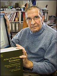 Said Mughabghab合作论文数National Nuclear Data Center, Brookhaven National Laboratory5U. Gruber合作论文数5Ch. Herfarth合作论文数5u heim合作论文数4
 Wojciech Warchoł合作论文数4H Bindewald合作论文数4l wimmer合作论文数4Edward T. Jurney合作论文数4P. T. Prokofjev合作论文数4W. Mampe合作论文数4G VANDENPUT合作论文数4M. Balodis合作论文数4MR MACPHAIL合作论文数4T. D. MacMahon合作论文数4O. Wasson合作论文数4O Chalhoub合作论文数3g b orr合作论文数3G R Massoumi合作论文数3j j tambergs合作论文数3g l struble合作论文数3bernd p k maier合作论文数3g j smith合作论文数3Re Chrien合作论文数3Gw Cole合作论文数3GJ SMITH合作论文数3C Herfarth合作论文数3P Schlag合作论文数3b michaud合作论文数3




