Background: The purpose of this retrospective study was to investigate the efficacy, toxicity and mobilization rate after modified Magrath IVAC (mIVAC) chemotherapy regimen prescribed in relapsed disease (RD) or primary refractory disease (PRD) in aggressive non-Hodgkin lymphoma (NHL). Patients and methods: Twenty-four patients (16 males, 8 females) aged 18-59 years (median age 37 year) were analyzed. The most frequent histopathological subgroup was diffuse large B-cell lymphoma (DLCL-B) (n = 21/24), 13 (54%) were considered RD and 11 (46%) PRD. The mIVAC consisted of ifosfamide (IFM), high dose cytarabine and etoposide repeated every 28 days. Results: The overall response (OR) after three cycles of mlVAC was 66.6%. Among-the patients with PRD, OR was 45.5% (5 out of 11) and with RD was 86.4%, p > 0.05, however, it was observed in RD better complete response (CR) than PRD 53.8 x 9.1% (p < 0.05). Eighty-eight percent (14 Out Of 16) of patients with chemosensitive disease to mIVAC underwent autologous stern cell transplantation (ASCT). The median number of collected CD34+ cells was 2.86 x 10(6) (range 2.17 x 10(6) to 4.9 x 10(6)). The median overall survival rate (OS) for chemosensitive to mIVAC was 16.3 months, with a median follow-up of 16 months. Grades III-IV neutropenia was observed in 85.6% per cycles and grades III-IV thrombocytopenia in 87.5%. Grades III-IV febrile neutropenia was the most common nonhematological toxicity, it occurred in 28% of the cycles and no deaths by toxicity were observed. Discussion: Although a statistic comparative study was not carried out for these 24 patients, the rate of OR to mIVAC was alike the other second-line infusion regimens. The mobilization failure rate was 57.1% and it was similar to other regimens with high dose cytarabine, but it did not limit performed ASCT. (c) 2005 Elsevier Ltd. All rights reserved.
Background: Bone marrow stromall cells(BMC) when implanted into myocardial in animals can undergo mileu-dependent differentiation and express cardiomyogenic phenotypes. We hypothesized that BMC mobilized by granulocyte-colony stimulating factor (GCS-F) would home to the heart with cardiomyopathy and promote repair.
. The prevalence of GB virus C (GBV-C) varies widely throughout the world. A cross-sectional study was conducted in the city of São Paulo, Brazil, to estimate the prevalence of GBV-C infection and to identify associated risk factors, using a large sampling of the general population rather than blood donors or an illness-related group of subjects. GBV-C RNA was detected by reverse-transcriptase polymerase chain reaction using primers directed to the 5′ noncoding region (NCR) and nonstructural 5A region (NS5A) in serum samples from 1,039 healthy individuals 2 years of age or more. Fifty-two individuals were positive for both sets of primers and one was positive for NS5A only (prevalence of GBV-C infection, 5.1%; 95%CI, 3.9–6.7%). No child under 5 years of age was found positive. Among subjects aged 5 years or more, the prevalence of infection increased consistently with age, up to 30–39 years (8.3%), and decreased from then on. The number of sexual partners in the last 3 years (2 or more: OR, 2.6; 95%CI, 1.3–5.5) and history of contact with blood-sucking insects (OR, 2.5; 95%CI 1.2–5.4) were independently associated with GBV-C infection. In conclusion, the prevalence of GBV-C infection is high in São Paulo. In addition to parenteral transmission, another route, e.g. sexual or vertical, may be involved.
Despite the fact Mulattos (individuals resulting from admixture of Caucasian and Black individuals) represent one of the most common racial mixed individuals not only in Brazil but in many other countries, there is little information regarding the distribution of blood groups among them. We studied 2,462 blood donors classified as Caucasian, Mulattos e Blacks according to their anthropological characteristics as well as to their ancestry information. Phenotype frequencies were studied in the ABO, MNS, P, Rh, Lutheran, Kell, Lewis, Duffy e Kidd blood group systems. We did not find significant statistically difference between Blacks and Mulattos for the majority of the blood groups systems here reported, excepting of P1 positive, Dccee, Le(a-b-), Js(a+b+), Js(a-b+), Fy(a-b-), Fy(a+b+) e Fy(a-b+). On the other he there was a significant difference between Caucasian and Blacks for the following red blood cells phenotypes: A, B, M+N-S+s-, M+N-S-s+, P1 positivo, ddccee, Dccee, Dccee, DCCee, DccEe, K+k+, K-k+, Kp(a-b+), Kp(a+b+), Js(a-b+), Js(a+b-), Le(a-b+), Le(a-b-), Fy(a-b+), Fy(a+b+), Fy(a-b-), Jk(a+b-), Jk(a+b+) e Jk(a-b+). Conclusion: As expected, the results for the Mulattos group were intermediate between Caucasian and Blacks, with strong Negroid influence.
Genotypes 1, 2, and 3 of the hepatitis C virus (HCV) are widely distributed throughout Western countries and the Far East ( Japan, China, Taiwan, Thailand). Types 5 and 6 are mainly confined to South Africa and Southeast Asia, respectively, in contrast to type 4, which is predominant in the Middle East and Central Africa.1-3 To investigate the prevalence and distribution of HCV genotypes in Brazil, 348 subjects from four different populations were studied: 34 anti-HCV–positive blood donors, 23 hemophiliacs, 40 renal-transplant recipients, and 251 chronic hepatitis C (CH-C) patients. HCV genotyping was performed in serum samples of 239 (95%) out of 251 CH-C patients and of all blood-donor and hemophiliac samples by a reverse hybridization assay (Line Probe Assay – INNO-LiPA HCV or INNO-LiPA HCV II, second generation, Innogenetics, Ghent, Belgium), in which a reverse transcriptase-polymerase chain reaction (RT-PCR) product of the 58untranslated region (58UTR) is hybridized with probes from various HCV genotypes.2 Twelve samples, of the 251 sera from the CH-C patients, and all from the renal-transplant patients were tested by a serotyping assay (HCV Serotyping Assay 1-6, Murex Diagnostics, Dartford, UK).2 The genotyping results of the 348 HCV isolates, on the basis of the line probe and/or serotyping assays, are shown in Table 1. Four different types were found, and their overall prevalence was 63% for type 1, 4.3% for type 2, 31.3% for type 3, and 0.3% for type 4. Four subjects revealed mixed infections: two cases within types (1a 1 1b in a renaltransplant recipient and 2a 1 2b in a CH-C patient), and two cases across types (1b 1 3a in a blood donor and in a CH-C patient). A similar distribution was observed for each of the four populations, as a general rule; the exceptions were genotypes 2 for the renal-transplant recipients and 3 for the hemophiliacs, whose frequencies were found to be approximately double (13%) and half (15%), respectively, their values in the other populations. Data available from other Brazilian regions, showed corroborating figures for 114 blood donors in Rio de Janeiro, Southeast (1a, 42%; 1b, 34%; and 3a, 20%),2 and for 100 CH-C patients in Porto Alegre, South (type 1, 55%; and type 3, 37%).4 Moreover, as it is known, the current screening assays are based on epitopes derived from only genotypes 1a or 1b, causing variation in seroreactivity among different HCV genotypes.5,6 Therefore, the high prevalence hereby reported not only for genotypes 1a and 1b, but for others (3 in particular) should be of main concern and of much interest for research and development of serological screening assays. Finally, we have identified in a renal-transplant patient an unpublished genotype in Brazil: the 4a.
To better understand the origin of human T-cell leukemia virus type l (HTLV-l) in South America, we conducted a phylogenetic study on 27 new HTLV-ls in Brazil. These were obtained from Brazilians of various ethnic origins, such as Japanese immigrants, whites, blacks and mulattos. We amplified and sequenced proviral DNAs of a part of the long terminal repeats. Phylogenetic trees revealed that all but 6 of the new isolates were not only similar to each other but also similar to HTLV-ls of other South American countries, including those from Amerindians. However, the isolates differed from the HTLV-ls of Africa and Japan. The other six isolates were from Japanese immigrants and were phylogenetically almost identical to HTLV-ls in Japan but different from the majority of South American HTLV-ls, including the other new Brazilian HTLV-ls. These findings indicate that the recent introduction of HTLV-1 from Japan is limited to Japanese immigrants. In addition, the results do not support the prevailing hypothesis that HTLV-ls in South America were introduced by blacks who were brought from Africa as slaves. Rather, these results suggest that the majority of HTLV-1s prevailing in South America have spread from Amerindians, some of whom are likely to have possessed this human retrovirus from the beginning of their settlement in South America.
 F Dulley合作论文数6E. Sabino合作论文数4G Ribeiro-dos-Santos合作论文数4PE Dorlhiac-Llacer合作论文数4Fulvio Pileggi合作论文数4Dorlhiac P合作论文数4
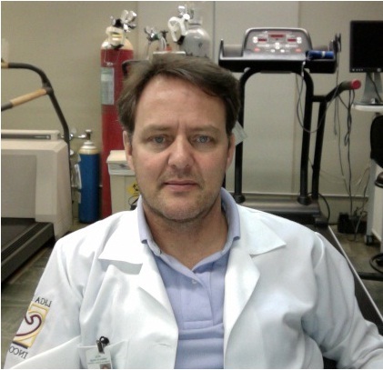 Guilherme Veiga Guimarães合作论文数University of Sao Paulo4Leda Bassit合作论文数4B Beitler合作论文数4M Hayami合作论文数3MC Resende合作论文数3R Medeiros合作论文数3Jf Ramires合作论文数3E Zambon合作论文数3
 Jose A F Ramires合作论文数Medical School, University of São Paulo3
 Sergio Bydlowski合作论文数Laboratory of Genetics and Molecular Hematology (LIM31), University of São Paulo Medical School3
 Fernando Bacal合作论文数. Río de Janeiro. Brasil3Julieta Re Moraes合作论文数3
 Amadeo Sáez-Alquezar合作论文数Programa Nacional de Controle de Qualidade3Ricardo Veronesi合作论文数3
 Giovanni Bellotti合作论文数From the Johns Hopkins Hospital3Mauricio Ostronoff合作论文数3
 Elbio Antonio D Amico合作论文数Hospital Sírio-Libanês Bela Vista;Instituto Central, Hospital das Clínicas, Universidade de São Paulo3E Bocchi合作论文数3S Mizukami合作论文数2TRF Rocha合作论文数2Rps Pugliese合作论文数2C Massumoto合作论文数2Ik Miura合作论文数2Vl Baggio合作论文数2Ra Cardoso合作论文数2Mf Campos合作论文数2Cm Massumoto合作论文数2Araci Massami Sakashita合作论文数2Mcz Novaretti合作论文数2Me Moraes合作论文数2
 Roberto Focaccia合作论文数2Solimene M C合作论文数2Nukui Youko合作论文数Faculty of Medicine, University of São Paulo2Villaça Paula Ribeiro合作论文数2
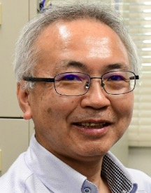 Tomoyuki Miura (三浦智行)合作论文数Institute for Life and Medical Sciences, Kyoto University;Institute for the Advanced Study of Human Biology, Faculty of Medicine Bldg. B, Kyoto University;Graduate School of Human and Environmental Studies, Kyoto University2
 Bruno Caramelli合作论文数São Paulo2M C A Macedo合作论文数2Shunro Sonoda合作论文数2Jaf Ramires合作论文数2Tatsuhiko Igarashi合作论文数2Gilda Porta合作论文数Instituto da Criança;Complexo Hospital das Clínicas da USP;Complexo Hospital das Clínicas da USP, Instituto da Criança2Nanci Salles合作论文数2
 Marcia Cristina Zago Novaretti合作论文数2




