Heritable genetic changes continually arise in cancer, especially in solid tumors where cells are sometimes compressed. Rare heritable losses of chromosomes in live cells are quantified here with chromosome reporters (ChReporters), which reveal losses only after imposing a threshold level of confinement. Compression to ∼60% of interphase height ruptures few nuclei compared to deeper compression but perturbs mitotic spindles and prolongs pro/metaphase. Chromosome mis-segregation into micronuclei is discovered only after release from modest confinement, but arrest and death predominate. All such effects are phenocopied by nocodazole washout, which generates a “memory” of prolonged mitosis. The effects also differ from the rapid induction of micronuclei by a spindle-assembly checkpoint inhibitor and by a clinical CDK4/6 inhibitor of cell-cycle entry. Single-cell RNA sequencing confirms chromosome loss days after confinement and reveals dysregulation of chromosome-segregation pathways. Chromosome losses as mitotic memories of confinement ultimately address knowledge gaps in mechanobiology and cancer evolution.
As with many cell types, macrophages are sometimes filled with micron-sized lipid droplets (LD's), but effects on phagocytosis of other cells, particulates, and microbes remain unclear. Here, we show that LDs restructure the cytoskeleton but remain round, consistent with a high interfacial tension; functionally, LD's impair actomyosin-driven uptake, which proves independent of target size. Engulfment of targets starts at the apical surface, but LD's displace apical actomyosin to the basal cortex. Partial rescue occurs tissue-relevant compressive stresses which activate actomyosin. Macrophages that are densely filled with LD's or pre-engulfed rigid beads likewise activate actomyosin, which again rescues phagocytosis relative to sparsely loaded cells. As further evidence of LD rigidity, both LD's and rigid beads impede macrophage migration through small pores, and LD's pressed into a nucleus cause rapid focal rupture independent of actin. LD rigidity thus disrupts cytoskeleton organization and nucleus integrity, suppressing motility processes unless actomyosin is activated by cell compression or stretching.
Integrating mechanobiological principles into disease pathogenesis, therapeutic development, and tissue engineering is reshaping our understanding of biological systems and accelerating the advancement of mechanotherapy and mechanohealth. This field reveals how mechanical cues regulate cellular behavior, such as force transmission along integrin-nucleus pathways and collective cell migration, and tissue functions. In doing so, mechanobiology connects fundamental research with clinical applications, from limiting cancer metastasis and fibrosis to promoting bone regeneration and maintaining vascular homeostasis. Building on discussion from the Inaugural International Conference on Mechanobiology (ICM) 2025, integrating mechanobiological research with clinical strategies offers new opportunities to address unmet needs, including personalized anti-fibrotic interventions or precision bone regenerative therapies. At the same time, it supports the broader concept of mechanohealth -- a paradigm focused on preserving the physiological mechanical balance within tissues.
Sequencing of many types of tumors shows that solid tumors are far more mutated than liquid tumors and soft tumors such as brain tumors, but it is unclear whether the differences are caused by induction of more mutations or subsequent selective pressures. Our pan-cancer analyses of clinical data shows increased chromosomal changes associate with collagen-I and further shows that genetic variance between patients also increases with the mean for a given tumor type. Using a 3D hydrogel-based model with tunable stiffness and of cancer spheroids, we discover that matrix stiffness increases mitotic aberrations including micronuclei and also increases chromosome loss as detected using live cell chromosome reporters (ChReporters). However, stiffness suppresses growth and size variation, which indicates more mistakes per division. Our approach is >10-fold more sensitive than sequencing approaches, and we quantify colonies of ChReporter-negative cells within cancer spheroids. Our results are consistent with Luria-Delbruck’s theory of heritable genetic changes which predicts inter-spheroid variance greatly exceeds Poisson statistics. Knockdown of myosin-II, a well-known mechanosensor, increases chromosomal loss and variance without affecting spheroid growth, consistent with a tumor suppressor role. Various drug strategies provide deeper insight into mechanisms, and use of clinically deployed CDK4/6-inhibitor reveals this drug on its own drives micronuclei formation and genetic variation. Dennis E. Discher, Markus Sprenger. Solid tumor microenvironments cause more mutations than liquid or soft tumor microenvironments -- even before selection [abstract]. In: Proceedings of the American Association for Cancer Research Annual Meeting 2025; Part 1 (Regular Abstracts); 2025 Apr 25-30; Chicago, IL. Philadelphia (PA): AACR; Cancer Res 2025;85(8_Suppl_1):Abstract nr 2669.
Macrophages often pervade solid tumors, and clusters of macrophages sometimes associate with longer survival of patients. However, clustering mechanisms and impacts on key functions such as phagocytosis remain obscure. Here, under conditions that maximize cancer cell phagocytosis within cohesive tumors, we uncover pathways that favor dynamic clusters and find a colocalization of tumor-intrusive pseudopodia which we term "intrudopodia." Cluster formation over hours on low-adhesion substrates occurs after macrophage induction to a state colloquially referred to as M1 after exposure to interferons and T cell-derived cytokines. Clusters prove fluid on timescales of minutes and also sort from interleukin-4-treated, so-called M2 macrophages that tend to disperse. M1 macrophages upregulate specific cell-cell adhesion receptors but suppress actomyosin contractility, with both pathways contributing to cluster formation. Decreased cortical tension was not only reflected in a low level of nuclear lamin-A that downregulates cytoskeletal targets of serum response factor and tends to soften the nucleus but was also predicted to unleash pseudopodia. Macrophage neighbors in tumor spheroids indeed coextend intrudopodia between cancer cell junctions-at least when phagocytosis conditions are maximized. Intrudopodia from neighbors help detach and individualize cancer cells for rapid engulfment. Juxtaposition of a macrophage cluster with tumor cell nests defines a broad interface that minimizes cancer cell nearest neighbor interactions and maximizes coordination of macrophage intrudopodia. Cooperative phagocytosis thus overcomes solid tumor cohesion-and might explain why the macrophage clustering factor ITGAL associates with patient survival.
Macrophages often pervade solid tumors, but observations that macrophage clusters might associate with patient survival have remained largely unexplored. We observe dynamic macrophage clusters in tumors under conditions that maximize cancer cell phagocytosis, and our reductionist approaches to cluster formation reveal pathways and roles for tumor-intrusive pseudopodia. Aggregates form over hours on low-adhesion substrates after ‘M1’ polarization of macrophages with interferons, including Tcell-derived cytokines, and yet clusters prove fluid on timescales of minutes. Clusters also sort from M2 macrophages which are induced by an interleukin and that disperse on the same substrates. M1’s upregulate cell-cell adhesion receptors but suppress actomyosin contractility, and while both pathways contribute to cluster formation, decreased cortical tension was predicted to unleash pseudopodia. Macrophage neighbors in tumor spheroids indeed extend intrusive pseudopodia or ‘intrudopodia’ in between adjacent cancer cell junctions - at least when phagocytosis conditions are maximized, and coordinated intrudopodia help detach and individualize cancer cells for rapid engulfment. Macrophage clusters thereby provide a cooperative advantage for phagocytosis to overcome solid tumor cohesion. Dennis E. Discher, Lawrence Dooling. Clustered macrophages cooperate to eliminate tumors via coordinated intrudopodia [abstract]. In: Proceedings of the American Association for Cancer Research Annual Meeting 2025; Part 1 (Regular Abstracts); 2025 Apr 25-30; Chicago, IL. Philadelphia (PA): AACR; Cancer Res 2025;85(8_Suppl_1):Abstract nr 7465.
Lipid droplets (LDs) are phase-separated liquids with a poorly understood interfacial tension γ that determines how LDs interact with and influence other organelles. Here, micron-sized LDs remain spherical as they indent peri-nuclear actomyosin and the nucleus, while causing local dilution of the nuclear lamins and sometimes triggering nuclear rupture. Focal accumulation of the cytosolic DNA sensor cGAS at the rupture site is accompanied by sustained mislocalization of DNA repair factors to cytoplasm, increased DNA damage, and delayed cell cycle.
Lipid nanoparticles (LNPs) have emerged as the dominant platform for RNA delivery, based on their success in the COVID-19 vaccines and late-stage clinical studies in other indications. However, we and others have shown that LNPs induce severe inflammation, and massively aggravate pre-existing inflammation. Here, using structure-function screening of lipids and analyses of signaling pathways, we elucidate the mechanisms of LNP-associated inflammation and demonstrate solutions. We show that LNPs' hallmark feature, endosomal escape, which is necessary for RNA expression, also directly triggers inflammation by causing endosomal membrane damage. Large, irreparable, endosomal holes are recognized by cytosolic proteins called galectins, which bind to sugars on the inner endosomal membrane and then regulate downstream inflammation. We find that inhibition of galectins abrogates LNP-associated inflammation, both in vitro and in vivo . We show that rapidly biodegradable ionizable lipids can preferentially create endosomal holes that are smaller in size and reparable by the endosomal sorting complex required for transport (ESCRT) pathway. Ionizable lipids producing such ESCRT-recruiting endosomal holes can produce high expression from cargo mRNA with minimal inflammation. Finally, we show that both routes to non-inflammatory LNPs, either galectin inhibition or ESCRT-recruiting ionizable lipids, are compatible with therapeutic mRNAs that ameliorate inflammation in disease models. LNPs without galectin inhibition or biodegradable ionizable lipids lead to severe exacerbation of inflammation in these models. In summary, endosomal escape induces endosomal membrane damage that can lead to inflammation. However, the inflammation can be controlled by inhibiting galectins (large hole detectors) or by using biodegradable lipids, which create smaller holes that are reparable by the ESCRT pathway. These strategies should lead to generally safer LNPs that can be used to treat inflammatory diseases.
Solid tumors generally exhibit chromosome copy number variation, which is typically caused by chromosomal instability (CIN) in mitosis. The resulting aneuploidy can drive evolution and associates with poor prognosis in various cancer types as well as poor response to T-cell checkpoint blockade in melanoma. Macrophages and the SIRPα-CD47 checkpoint are understudied in such contexts. Here, CIN is induced in poorly immunogenic B16F10 mouse melanoma cells using spindle assembly checkpoint MPS1 inhibitors that generate persistent micronuclei and diverse aneuploidy while skewing macrophages toward a tumoricidal ‘M1-like’ phenotype based on markers and short-term anti-tumor studies. Mice bearing CIN-afflicted tumors with wild-type CD47 levels succumb similar to controls, but long-term survival is maximized by SIRPα blockade on adoptively transferred myeloid cells plus anti-tumor monoclonal IgG. Such cells are the initiating effector cells, and survivors make de novo anti-cancer IgG that not only promote phagocytosis of CD47-null cells but also suppress tumor growth. CIN does not affect the IgG response, but pairing CIN with maximal macrophage anti-cancer activity increases durable cures that possess a vaccination-like response against recurrence.
Polymer network properties such as stiffness often exhibit characteristic power laws in polymer density and other parameters. However, it remains unclear whether diverse animal tissues, composed of many distinct polymers, exhibit such scaling and how cell and molecular mechanisms contribute towards homeostatic differences among tissues. Here, we examined many diverse tissues from adult mouse and embryonic chick to determine if stiffness (Etissue) follows a power law in relation to the most abundant animal protein, collagen-I, even with molecular perturbations.
Macrophages are potential effector cells in immunotherapy against solid tumors, but any phagocytosis requires macrophage-cancer cell interactions to out-compete cohesive interactions between tumor cells (e.g., cadherins). It also requires overcoming a "don't eat me" signal from CD47. We engineered conditionally immortalized macrophages (CIMs) with deletion of the CD47-binding inhibitory receptor SIRPα, and we first show such SIRPαKO-CIMs readily engulf added suspensions of IgG-opsonized melanoma cells in a standard 2D assay.
Abstract Solid tumors generally exhibit chromosome copy number variation caused by chromosomal instability (CIN) in mitosis, and the resulting aneuploidy associates with poor prognosis in various cancer types and poor Tcell checkpoint blockade response in melanoma. In such contexts, however, macrophages and the SIRPa-CD47 checkpoint are understudied. Here, CIN is induced pharmacologically in poorly immunogenic B16F10 mouse melanoma cells, generating micronuclei plus diverse aneuploidy and skewing macrophages towards an anti-cancer phenotype based on markers and short-term tumor studies. Mice bearing CIN-afflicted tumors with wild-type CD47 levels survive only slightly longer compared to chromosomally stable controls, but long-term survival can be maximized when anti-tumor IgG opsonization is combined with adoptive transfer of macrophages with SIRPa blockade or with CD47 knockout of the B16F10. Multi-epitope, de novo anti-cancer IgG in survivors promote phagocytosis of CD47 knockout B16F10 cells by macrophages and suppress tumoroids in vitro and growth of tumors in vivo. An unexpected benefit of pairing CIN with maximal macrophage anti-cancer activity is thus an anti-cancer vaccination-like response that can lead to durable cures and potentiate cell-mediated acquired immunity. Citation Format: Dennis E. Discher. Chromosomal instability leads to durable tumor suppression upon macrophage-checkpoint disruption, with induction of anti-tumor IgG [abstract]. In: Proceedings of the American Association for Cancer Research Annual Meeting 2024; Part 1 (Regular Abstracts); 2024 Apr 5-10; San Diego, CA. Philadelphia (PA): AACR; Cancer Res 2024;84(6_Suppl):Abstract nr 417.
Cancer progression is highly associated with chromosome instability arising from mitotic errors, but the underlying mechanisms are difficult to explain simply by mutations in oncogenes or tumor suppressor genes. Physical constraints, such as extracellular constraints by a stiff tumor microenvironment, can also cause mitotic errors independent of genetic defects. Hepatic cells provide a natural system to study the impacts of physical constraints on mitosis because they deposit intracellular lipid droplets and extracellular matrices during cancer progression.
Macrophages often pervade solid tumors, but their nearest neighbor organization is understudied and potentially enables key functions such as phagocytosis. Here, we observe dynamic macrophage clusters in tumors under conditions that maximize cancer cell phagocytosis and use reductionist approaches to uncover pathways to cluster formation and roles for tumor-intrusive pseudopodia, which we term 'intrudopodia'. Macrophage clusters form over hours on low- adhesion substrates after M1 polarization with interferons, including T cell-derived cytokines, and yet clusters prove fluid on timescales of minutes. Clusters also sort from M2 macrophages that disperse on the same substrates. M1 macrophages upregulate specific cell-cell adhesion receptors but suppress actomyosin contractility, and while both pathways contribute to cluster formation, decreased cortical tension was predicted to unleash pseudopodia. Macrophage neighbors in tumor spheroids indeed extend intrudopodia between adjacent cancer cell junctions - at least when phagocytosis conditions are maximized, and coordinated intrudopodia help detach and individualize cancer cells for rapid engulfment. Macrophage clusters thereby provide a cooperative advantage for phagocytosis to overcome solid tumor cohesion.
Differential adhesion and contractility drive cell sorting in diverse systems such as embryonic germ layer cells and cancer cell mixtures, but similar behavior among immune cells is underexplored despite major alterations in adhesive protein repertoires and cytoskeleton when activated. We observed a strong tendency of macrophages to aggregate and segregate from melanoma cells in cohesive tumoroids, but only under pro-phagocytic conditions in which macrophage Fc receptors were activated by IgG opsonization of the melanoma cells and the "don't eat me" CD47-SIPRα checkpoint was inhibited.
Extremely soft tissues such as developing hearts or adult brain contain far less collagen than highly stiff adult tissues such as tendons, but cell and molecular mechanisms for such homeostatic differences remain unclear. We hypothesized that cell-generated or exogenous forces combine with tension-suppressed collagen degradation in order to sculpt extracellular matrix (ECM) collagen levels in tissues. For various mature mice tissues and beating embryonic chick hearts, we find collagen-sensitive second harmonic generation (SHG) image intensity scales non-linearly versus tissue stiffness, aligning well with the results from cellularized gels of collagen. Chick hearts beating at ∼5% strain maintain collagen levels until their contractile strain is suppressed by myosin-II inhibition and endogenous matrix metalloproteinases (MMPs) then degrade collagens within ∼30-60 minutes - based on SHG and mass spectrometry proteomics. Although tendons composed of oriented collagen fibrils exhibit heterogeneous strain distributions upon deformation, the addition of exogenous MMP or bacterial collagenase suppresses collagen degradation for strains within physiological limits (i.e., up to ∼5-8%). Sequestration of collagen cleavage sites by tissue strain is a likely mechanism because molecular permeation and mobility prove strain-independent whereas artificial collagen cross-links accelerate strain-dependent collagen degradation via collagen molecular unfolding. Tension-suppressed degradation of collagen thus underlies tissue stiffness scaling.
Polymer network properties such as stiffness often exhibit characteristic power laws in polymer density and other parameters. However, it remains unclear whether diverse animal tissues, composed of many distinct polymers, exhibit such scaling. Here, we examined many diverse tissues from adult mouse and embryonic chick to determine if stiffness ( E tissue ) follows a power law in relation to the most abundant animal protein, Collagen-I, even with molecular perturbations. We quantified fibrillar collagen in intact tissue by second harmonic generation (SHG) imaging and from tissue extracts by mass spectrometry (MS), and collagenase-mediated decreases were also tracked. Pan-tissue power laws for tissue stiffness versus Collagen-I levels measured by SHG or MS exhibit sub-linear scaling that aligns with results from cellularized gels of Collagen-I but not acellular gels. Inhibition of cellular myosin-II based contraction fits the scaling, and combination with inhibitors of matrix metalloproteinases (MMPs) show collagenase activity is strain - not stress- suppressed in tissues, consistent with past studies of gels and fibrils. Beating embryonic hearts and tendons, which differ in both collagen levels and stiffness by >1000-fold, similarly suppressed collagenases at physiological strains of ∼5%, with fiber-orientation regulating degradation. Scaling of E tissue based on 'use-it-or-lose-it' kinetics provides insight into scaling of organ size, microgravity effects, and regeneration processes while suggesting contractility-driven therapeutics.
Phagocytic elimination of solid tumors by innate immune cells seems attractive for immunotherapy, particularly because of the possibilities for acquired immunity. However, the approach remains challenging, with blockade of the macrophage checkpoint CD47 working in immunodeficient mice and against highly immunogenic tumors but not in the clinic where tumors are poorly immunogenic. Even when mouse tumors of poorly immunogenic B16F10 melanoma are opsonized to drive engulfment with a suitable monoclonal antibody (mAb), anti-CD47 blockade remains insufficient. Using both in vitro immuno-tumoroids and in vivo mouse models, we show with CRISPR interference (CRISPRi) that a relatively uniform minimum repression of CD47 by 80% is needed for phagocytosis to dominate net growth when combined with an otherwise ineffective mAb (anti-Tyrp1). Heterogeneity enriches for CD47-high cells, but mice that eliminate tumors generate prophagocytic IgGs that increase in titer with CD47 repression and with tumor accumulation of macrophages, although deeper repression does not improve survival. Given well-known limitations of antibody permeation into solid tumors, our studies clarify benchmarks for CD47 disruption that should be more clinically feasible and safer but just as effective as complete ablation. Additionally, safe but ineffective opsonization in human melanoma trials suggests that combinations with deep repression of CD47 could prove effective and initiate durable immunity.
The nucleus in many cell types is a stiff organelle, and yet fat-filled lipid droplets (FD's) in the cytoplasm can indent and displace the nucleus. FD's are phase-separated liquids with a poorly understood interfacial tension that determines how FD's interact with other organelles. Here, micron-sized FD's remain spherical while deforming both the nuclear lamina and peri-nuclear actomyosin. Nuclear lamins are intermediate filaments with a persistence length in the range of ∼0.2-1.5 μm with Lamin-B1 directly attached to the inner nuclear membrane via farnesylation groups that are lacking in mature lamin-A,C. Such rigidity can compromise the attachment of Lamin-B1 to the membrane in cases of high Gaussian curvature as seen through local dilution of Lamin-B1 at sites of indentation which is independent of Lamin-A,C. A single filament model of curvature-driven membrane detachment fits to Lamin-B1's relative density with the association energy being the key parameter to fit the experimental data. Lamin-B1 depletion triggers rupture as indicated by persistent, local accumulation of cytosolic DNA sensor cGAS at the nuclear boundary. FD-nucleus interactions also initiate rapid mis-localization of the DNA repair factor KU80, confirming nuclear rupture, and associate with heightened DNA damage and perturbed cell cycle. Similar results are evident in FD-laden cells after constricted 3D-migration, which is impeded by FD's. Spherical shapes of small FD's are consistent with a high interfacial tension that we measure for FD's mechanically isolated from fresh adipose tissue as ∼40 mN/m - which is far higher than other liquid condensates, but typical of oils in water and sufficiently rigid to disrupt cell structures.
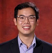 Irianto Jerome合作论文数Univ Penn, Philadelphia, PA 19104 USA63
 Joe Swift合作论文数Division of Cell Matrix Biology and Regenerative Medicine, School of Biological Sciences, Faculty of Biology, Medicine and Health, The University of Manchester56
 Irena Ivanovska合作论文数55
 Charlotte R. Pfeifer合作论文数Mol & Cell Biophys Lab, Univ Penn47
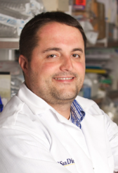 Adam J. Engler合作论文数Shu Chien-Gene Lay Department of Bioengineering, Jacobs School of Engineering, University of California, San Diego;Glycobiology Research and Training Center, University of California, San Diego;Moores Cancer Center, University of California, San Diego41
 Yuntao Xia合作论文数Physical Science Oncology Center, Penn40
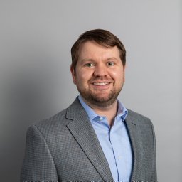 Lawrence J Dooling合作论文数University of Pennsylvania37Paul Dalhaimer合作论文数33
 Frank Bates合作论文数Department of Chemical Engineering and Materials Science, College of Science and Engineering, University of Minnesota32
 Florian Rehfeldt合作论文数University of Göttingen32
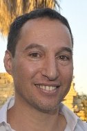 Amnon Buxboim合作论文数Graduate Group of Physics & Astronomy and Biophysical Engineering Lab, University of Pennsylvania30
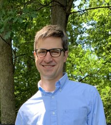 David A. Christian合作论文数Molecular Biology of Disease Resistance Laboratory, Washington State University29
 Sangkyun Cho合作论文数Cardiovascular Institute, School of Medicine, Stanford University28
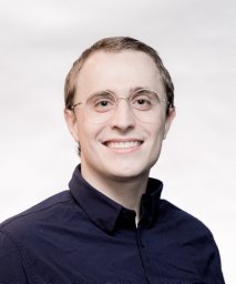 Jason C Andrechak合作论文数Univ Penn, Biophys Engn Labs, Philadelphia, PA 19104 USA27
 Michael L. Klein合作论文数Department of Chemistry, College of Science and Technology, Temple University;Institute for Computational Molecular Science, College of Science and Technology, Temple University27Kyle R. Spinler合作论文数27
 David W. Speicher合作论文数The Speicher laboratory, The Wistar Institute;Biochemistry and Molecular Biophysics Graduate Group, University of Pennsylvania25Takamasa Harada合作论文数25J-W. Shin合作论文数24André E. X. Brown合作论文数23Manu Tewari合作论文数23
 Brandon Hastings Hayes合作论文数22Cory Alvey合作论文数22Michael P Tobin合作论文数22
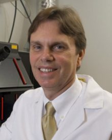 Lee Sweeney合作论文数Department of Pharmacology & Therapeutics, College of Medicine, University of Florida22
 Manasvita Vashisth合作论文数Molecular & Cell Biophysics Lab, University of Pennsylvania, Philadelphia, PA 19104, USA21Richard K. Tsai合作论文数21
 Dingal P C Dave P合作论文数20
 Andrea Liu合作论文数Department of Physics & Astronomy, College of Arts & Sciences, University of Pennsylvania20Matthew Raab合作论文数Department of Bioengineering;Pennsylvania State University;Department of Bioengineering, Pennsylvania State University18
 Kris Dahl合作论文数Thornton Tomasetti18Shenshen Cai合作论文数18
 Roger A. Greenberg合作论文数Department of Cancer Biology, Perelman School of Medicine, University of Pennsylvania17Harry Bermudez合作论文数Department of Polymer Science and Engineering, University of Massachusetts Amherst17
 Bohdana M Discher合作论文数The Johnson Research Foundation and the Department of Biochemistry and Biophysics, the University of Pennsylvania17
 Sharon M. Loverde合作论文数17
 Daniel Hammer合作论文数Department of Chemical and Biomolecular Engineering, School of Engineering and Applied Science, University of Pennsylvania16
 Stephanie Majkut合作论文数Biophysical Engineering Laboratory, University of Pennsylvania;Physics and Astronomy Graduate Group, University of Pennsylvania;University of Pennsylvania, University of Pennsylvania16
 Narla Mohandas合作论文数Laboratory of Red Cell Physiology, New York Blood Center Enterprises15
 Jae-Won Shin合作论文数Molecular &Cell Biophysics Laboratory and Graduate Group in Pharmacological Science, University of Pennsylvania;Molecular & Cell Biophysics Laboratory and Graduate Group in Pharmacological Science, University of Pennsylvania15J. David Pajerowski合作论文数15
 Peter J. Photos合作论文数School of Engineering and Applied Science, University of Pennsylvania15Nishant Bhasin合作论文数Department of Chemical and Biomolecular Engineering;Uni Versity of PennsylVania14K. Rajagopal合作论文数14
 Shamik Sen合作论文数Department of Bioengineering, University of California14Mai Wang合作论文数13Nair Praful合作论文数13Manorama Tewari合作论文数13Jcm Lee合作论文数12




