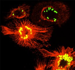
The Sec61 complex sits between the distinct environments of the cytosol and the ER lumen and it's appropriate gating is essential to prevent the deleterious flux of molecules and ions between them. Using the sss1-7 mutant we show that actin dynamics is grossly perturbed when translocon gating is defective. Importantly, normal actin morphology is restored when sss1-7 translocon gating defects are suppressed or when these cells are treated with cell-permeable Ca2+ chelators. Our findings underscore the importance of translocon gating, particularly in regulating Ca2+ homeostasis, in the overall regulation and functional distribution of the actin cytoskeleton.
Fetal skin has an intrinsic regenerative capability to restore an injured site’s architecture and functionality. This is preserved until the third trimester, when wound healing transitions to a scarring reparative response. This change coincides with the dynamic remodelling of dermal extracellular matrix (ECM). Here, we used primary human fetal or adult dermal fibroblast (fHDF and aHDF)-derived ECMs to demonstrate that different extrinsic signals from these ECMs dramatically altered gene expression in a primary human keratinocyte population grown on these matrices. Gene array data revealed keratinocytes grown on fHDF ECM markedly upregulated expression of cell-cycle genes, whereas on aHDF ECM expression of differentiation genes was favoured. Detailed proteomic analyses indicated compositionally distinct ECMs were deposited by aHDFs and fHDFs. Moreover, aHDFs and fHDFs contained subpopulation(s) that differentially expressed CD90, CD146 and CD26. On fHDFs the extracellular domain of CD26 was shed whereas on aHDFs full-length CD26 dominated. The proteomic and gene array data supported the fine-tuning of BMP/TGFβ/SMAD signalling pathways being a mechanism by which fetal matrices promote keratinocyte self-renewal. Collectively, these findings revealed that a fundamental aspect of skin development is dictated by the ECM of the dermis, specifically extrinsic signals from dermal fibroblast ECM direct keratinocyte self-renewal or differentiation.
Pancreatic ductal adenocarcinoma (PDAC) is a type of cancer alarmingly expanding in our modern societies that is still proving to be very challenging to counteract. This disease constitutes a quintessential example of the multiple interactions existing between the tumour and its surrounding microenvironment. In particular, PDAC is characterized by a very immunosuppressive environment that favours cancer growth and makes this cancer type very resistant to immunotherapy. The primary tumour releases many factors that influence both the microenvironment and the immune landscape. Extracellular vesicles (EVs), recently identified as indispensable entities ensuring cell-to-cell communication in both physiological and pathological processes, seem to play a pivotal function in ensuring the delivery of these factors to the tumour-surrounding tissues. In this review, we summarize the present understanding on the crosstalk among tumour cells and the cellular immune microenvironment emphasizing the pro-malignant role played by extracellular vesicles. We also discuss how a greater knowledge of the roles of EVs in tumour immune escape could be translated into clinical applications.
This study investigates the nanostructural mechanisms that lie behind load transmission in tendons and the role of glycosaminoglycans (GAGs) in the transmission of force in the tendon extracellular matrix. The GAGs in white New Zealand rabbit Achilles tendons were enzymatically depleted, and the tendons subjected to cyclic loading at 6% strain for up to 2 hr. A nanoscale morphometric assessment of fibril deformation under strain was linked with the decline in the tendon macroscale mechanical properties. An atomic force microscope (AFM) was employed to characterize the D-periodicity within and between fibril bundles (WFB and BFB, respectively). By the end of the second hour of the applied strain, the WFB and BFB D-periodicities had significantly increased in the GAG-depleted group (29% increase compared with 15% for the control, p < .0001). No statistically significant differences were found between WFB and BFB D-periodicities in either the control or GAG-depleted groups, suggesting that mechanical load in Achilles tendons is uniformly distributed and fairly homogenous among the WFB and BFB networks. The results of this study have provided evidence of a cycle-dependent mechanism of damage accumulation. The accurate quantification of fibril elongation (measured as the WFB and BFB D-periodicity lengths) in response to macroscopic applied strain has assisted in assessing the complex structure-function relationship in Achilles tendon.
Tendinopathy of the Achilles tendon contributes to a large range of disorders, including mechanical damage and degenerative diseases. Glycosaminoglycans (GAGs), are thought to play a role in the mechanical strength of tendons by forming cross-links between collagen molecules and allowing the transmission of forces between fibrils. This study assessed the response of GAG-depleted tendons to damage induced by fatigue loading, investigating the mechanical damage (stiffness, hysteresis and maximum load), macrostructural changes (tenocyte morphology, fiber anisotropy and waviness) assessed by confocal imaging and nanostructural changes (fibril D-periodicity length) within the same non-viable intact tendons. Changes in fiber waviness and tenocyte shape are strongly correlated to mechanical and nano-structural (D-periodicity elongation) properties in both Control and GAG-depleted tendons. This study supports firstly, the vital role GAGs play as mechanical connectors facilitating the load transfer between the fibrils and their hydrophilic role in facilitating fibril sliding. Secondly, that observed changes in tenocyte shape and fiber waviness correlate with tendon stiffness and other mechanical profiles.
Older age is associated with deteriorating health, including escalating risk of diseases such as cancer, and a diminished ability to repair following injury. This rise in age-related diseases/co-morbidities is associated with changes to immune function, including in myeloid cells, and is related to immunosenescence. Immunosenescence reflects age-related changes associated with immune dysfunction and is accompanied by low-grade chronic inflammation or inflammageing. This is characterised by increased levels of circulating pro-inflammatory cytokines such as tumor necrosis factor (TNF), interleukin (IL)-1β and IL-6. However, in healthy ageing, there is a concomitant age-related escalation in anti-inflammatory cytokines such as transforming growth factor-β1 (TGF-β1) and IL-10, which may overcompensate to regulate the pro-inflammatory state. Key inflammatory cells, macrophages, play a role in cancer development and injury repair in young hosts, and we propose that their role in ageing in these scenarios may be more profound. Imbalanced pro- and anti-inflammatory factors during ageing may also have a significant influence on macrophage function and further impact the severity of age-related diseases in which macrophages are known to play a key role. In this brief review we summarise studies describing changes to inflammatory function of macrophages (from various tissues and across sexes) during healthy ageing. We also describe age-related diseases/co-morbidities where macrophages are known to play a key role, focussed on injury repair processes and cancer, plus comment briefly on strategies to correct for these age-related changes.
Background: Extensive research has reported that extracellular ADP in the tumour microenvironment can stimulate platelets through interaction with the platelet receptor P2Y12. In turn, activated platelets release biological factors supporting cancer progression. Experimental data suggest that the tumour microenvironment components, of which platelets are integral, can promote chemotherapy resistance in pancreatic ductal adenocarcinoma (PDAC). Thus, overcoming chemoresistance requires combining multiple inhibitors that simultaneously target intrinsic pathways in cancer cells and extrinsic factors related to the tumour microenvironment. We aimed to determine whether ticagrelor, an inhibitor of the ADP–P2Y12 axis and a well-known antiplatelet drug, could be a therapeutic option for PDAC. Methods: We investigated a functional P2Y12 receptor and its downstream signalling in a panel of PDAC cell lines and non-cancer pancreatic cells termed hTERT-HPNE. We tested the synergistic effect of ticagrelor, a P2Y12 inhibitor, in combination with chemotherapeutic drugs (gemcitabine, paclitaxel and cisplatin), in vitro and in vivo. Results: Knockdown studies revealed that P2Y12 contributed to epidermal growth factor receptor (EGFR) activation and the expression of SLUG and ZEB1, which are transcriptional factors implicated in metastasis and chemoresistance. Studies using genetic and pharmacological inhibitors showed that the P2Y12–EGFR crosstalk enhanced cancer cell proliferation. Inhibition of P2Y12 signalling significantly reduced EGF-dependent AKT activation and promoted the anticancer activity of anti-EGFR treatment. Importantly, ticagrelor significantly decreased the proliferative capacity of cancer but not normal pancreatic cells. In vitro, synergism was observed when ticagrelor was combined with several chemodrugs. In vivo, a combination of ticagrelor with gemcitabine significantly reduced tumour growth, whereas gemcitabine or ticagrelor alone had a minimal effect. Conclusions: These findings uncover a novel effect and mechanism of action of the antiplatelet drug ticagrelor in PDAC cells and suggest a multi-functional role for ADP-P2Y12 signalling in the tumour microenvironment.
The long-term expansion of keratinocytes under conditions that avoid xenogeneic components (i.e. animal serum- and feeder cell-free) generally causes diminished proliferation and increased terminal differentiation. Here we present a culture system free of xenogeneic components that retains the self-renewal capacity of primary human keratinocytes. In vivo the extracellular matrix (ECM) of the tissue microenvironment has a major influence on a cell's fate. We used ECM from human dermal fibroblasts, cultured under macromolecular crowding conditions to facilitate matrix deposition and organisation, in a xenogeneic-free keratinocyte expansion protocol. Phospholipase A2 decellularisation produced ECM whose components resembled the core matrix composition of natural dermis by proteome analyses. Keratinocytes proliferated rapidly on these matrices, retained their small size, expressed p63, lacked keratin 10 and rarely expressed keratin 16. The colony forming efficiency of these keratinocytes was enhanced over that of keratinocytes grown on collagen I, indicating that dermal fibroblast-derived matrices maintain the in vitro expansion of keratinocytes in a stem-like state. Keratinocyte sheets formed on such matrices were multi-layered with superior strength and stability compared to the single-layered sheets formed on collagen I. Thus, keratinocytes expanded using our xenogeneic-free protocol retained a stem-like state, but when triggered by confluence and calcium concentration, they stratified to produce epidermal sheets with a potential clinical use.
Increasing life expectancy is associated with increased cancer incidence, yet the effect of cancer and anti-cancer treatment on elderly patients and their immune systems is not well understood. Declining T cell function with aging in response to infection and vaccination is well documented, however little is known about aged T cell responses to tumor antigens during cancer progression or how these responses are modulated by standard chemotherapy. We examined T cell responses to cancer in aged mice using AE17sOVA mesothelioma in which ovalbumin (OVA) becomes a spy' tumor antigen containing one dominant (SIINFEKL) and two subdominant (KVVRFDKL and NAIVFKGL) epitopes. Faster progressing tumors in elderly (22-24months, cf. 60-70 human years) relative to young (2-3months, human 15-18years) mice were associated with increased pro-inflammatory cytokines and worsened cancer cachexia. Pentamer staining and an in-vivo cytotoxic T lymphocyte (CTL) assay showed that whilst elderly mice generated a greater number of CD8(+) T cells recognizing all epitopes, they exhibited a profound loss of function in their ability to lyse targets expressing the dominant, but not subdominant, epitopes compared to young mice. Chemotherapy was less effective and more toxic in elderly mice however, similar to young mice, chemotherapy expanded CTLs recognizing at least one subdominant epitope in tumors and draining lymph nodes, yet treatment efficacy still required CD8(+) T cells. Given the significant dysfunction associated with elderly CTLs recognizing dominant epitopes, our data suggest that responses to subdominant tumor epitopes may become important when elderly hosts with cancer are treated with chemotherapy.
The ability to grow C2C12 myoblasts in a completely defined, serum free medium enables researchers to investigate the role of specific factors in myoblast proliferation, migration, fusion, and differentiation without the confounding effects of serum. The use of defined, animal free in vitro culture systems will improve reproducibility between research groups and may also enhance translation of tissue engineering techniques into clinical applications. Here, we describe the use and characterization of a serum free culture system for C2C12 myoblasts using standard tissue culture medium and readily available, defined growth factors and supplements.
Most cancers emerge in the elderly, including lung cancer and mesothelioma, yet the elderly remain an underrepresented population in pre-clinical cancer studies and clinical trials. The immune system plays a critical role in the effectiveness of many anticancer therapies in young hosts via tumor-specific T cells. However, immunosuppressive macrophages can constitute up to 50% of the tumor burden and impair anti-tumor T cell activity. Altered macrophage phenotype and function during aging may further impact anti-tumor T cell responses. Yet, the impact of macrophages on anti-tumor T cell responses and immunotherapy in the elderly is unknown. Therefore, we examined macrophages and their interaction with T cells in young (3 months) and elderly (20-24 months) AE17 mesothelioma-bearing female C57BL/6J mice during tumor growth. Mesothelioma tumors grew faster in elderly compared with young mice, and this corresponded with an increase in tumor-associated macrophages. During healthy aging, macrophages increase in bone marrow and spleens suggesting that these sites have an increased potential to supply cancer-promoting macrophages. Interestingly, in tumor-bearing mice, bone marrow macrophages increased proliferation whilst splenic macrophages had reduced proliferation in elderly compared with young mice, and macrophage depletion using the F4/80 antibody slowed tumor growth in young and elderly mice. We also examined responses to treatment with intra-tumoral IL-2/anti-CD40 antibody immunotherapy and found it was less effective in elderly (38% tumor regression) compared to young mice (90% regression). Tumor-bearing elderly mice decreased in vivo anti-tumor cytotoxic T cell activity in tumor draining lymph nodes and spleens. Depletion of macrophages using F4/80 antibody in elderly, but not young mice, improved IL-2/anti-CD40 immunotherapy up to 78% tumor regression. Macrophage depletion also increased in vivo anti-tumor T cell activity in elderly, but not young mice. All the tumor-bearing elderly (but not young) mice had decreased body weight (i.e., exhibited cachexia), which was greatly exacerbated by immunotherapy; whereas macrophage depletion prevented this immunotherapy-induced cachexia. These studies strongly indicate that age-related changes in macrophages play a key role in driving cancer cachexia in the elderly, particularly during immunotherapy, and sabotage elderly anti-tumor immune responses.
The average age of the human population is rising, leading to an increasing burden of age-related diseases, including increased susceptibility to infection. However, immune function can decrease with age which could impact on processes that require a functional immune system. Aging is also characterized by chronic low-grade inflammation which could further impact immune cell function. While changes to neutrophils in blood during aging have been described, little is known in aging lymphoid organs. This study used female C57BL/6J mice comparing bone marrow (BM), spleen and lymph nodes from young mice aged 2–3 months (equivalent to 18 human years) with healthy elderly mice aged 22–24 months (equivalent to 60–70 human years). Neutrophil proportions increased in BM and secondary lymphoid organs of elderly mice relative to their younger counterparts and presented an atypical phenotype. Interestingly, neutrophils from elderly spleen and lymph nodes were long lived (with decreased apoptosis via Annexin V staining and increased proportion of BrdU neg mature cells) with splenic neutrophils also demonstrating a hypersegmented morphology. Furthermore, splenic neutrophils of elderly mice expressed a mixed phenotype with increased expression of activation markers, CD11b and ICAM-1, increased proinflammatory TNFα, yet increased anti-inflammatory transforming growth factor-beta. Elderly splenic architecture was compromised, as the marginal zone (required for clearing infections) was contracted. Moreover, neutrophils from elderly but not young mice accumulated in lymph node and splenic T- and B-cell zones. Overall, the expansion of functionally compromised neutrophils could contribute to increased susceptibility to infection observed in the elderly.
This article has been withdrawn by the authors as part of this review overlapped with the contents of Pietrangelo A and Ridgway ND. 2018. Cellular and Molecular Life Sciences. 75; 3079-98.
The average age of the human population is rising, leading to an increasing burden of age‐related diseases, including increased susceptibility to infection. However, immune function can decrease with age which could impact on processes that require a functional immune system. Aging is also characterized by chronic low‐grade inflammation which could further impact immune cell function. While changes to neutrophils in blood during aging have been described, little is known in aging lymphoid organs. This study used female C57BL/6J mice comparing bone marrow (BM), spleen and lymph nodes from young mice aged 2–3 months (equivalent to 18 human years) with healthy elderly mice aged 22–24 months (equivalent to 60–70 human years). Neutrophil proportions increased in BM and secondary lymphoid organs of elderly mice relative to their younger counterparts and presented an atypical phenotype. Interestingly, neutrophils from elderly spleen and lymph nodes were long lived (with decreased apoptosis via Annexin V staining and increased proportion of BrdU neg mature cells) with splenic neutrophils also demonstrating a hypersegmented morphology. Furthermore, splenic neutrophils of elderly mice expressed a mixed phenotype with increased expression of activation markers, CD11b and ICAM‐1, increased proinflammatory TNFα, yet increased anti‐inflammatory transforming growth factor‐beta. Elderly splenic architecture was compromised, as the marginal zone (required for clearing infections) was contracted. Moreover, neutrophils from elderly but not young mice accumulated in lymph node and splenic T‐ and B‐cell zones. Overall, the expansion of functionally compromised neutrophils could contribute to increased susceptibility to infection observed in the elderly.
 Deirdre R. Coombe合作论文数Department of Zoology and Department of Microbiology and Immunology, The University of Adelaide14
 Miranda D Grounds合作论文数School of Human Sciences, University of Western Australia9Delia J. Nelson合作论文数9
 Connie Jackaman合作论文数School of Biomedical Sciences;Curtin University;School of Biomedical Sciences, Curtin University9Kinnear Beverley F合作论文数6Pat Metharom合作论文数6Radley-Crabb Hannah G合作论文数5
 Vishal Chaturvedi合作论文数Centre for Cellular and Molecular Biology, Hyderabad, India.5
 Nigel Laing合作论文数UWA Centre for Medical Research, Harry Perkins Institute of Medical Research;Medical School, The University of Western Australia5
 Helen Leonard合作论文数Telethon Kids Institute;Child Disability Group, Centre for Child Health Research, University of Western Australia;Disability Services, Western Australia5Peter O'Leary合作论文数Div Obstet & Gynaecol, Univ Western Australia4S Fyfe合作论文数4Emma Glasson合作论文数4Jack Goldblatt合作论文数4
 Federica Tomay合作论文数Dipartimento di Scienze e Tecnologie Biomediche, Università di Udine4P Yanakiev合作论文数3
 Lelinh Duong合作论文数Curtin Health Innovation Research Institute, Curtin University3S Leonard合作论文数3
 Frank Baas合作论文数Graviton Bioscience3Maxwell Susannah J合作论文数Office of Population Health Genomics, Department of Health, Perth, Western Australia, Australia.3
 Marco Falasca合作论文数Curtin Medical School, Faculty of Health Sciences, Curtin University3Gardner Joanne K合作论文数3
 Kathryn North合作论文数Murdoch Children's Research Institute;Division of Medicine, Dentistry and Health Sciences, The University of Melbourne3Kate J. Brameld合作论文数3Luba Kalaydjieva合作论文数3D Angelicheva合作论文数2Norbaini Binti Abdol Razak合作论文数2Elizabeth Grenik合作论文数2Anas K Al Makhzoomi合作论文数2Fiona Pixley合作论文数2
 Garry Allison合作论文数GP Down South;Research Office, Graduate Research School, Curtin University2Shavlakadze Tea合作论文数2
 Naskar Deboki合作论文数Dept Biotechnol, Indian Inst Technol2Wells Kelsi合作论文数2
 Kirk Thomas Brett合作论文数2Wong Chee-Wai合作论文数2
 Subhas C. Kundu合作论文数3Bs Research Group, I3bs-Research Institute on Biomaterials, Biodegradables, biomimetics, AvePark, University of Minho2P. K. Thomas合作论文数2Nigel F. Clarke合作论文数2
 Alan Bittles合作论文数Centre for Comparative Genomics, Murdoch University;School of Medical and Health Sciences, Edith Cowan University2
 Simon A. Fox合作论文数University Department of Medicine and Department of Gastroenterology, Sir Charles Gairdner Hospital2John C. Sparrow合作论文数2Nicole Monnier合作论文数2
 Alexey Savov合作论文数2Andrew Grierson合作论文数Department of Neuroscience, The Medical School, Faculty of Medicine, Dentistry & Health, The University of Sheffield2
 Carl J. Mousley合作论文数School of Medicine, University of North Carolina at Chapel Hill2Ivailo Tournev合作论文数Medical University2B. Keats合作论文数1I ZORN合作论文数1




