Previous work described 3 cases of CD4+ T-cell lymphomas harboring Merkel cell polyomavirus (MCPyV), as demonstrated by in situ hybridization (ISH), next generation sequencing, and polymerase chain reaction. Two of these cases were cutaneous T-cell lymphomas compatible with mycosis fungoides in postcardiac transplant patients, while the third was a peripheral T-cell lymphoma, not otherwise specified (PTCL-NOS), in a patient without known immunosuppression. In this study, we assess an expanded cohort of T-cell neoplasms from 4 institutions for expression of MCPyV by ISH. This cohort includes 9 T-cell lymphomas occurring in the post solid organ transplant setting, of which 5 (60%) were positive for MCPyV by ISH. Four of these cases were classified as PTCL-NOS and the remaining case as nodal T follicular helper cell lymphoma, angioimmunoblastic type. In addition, we performed MCPyV ISH on a variety of neoplastic and non-neoplastic lymphoid tissues from both transplant and nontransplant patients. No evidence of MCPyV infection was found in lymphoid tissues in the absence of T-cell lymphoma, suggesting that the presence of MCPyV is specific to this rare subset of T cell lymphomas. Furthermore, all MCPyV-positive T-cell lymphomas (8 of 8) were CD4+ and the large majority were observed in the post-transplant setting (7 of 8). Multiplex single-molecule fluorescence ISH confirmed the presence of MCPyV in CD4+ T-cells. These findings support the hypothesis that MCPyV infection is specifically associated with rare CD4+ T-cell lymphomas, particularly in transplant recipients.
Digital Pathology (DP) is a fast-emerging branch of pathology focused on digitizing pathology data. A key challenge of DP usage for pathology laboratories, especially mid- to small-sized clinical labs, are the upfront costs associated with instrumentation and the logistical challenges of implementation. In the current project, we built an end-to-end DP solution using low-cost, open-source components that is user-friendly at a small scale. We repurposed readily available microscopy components in a pathology lab to assemble a fully functional DP pipeline for translational research applications. We tested multiple low-cost complementary metal-oxide semiconductor (CMOS) cameras in this project and chose a user-friendly Canon camera for image acquisition. An open-source DP server solution, OMERO v.5.6.4, was used as the image management system (IMS) to host and serve the WSIs on an Ubuntu 22.04 operating system. The server-hosted WSI images were evaluated remotely and asynchronously by multiple pathologists physically situated in Albuquerque, NM; Salt Lake City, UT; and Palo Alto, CA. Each pathologist assessed the quality of the WSI pipeline, image quality, and WSI interaction experience using a 23-question survey. Overall, the custom, low-cost WSI pipeline was noted to be a robust and user-friendly experience by the pathologists. The current DP setup is unlikely to be useful as a commercial, scalable DP pipeline for large-scale clinical applications. However, it demonstrates the feasibility of creating customized, small-scale DP solutions (at a low price point) for asynchronous translational pathology research applications. Additionally, building customized DP pipelines provides excellent educational opportunities for pathology residents to gain in-depth knowledge of the various technical elements of a DP workflow. In summary, we have established a low-cost, end-to-end WSI DP pipeline useful for spatiotemporally asynchronous translational pathology research, in an academic setting.
Next-generation sequencing-based tests have advanced the field of medical diagnostics, but their novelty and cost can lead to uncertainty in clinical deployment. The Heme-STAMP is one such assay that tracks mutations in genes implicated in hematolymphoid neoplasms. Rather than limiting its clinical usage or imposing rule-based criteria, we propose leveraging machine learning to guide clinical decision-making on whether this test should be ordered. We trained a machine learning model to predict the outcome of Heme-STAMP testing using 3472 orders placed between May 2018 and September 2021 from an academic medical center and demonstrated how to integrate a custom machine learning model into a live clinical environment to obtain real-time model and physician estimates. The model predicted the results of a complex next-generation sequencing test with discriminatory power comparable to expert hematologists (AUC score: 0.77 [0.66, 0.87], 0.78 [0.68, 0.86] respectively) and with the capacity to improve the calibration of human estimates.
Patients with inborn errors of immunity/primary immunodeficiency (IEI/PID) frequently present with reactive lymphadenopathy which is biopsied to rule out lymphoma or infection. We asked whether reactive lymphoid tissue from an international cohort of 35 patients with IEI/PID contains diagnostic clues to the underlying immune dysfunction as compared to 13 control pediatric patients. To this end, we investigated abnormalities of B-cell follicle architecture and Immunoglobulin G (IgG) + class-switched (CS) versus IgM/IgD + non-IgG-CS Ig production. Abnormalities of B-cell follicles including absent or naked germinal centers (GCs) and/or increased T follicular helper(TFH) cells within GCs were seen in 45.7
Pediatric leukemias are commonly driven by chromosomal translocations which create gene fusions involving hematopoietic transcription factors (TFs) in progenitor lymphoid and myeloid cell populations. Although driver gene fusions and altered signaling pathways across leukemia subtypes have been extensively cataloged, the regulatory mechanisms enabling TF fusions to reprogram the epigenome and arrest hematopoietic differentiation remain unclear. In this study, we generated a single nucleus multiomic atlas from pediatric leukemia patient samples and leveraged deep learning models of regulatory DNA sequence to decipher the regulatory logic linking mutant TFs and regulatory elements to downstream genes and pathways in a sample and cell type-specific manner. We profiled 22 bone marrow specimens from major diagnostic categories (T-ALL, B-ALL, AML) and including recurrent gene fusions such as ETV6-RUNX1 and RUNX1-RUNX1T1. Using whole genome sequencing (WGS) and multiplexed 10X multiome profiling (single-nucleus RNA+ATAC-seq), we simultaneously profiled gene expression and chromatin accessibility for over 70,000 cells. Unsupervised clustering and cell type annotation revealed 21 distinct clusters, including leukemic and healthy cell populations. Demultiplexing using SNPs from WGS allowed us to recover sample identities and distinguish between malignant and healthy cell populations. To determine the sequence basis and downstream functional effects of TF rewiring in leukemia, we trained and interpreted ChromBPNet, a fully convolutional neural network, on ATAC-seq data from healthy and malignant B cell clusters. The model discovered enriched motifs for hematopoietic transcription factors, including RUNX1, ETV6, PAX5, and ERG, in ATAC peak regions of ETV6-RUNX1 B-ALL samples. This integrative approach provides new insights into how oncogenic fusions confer blocked differentiation, survival, and proliferation to leukemia cells. Future work will leverage these models to identify novel motifs for TF fusions and fine-map germline variants to identify functional mutations that perturb TF binding and accessibility through motif disruption.
Post-transplant lymphoproliferative disorders (PTLDs) remain a feared complication of transplantation, with significant morbidity and mortality. The oncogenic Epstein-Barr virus (EBV) is a key pathogenic driver in 50%-80% of cases. Numerous prognostic indices, comprising multiple clinical, epidemiological and tumor characteristics, including EBV tumor positivity, do not consistently associate with worse patient survival, suggesting a potential role for EBV genome variants in determining outcome. However, the precision medicine tools for determining if a viral genome variant is pathogenic are very limited compared with human genome variants. Further, targeted studies have not implicated a specific viral etiological agent in EBV-negative PTLD. Using novel cutting-edge technologies, we are extracting viral nucleic acids from formalin-fixed, paraffin-embedded archived, or frozen PTLD tissues or plasma, to test for all vertebrate viruses simultaneously in an unbiased fashion, using metagenomic shotgun sequencing (MSS). We are collecting such samples from multiple transplant centers to address the following specific aims and close the following knowledge gaps: (1) Validate our novel observation that PTLD tissue positivity by MSS for anellovirus (and confirmed by PCR) serves as a biomarker for higher transplant recipient mortality after the diagnosis of PTLD; (2) determine the role of other oncogenic viruses in EBV-negative PTLD by unbiased MSS of multiple viral groupings, confirmed by other techniques; and (3) develop the necessary computational, algorithmic and software analytic tools required to determine association of EBV genome variants with worse presentations or outcomes in PTLD. Study completion will contribute to better patient care and may provide avenues for novel therapies.
OBJECTIVES:Measurable residual disease flow cytometry (MRD-FC) and molecular studies are the most sensitive methods for detecting residual malignant populations after therapy for TP53-mutated acute myeloid leukemia and myelodysplastic neoplasms (TP53+ AML/MDS). However, their sensitivity is limited in suboptimal aspirates or when the immunophenotype of the neoplastic blasts overlaps with erythroids or normal maturing myeloid cells. In this study, we set out to determine if p53 immunohistochemistry (IHC) correlates with MRD-FC and next-generation sequencing (NGS) in the posttherapy setting and to determine the utility of p53 IHC to detect residual disease in the setting of negative or equivocal MRD-FC. METHODS:We retrospectively identified 28 pre- and posttherapy bone marrow biopsy specimens from 9 patients with TP53+ AML/MDS and a p53 overexpressor phenotype by IHC (strong 3+ staining at initial diagnosis). Next-generation sequencing and/or MRD-FC results were collected for each specimen. RESULTS:Using a threshold of more than ten 2-3+ cells in any one 400× field, p53 IHC detected residual disease with a sensitivity of 94% and a specificity of 89%. The threshold used in this study showed a high degree of concordance among 6 blinded pathologists (Fleiss κ = 0.97). CONCLUSIONS:Our study suggests that p53 IHC can be used as a rapid tool (within 24 hours) to aid in the detection of residual disease that may complement MRD-FC or NGS in cases in which the flow cytometry immunophenotype is equivocal and/or the bone marrow aspirate is suboptimal.
In pathology, the deployment of artificial intelligence (AI) in clinical settings is constrained by limitations in data collection and in model transparency and interpretability. Here we describe a digital pathology framework, nuclei.io, that incorporates active learning and human-in-the-loop real-time feedback for the rapid creation of diverse datasets and models. We validate the effectiveness of the framework via two crossover user studies that leveraged collaboration between the AI and the pathologist, including the identification of plasma cells in endometrial biopsies and the detection of colorectal cancer metastasis in lymph nodes. In both studies, nuclei.io yielded considerable diagnostic performance improvements. Collaboration between clinicians and AI will aid digital pathology by enhancing accuracies and efficiencies.
Entrustable professional activities (EPAs) are observable clinical skills and/or procedures that have been introduced into medical education at the student and resident levels in most specialties to determine readiness to advance into residency or independent practice, respectively. This publication describes the process and outcomes of a pilot study looking at the feasibility of using two anatomic pathology and two clinical pathology EPAs in pathology residency in 6 pathology residency programs that volunteered for the study. Faculty development on EPAs and their assessment was provided to pilot program faculty, and EPA assessment tools were developed and used by the pilot programs. Pre- and post-study surveys were given to participating residents, faculty, and program directors to gauge baseline practices and to gather feedback on the EPA implementation experience. Results demonstrated overall good feasibility in implementing EPAs. Faculty acceptance of EPAs varied and was less than that of program directors. Residents reported a significant increase in the frequency with which faculty provided formative assessments that included specific examples of performance and specific ways to improve, as well as increased frequency with which faculty provided summative assessments that included specific ways to improve. EPAs offered the most benefit in setting clear expectations for performance of each task, for providing more specific feedback to residents, and in increasing Program director's understanding of resident strengths abilities and weaknesses.
Entrustable professional activities (EPAs) are observable activities that define the practice of medicine and provide a framework of evaluation that has been incorporated into US medical school curricula in both undergraduate and graduate medical education. This manuscript describes the development of an entrustment scale and formative and summative evaluations for pathology EPAs, outlines a process for faculty development that was employed in a pilot study implementing two Anatomic Pathology and two Clinical Pathology EPAs in volunteer pathology residency programs, and provides initial validation data for the proposed pathology entrustment scales. Prior to implementation, faculty development was necessary to train faculty on the entrustment scale for each given activity. A “train the trainer” model used performance dimension training and frame of reference training to train key faculty at each institution. The session utilized vignettes to practice determination of entrustment ratings and development of feedback for trainees as to strengths and weaknesses in the performance of these activities. Validity of the entrustment scale is discussed using the Messick framework, based on concepts of content, response process, and internal structure. This model of entrustment scales, formative and summative assessments, and faculty development can be utilized for any pathology EPA and provides a roadmap for programs to design and implement EPA assessments into pathology residency training.
Background The risk of second tumors after chimeric antigen receptor (CAR) T-cell therapy, especially the risk of T-cell neoplasms related to viral vector integration, is an emerging concern.Methods We reviewed our clinical experience with adoptive cellular CAR T-cell therapy at our institution since 2016 and ascertained the occurrence of second tumors. In one case of secondary T-cell lymphoma, a broad array of molecular, genetic, and cellular techniques were used to interrogate the tumor, the CAR T cells, and the normal hematopoietic cells in the patient.Results A total of 724 patients who had received T-cell therapies at our center were included in the study. A lethal T-cell lymphoma was identified in a patient who had received axicabtagene ciloleucel therapy for diffuse large B-cell lymphoma, and both lymphomas were deeply profiled. Each lymphoma had molecularly distinct immunophenotypes and genomic profiles, but both were positive for Epstein-Barr virus and were associated with DNMT3A and TET2 mutant clonal hematopoiesis. No evidence of oncogenic retroviral integration was found with the use of multiple techniques.Conclusions Our results highlight the rarity of second tumors and provide a framework for defining clonal relationships and viral vector monitoring. (Funded by the National Cancer Institute and others.) Second tumors after CAR T-cell therapy are rare, and in one case of a secondary T-cell lymphoma, detailed analysis showed that the genetic construct that was used to make the CAR T cells was unrelated to the second tumor.
IntroductionFluorescence in situ hybridization (FISH) is an essential ancillary study used to identify clinically aggressive subsets of large B-cell lymphomas that have MYC, BCL2, or BCL6 rearrangements. Small-volume biopsies such as fine needle aspiration biopsy (FNAB) and core needle biopsy (CNB) are increasingly used to diagnose lymphoma and obtain material for ancillary studies such as FISH. However, the performance of FISH in small biopsies has not been thoroughly evaluated or compared to surgical biopsies.MethodsWe describe the results of MYC, BCL2, and BCL6 FISH in a series of 222 biopsy specimens, including FNAB with cell blocks, CNBs, and surgical excisional or incisional biopsies from 208 unique patients aggregated from 6 academic medical centers. A subset of patients had FNAB followed by a surgical biopsy (either CNB or excisional biopsy) obtained from the same or contiguous anatomic site as part of the same clinical workup; FISH results were compared for these paired specimens.ResultsFISH had a low hybridization failure rate of around 1% across all specimen types. FISH identified concurrent MYC and BCL2 rearrangements in 20 of 197 (10%) specimens and concurrent MYC and BCL6 rearrangements in 3 of 182 (1.6%) specimens. The paired FNAB and surgical biopsy specimens did not show any discrepancies for MYC or BCL2 FISH; of the 17 patients with 34 paired cytology and surgical specimens, only 2 of the 49 FISH probes compared (4% of all comparisons) showed any discrepancy and both were at the BCL6 locus. One discrepancy was due to necrosis of the CNB specimen causing a false negative BCL6 FISH result when compared to the FNAB cell block that demonstrated a BCL6 rearrangement.DiscussionFISH showed a similar hybridization failure rate in all biopsy types. Ultimately, MYC, BCL2, or BCL6 FISH showed 96% concordance when compared across paired cytology and surgical specimens, suggesting FNAB with cell block is equivalent to other biopsy alternatives for evaluation of DLBCL or HGBCL FISH testing.
Despite the success of chimeric antigen receptor (CAR) T-cell therapies, concerns over toxicity remain. Recent reports indicate development of post-infusion T-cell lymphoma (TCL) after CAR therapy. There is minimal data regarding TCL development after commercially available CAR19 products. We analyzed 234 cases of lymphoma (189 LBCL, 20 FL, 25 MCL) treated with commercial CAR19 (100% axi-cel and brexu-cel). One patient, a 59-year-old female with CD19+/CD20+/EBV+ DLBCL, developed a post-infusion CD3+/CD4+/EBV+ T-cell lymphoma (Fig 1) diagnosed in the bone marrow (BM) on day 55 (D55). We comprehensively characterized this patient using longitudinal cfDNA samples (CAPP-seq) and single cell RNA sequencing (scRNA).Peripheral blood viral profiling by qPCR and cell free DNA (cfDNA) sequencing demonstrated post-infusion EBV viral expansion (Fig 2A). Despite this EBV expansion, Clonoseq MRD analysis for the original tumor immunoglobulin clonotype promptly became negative in the blood and bone marrow after CAR19 (Fig 2B). cfDNA analysis suggested retraction followed by subsequent re-expansion of circulating tumor DNA (ctDNA) with good expansion of the axi-cel vector (Fig 2C). A TCR clone first detectable at D28 then dominated the cfDNA at D55 consistent with a new TCL (Fig 2D). Copy number profiling of ctDNA demonstrated novel amplification of chr1q and deletion of chr6q in the D55 sample that was not present in the pre-infusion sample possibly indicating development of a novel driver mutation post-infusion (D0, Fig 2E).To better define the biology of the post-CAR19 TCL we performed scRNA-Seq profiling of the bone marrow in the index patient and 4 healthy controls (Fig 3A). The post-CAR TCL demonstrated the presence of a highly clonal T-cell population (Fig 3B) with TCR sequence consistent with that found in the peripheral blood (Fig 3C). This tumor was universally negative for CAR19 RNA (Fig 3D). Inferred copy number variation analysis revealed novel amplification of chromosome 1q and deletion of chromosome 6q associated with the malignant clone (Fig 3E).This study highlights what is to our knowledge the first comprehensive genomic profiling of a post-CAR TCL after commercial CAR19. We find a novel TCL population that does not demonstrate evidence of CAR19 mRNA expression. Instead, the likely pre-existent TCL clone arises on D28 with novel amplification of chr1q, deletion of ch6q, and expansion of EBV virus in the peripheral blood. Additional profiling of the B- and T-cell tumors is ongoing and will be presented to discern a shared lineage implicating infidelity or trans-differentiation versus an unrelated secondary malignancy. These additional studies in progress include direct DLBCL vs TCL comprehensive genotyping, scDNA sequencing of marrow lymphocytes and progenitors, EBV genotyping and terminal repeat analysis, and retroviral insertion site mapping.
 Yasodha Natkunam合作论文数Department of Pathology, Stanford University55
 Daphne De Jong合作论文数Department of Pathology, Amsterdam University Medical Center (UMC), VU University Medical Center19
 Joshua Menke合作论文数Science Department, Computer;Brigham Young University;Computer Science Department, Brigham Young University19
 Amy Chadburn合作论文数Department of Pathology and Laboratory Medicine, Weill Cornell Medicine, Cornell University17
 Jonathan W. Said合作论文数Center for the Health Sciences, Department of Pathology, University of California, Los Angeles;Santa Monica Medical Center, University of California, Los Angeles;David Geffen School of Medicine, University of California, Los Angeles;Department of Pathology and Laboratory Medicine, University of California, Los Angeles17
 Elaine S. Jaffe合作论文数Laboratory of Pathology, Center for Cancer Research, National Cancer Institute, National Institutes of Health15
 Ranjana Advani合作论文数Division of Oncology, Department of Medicine, Stanford University;Stanford Cancer Institute14
 Steven Long合作论文数13J R Goodlad合作论文数13McCloskey Cindy B合作论文数13
 Brent Tan合作论文数Department of Pathology, School of Medicine, Stanford University;Division of Hematology, School of Medicine, Stanford University13John K. C. Chan合作论文数13Brissette Mark D合作论文数12Hoffman Robert D合作论文数11
 Ruiz-Cordero Roberto合作论文数Department of Gastroenterology, Instituto Nacional de Ciencias Médicas y Nutrición Salvador Zubirán11
 Rob Tibshirani合作论文数Department of Statistics, Stanford University;Department of Biomedical Data Science, Stanford University11
 Izidore S. Lossos合作论文数Division of Hematology, Department of Medicine, Sylvester Comprehensive Cancer Center, University of Miami10Shuchun Zhao合作论文数10
 Oscar Lin合作论文数Department of Pathology and Laboratory Medicine, Memorial Sloan Kettering Cancer Center10
 Scott D. Boyd合作论文数Department of Pathology, Stanford University10
 Kenneth Weinberg合作论文数Division of Pediatric Hematology, Oncology, Stem Cell Transplantation & Regenerative Medicine, Stanford School of Medicine, Stanford University;Maternal & Child Health Research Institute, Stanford University10
 Amyn Rojiani合作论文数H. Lee Moffitt Cancer Center and Research Institute, University of South Florida10Armstrong Brian合作论文数9Richard M. Conran合作论文数9Charles Timmons合作论文数9
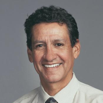 Carlos O. Esquivel合作论文数Division of Gastroenterology, Hepatology, and Nutrition, Department of Podiatric, School of Medicine, Stanford University;Department of Surgery, School of Medicine, Stanford University9
 Olivia M. Martinez合作论文数Department of Surgery/Division of Transplantation and Program in Immunology, Stanford University School of Medicine9
 Sheri Krams合作论文数Stanford University9Srishti Gupta合作论文数8Kristen A Johnson合作论文数8
 Peter Greenberg合作论文数Medicine Department, Stanford University8
 Youn H. Kim合作论文数Department of Dermatology, School of Medicine, Stanford University;Stanford Cancer Institute, Stanford University8
 Suzanne Powell合作论文数Department of Pathology and Laboratory Medicine, Weill Cornell Medical College, Cornell University;Department of Pathology & Genomic Medicine, The Methodist Hospital;Texas A and M University System Health Science Center8
 Robert Hasserjian合作论文数Department of Pathology, Massachusetts General Hospital;Harvard Medical School;Dana-Farber / Harvard Cancer Center8John M Childs合作论文数7
 Sara Zadeh合作论文数7
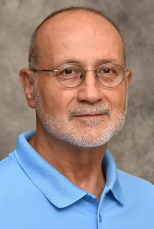 Joseph Madri合作论文数Pathology Faculty, Yale School of Medicine, Yale University7
 Ronald M. Levy合作论文数Department of Chemistry, College of Science and Technology, Temple University7Twist Clare J合作论文数7George Melissa R合作论文数7
 Daniel Bernstein合作论文数Pediatrics Department, Stanford University7Lisa R Dixon合作论文数6Raheem Peerani合作论文数6
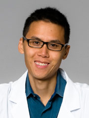 Matthew Cheung合作论文数Division of Hematology, Department of Medicine, University of Toronto;Sunnybrook Research Institute6Venick Robert S合作论文数6
 Jean S. Oak合作论文数Department of Pathology, Stanford University6
 Sebastian Fernandez Pol合作论文数Department of Pathology, Stanford University6
 Henning Stehr合作论文数Department of Pathology, Stanford University6Thomas M. Fishbein合作论文数6




