We made quantitative analysis of seizure frequency 1 week and 6, 12, and 24 months after seizure surgery. Seizure recurrence was significantly higher when seizures occurred in the first postoperative week. Seizure recurrence increased progressively with longer follow-ups, but the 6 month postoperative follow-up period was an excellent index of long-term outcome. In operative follow-up studies, seizure frequency should be reported at fixed follow-up periods, e.g., at 6 months and 1, 2, 5, and 10 years. Meaningful comparison of outcomes between different studies is possible only when reports include outcome at fixed postoperative follow-up periods (as opposed to ranges of follow-up periods).
We evaluated the accuracy and interobserver variability of selected ictal and postictal behavioral changes. Three observers, blinded to clinical history, EEG, and side of surgical resection, analyzed videotapes of 166 seizures in 38 patients, looking for lateralizing signs. Twenty-seven patients with temporal lobe resections were seizure-free for > or = 1 year postoperatively, and 11 with extratemporal resections had at least 90% reduction in seizures > or = 1 year postsurgery. The epileptogenic region (ER) was lateralized by analyzing lateralizing signs in 78% of patients; positive predictive value (PPV) was 94% (90% CI = 87% to 100%). Overall kappa was 0.68. Signs were considered present if seen by two or more observers. Forty-five percent had version, ie, forced and sustained head deviation (kappa = 0.76, PPV = 94%); 37% had dystonic posturing of the upper extremity (kappa = 0.47, PPV = 93%); and 34% had unilateral mouth deviation (kappa = 0.83, PPV = 92%). These signs indicated a contralateral ER. Twenty-one percent had unilateral upper extremity automatisms, all ipsilateral to the ER (kappa = 0.65, PPV = 100%); 21% had postictal dysnomia, indicating a dominant-hemisphere ER (kappa = 0.89, PPV = 100%); and 16% had ictal speech, usually indicating a nondominant-hemisphere ER (kappa = 0.75, PPV = 83%). Dystonic posturing, postictal dysnomia, ictal speech, and unilateral upper extremity automatisms may indicate a higher probability of temporal lobe epilepsy. Analysis of lateralizing signs shows good interobserver agreement and provides useful clinical information.
Eighty-eight patients had bilateral intracarotid amobarbital (Wada) testing to determine hemispheric dominance for language in preparation for epilepsy surgery, as well as unilateral extraoperative cortical electrical stimulation using subdural electrode arrays. In none of the patients with left dominance by Wada testing were language areas found with right-sided stimulation, but two patients with right dominance by Wada testing had language areas mapped on the left side. These findings suggest that left dominance by Wada testing is strong evidence for exclusive lateralization of language function in the left hemisphere, but there is concern about the ability of the Wada test to exclude the possibility of some left-sided language function despite apparent right-sided dominance. Patients with left dominance on Wada testing do not need cortical stimulation before extensive right temporal lobectomy, but we believe that patients with right or bilateral dominance on Wada testing should have cortical stimulation for localization of language areas if extensive left or right temporal or frontal resection is planned.
In our patient population that had undergone antero-temporal lobectomy, we found 20 patients with a unilateral sphenoidal/antero-temporal interictal focus. All patients had normal computed tomography (CT) scans. Invasive recordings with subdural electrode arrays placed over and under the temporal lobe were used in every patient. We found that the scalp interictal focus predicted for all patients that both the interictal sharp waves and ictal onset would be mesiobasal/anterotemporal in location on the subdural arrays. Seventy-five percent of these patients had an excellent outcome with temporal lobectomy.
We retrospectively analyzed the presence of sharp waves in 2-h EEGs performed 6 months after epilepsy surgery in 59 patients. To study the significance of the postoperative interictal epileptiform activity in the tissue remaining after resection, we included only patients with a single epileptic focus (as defined preoperatively by prolonged video/EEG recordings and subdural electrode arrays studies) and no progressive structural lesions. Temporal lobectomy was performed in 51 patients (86%); extratemporal resections were performed in the remainder. The epileptogenic focus was completely resected in 26 patients (44%). The immediate postoperative electrocorticograms (EcoG) showed spikes in 13 patients (22%). At 6-month follow-up, 43 patients (73%) were seizure-free or had auras only and 12 patients (20%) had epileptiform activity on EEG. A significant correlation was noted between presence of sharp waves in the 6-month postoperative EEG and recurrence of seizures (Fisher's exact test p = 0.011) and also with the extent of the resection (complete vs. incomplete p = 0.042). We noted no correlation between postoperative epileptiform activity and location of the resection (temporal vs. extratemporal), presence of spikes in immediate postoperative EcoG, or occurrence of auras only at 6-month follow-up.
 Elaine Wyllie合作论文数Cleveland Clinic Lerner College of Medicine;Epilepsy Center, Cleveland Clinic7
 H Lüders合作论文数6DS DINNER合作论文数5HH MORRIS合作论文数5Godoy J合作论文数5
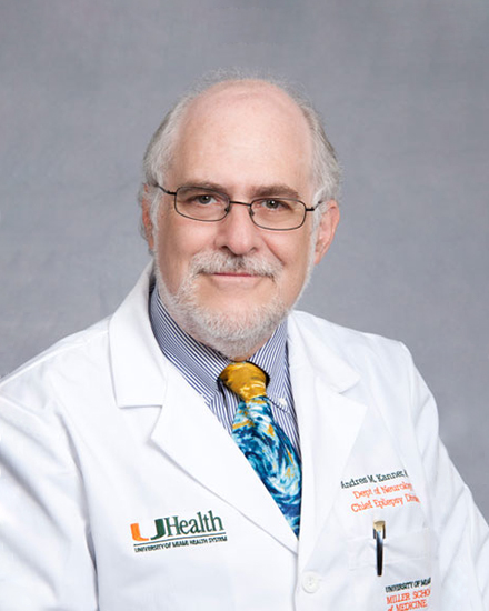 Andres M. Kanner合作论文数Division of Epilepsy, Department of Neurology, Miller School of Medicine, University of Miami;Comprehensive Epilepsy Center, Department of Neurology, Miller School of Medicine, University of Miami3
 Prakash Kotagal合作论文数Pediatric Epilepsy Section, Cleveland Clinic Epilepsy Center;School of Medicine, Case Western Reserve University3D Dinner合作论文数2H Morris合作论文数2Rp Lesser合作论文数1R Lesser合作论文数1
 Issam A. Awad合作论文数Center of Excellence for Cerebral Cavernous Malformations, The University of Chicago Medicine1A. David Rothner合作论文数Pediatric Neurology, Cleveland Clinic1
 Michael Chee合作论文数Centre for Sleep and Cognition, Yong Loo Lin School of Medicine, National University of Singapore1
 Hans Lüders合作论文数Department of Neurology, School of Medicine, Case Western Reserve University;University Hospitals Cleveland Medical Center1P C Van Ness合作论文数1




