Significant progress has been made in the development of magnetic micromanipulation for minimally invasive surgery. The development of systems to localize millimeter-sized robots during magnetic manipulation without line-of-sight detection remains, however, a challenging task. In this study, we focused on the development of a tracking system aiming to fill this gap. A robot which consists of a cylindrical magnet of 1 mm diameter is localized using a 2D array of 3D magnetoresistive sensors. The system, also called magnetic field camera, provides tracking of the robot with a refresh rate of 2 Hz. The developed tracking algorithm reaches a mean absolute error for the position and the orientation of, respectively 0.56 mm and 5.13° in 2D. This system can be added to existing magnetic manipulation systems allowing closed loop control of the navigation. The performances of the magnetic field camera are not affected by an exposure to strong magnetic fields. Exposures up to 3 T have been validated. Increasing the integrability of the magnetic field camera into magnetic manipulation systems. The presented tracking system makes it possible to target applications such as minimally invasive eye surgery or drug delivery. The high spatial and magnetic resolutions allow the tracking of magnetic particles, down to 200 μm diameter, when placed close to the surface. The system could also be suitable for the localization of small objects for 2D biomanipulation.
Significant progress has been made in the development of magnetic micromanipulation for minimally invasive surgery. The development of systems to localize millimetric size robots during magnetic navigation and without line of sight remains however a challenging task. In this study, we focused on the development of a tracking system aiming to fill this gap. A robot which consists of a cylindrical magnet of 1 mm diameter is localized using a 2D array of 3D magneto resistive sensors. The system provides a tracking of the robot with a refreshing rate of 2 Hz. The developed tracking algorithm reaches a mean absolute error for the position and the orientation of, respectively 0.56 mm and 5.13° in 2D. This system can be added to existing magnetic navigation systems allowing closed loop control of the navigation. The presented tracking system makes it possible to target applications such as minimally invasive ocular drug delivery.
A robust automated system to collect protein crystals for X-ray crystallography is presented. This system uses an ultraviolet imaging system based on commercial off-the-shelf components, a magnetically manipulated tool, and a resilient behavior-based controller. The system is validated by collecting over 350 polystyrene beads, used as crystal emulators, and transporting them 2 mm to a predefined goal in a 14-h period without human intervention. The average time to identify, collect, transport, and deliver a crystal emulator is 2.4 min, similar to an expert operator. This is the first demonstration of a completely automated robust system for protein crystal harvesting.
We introduce the modelling and control of a rolling microrobot. The microrobot is capable of manipulating micro-objects through the use of a magnetic visual control system. This system consists of a rod-shaped microrobot, a magnetic actuation system and a visual control system. Motion of the rolling microrobot on a supporting surface is induced by a rotating magnetic field. As the robot is submerged in a liquid this motion creates a rising flow in front, a sinking flow behind, and a vortex above the robot, thus enabling non-contact transportation of micro-objects. Besides this fluid-vortex approach, the microrobot is also able to manipulate micro-objects via a pushing strategy. We present the design and modelling of the 50×60×300 μm micro-agent, the visual control system, and an experimental analysis of the micromanipulation and control methods.
A new micro-agent is proposed to assist in automated protein crystal harvesting. The microrobot, named the RodBot, is a wireless mobile device driven by rotating magnetic fields (field strength 5–10 mT). When the RodBot rolls on a substrate in a low Reynolds number liquid environment, it generates flows to lift up and trap crystals in a vortex above itself. The gentle fluidic force acting on the crystals is in the range of a few nanonewtons to tens of nanonewtons and is spread over the whole surface of the crystal. The RodBot is capable of trapping protein crystals ranging from a few micrometres to sub-millimetre size. The trapped crystal can be transported to and deposited onto a loop positioned to accept it, obviating the need for more complicated `fishing' systems dependent on particular motions of the loop, the presence of further manipulators or the use of mechanical grippers. The RodBot can be driven in 24- and 96-well plates or in a crystal soaking dish, making the system compatible with existing crystallization hardware.
In this work, a magnetic visual control system for automated protein crystal harvesting is proposed. The system consists of a rod-shaped microrobot, a magnetic actuation system and a visual control system. A rotating magnetic field induces the microrobot to roll on the supporting surface, thereby creating a vortex in a liquid environment. This vortex enables the robot to trap and transport even delicate objects in a non-contact manner to a pre-defined position. We present the micro-agent, the actuation system and the visual control system to achieve this automated procedure.
Automated crystal harvesting is the main gap in the otherwise highly automated process of structure determination by X-ray crystallography. Many approaches have been presented, but few have proceeded beyond the initial, developmental stage. We recently introduced a rod-shaped microrobot1 (the "RodBot") to assist in the harvesting process. Driven by rotating magnetic fields to roll on a substrate, RodBots induce fluid flows that can lift crystals off the surface and trap them in a cylindrical vortex that travels with the RodBot. The gentle, fluidic force acting on the crystals is in the range of a few nanoNewtons to tens of nanoNewtons, and is spread over the whole surface of the crystal. Forces of this magnitude enable the RodBot to safely manipulate crystals ranging from a few microns to sub-millimeter size. With this technique individual crystals can be selected and brought to a loop positioned in the growth droplet to accept it. Harvesting and flash-cooling is then possible using a simple mechanical linkage. In this way the whole operation of crystal selection, harvesting and flash-cooling is remotely and gently carried out without the operator jitter or application of excessive strain that lead to high late-stage failure rates in crystal harvesting. Guidance is provided by the driving magnetic field, and can involve either manual input with a joystick or fully automated algorithms with feedback control. Because of this option of remote operation, RodBots can also be used for harvesting in hostile, sensitive or inconvenient environments such as anaerobic chambers, controlled humidity environments or cold rooms. The system is compatible with existing crystallization hardware and can be integrated readily into typical laboratory setups or high-throughput platforms.
A type of magnetic, wireless microrobot has been designed for non-contact manipulation of micro-objects in liquids. The agent, named the RodBot, has typical dimensions of 300 μm × 60 μm × 50 μm. The RodBot is transversely magnetized and rolls around its long axis on a surface in a rotating external magnetic field. In liquid environments, the RodBot generates a rising flow in front of it and a vortex above its body. The flow and vortex are efficient for picking-up and trapping micro-objects of sizes ranging from microns to one millimeter. In viscous solutions, a RodBot can transport objects many times its own size and weight.
Yeast ISW1a is classified as a member of the Imitation SWItch (ISWI) subgroup of the SWI2/SNF2 superfamily of ATP-dependent chromatin remodelers.The main function of ISW1a is the repression of gene expression through modulation of nucleosome positioning.ISW1a is a hetero-dimeric complex comprised of the 135 KDa Isw1 subunit and the 94 KDa Ioc3 subunits.Isw1 contains an N-terminal ATPase domain connected by a linker to a C-terminal domain which is composed of three consecutive subdomains HAND, SANT and SLIDE (HSS).The Ioc3 protein does not share any significant sequence homology to any structually characterized domain.We have solved the crystal structure of ISW1a (DATPase) complex both with and without bound duplex DNA at resolutions of 3.60 (P6 5 22) and 3.25 (H32) Å, respectively [1].In both structures, there is one ISW1a complex in the asymmetric unit.In terms of biological significance, these structures revealed not only an entirely new protein fold architecture of Ioc3 carrying a novel recognition motif for a specific DNA sequence, but also SANT and SLIDE were proven to take topologically unique DNA binding forms.Of interest to crystallographers, the ISW1a crystal in the absence of DNA displayed resolution improvements after introducing point mutations which increased salt bridging in the crystal packing interface, and ISW1a-DNA crystal also exhibited the improvements only after applying dehydration and slow cooling techniques during the post crystal treatment.Taking both these results into account, the introduction of a point mutation at the crystal packing interface and the dehydration and slow cooling methodologies could represent more general tools to improve resolution for poorly diffracting crystals.
Microsymposia C49 MScomposite microstructure of the building units, which we imaged by EBSD down to the submicron scale and which is essential for the mechanical strength and self-sharpening ability of the calcite teeth.There is direct as well as indirect evidence supporting the paradigm [6] that most if not all of the biocalcites grow from amorphous precursors.Crystallographic textures vary from weakly cylindrical (in coralline red algea) via the very frequent strongly cylindrical textures in shell valves to 3-dimensional single crystal-like coherence.
Site-specific recognition of DNA in eukaryotic organisms depends on the arrangement of nucleosomes in chromatin. In the yeast Saccharomyces cerevisiae , ISW1a and related chromatin remodelling factors are implicated in establishing the nucleosome repeat during replication and altering nucleosome position to affect gene activity. Here we have solved the crystal structures of S. cerevisiae ISW1a lacking its ATPase domain both alone and with DNA bound at resolutions of 3.25 Å and 3.60 Å, respectively, and we have visualized two different nucleosome-containing remodelling complexes using cryo-electron microscopy. The composite X-ray and electron microscopy structures combined with site-directed photocrosslinking analyses of these complexes suggest that ISW1a uses a dinucleosome substrate for chromatin remodelling. Results from a remodelling assay corroborate the dinucleosome model. We show how a chromatin remodelling factor could set the spacing between two adjacent nucleosomes acting as a ‘protein ruler’.
Chromatin organization The fundamental level of DNA organization was revealed in 1997 with the determination of the nucleosome core structure. The crystal structure of a tetranucleosome has now been determined at 9Å resolution, showing how nucleosomes are arranged at the next level up, in the chromosomes that are the substrate for DNA replication. The nucleosomes form a two-start helix, and their geometry in the chromatin matrix is best described by the ‘crossed-linker’ model.
Previous authours have suggested that the Type I respiratory enhancement in Chlorella was the result of an increased supply of a respiratory substrate or intermediate, or a change in activity of a respiratory enzyme. Our studies with respiratory inhibitors show that the Type I effect is not a general respiratory enhancement, as would be expected from an increase in available substrate, but rather is specifically associated with the tricarboxylic acid cycle and the electron transport chain. The feedback controls on these two processes are such that changes in activities of the component enzymes or in concentrations of carbohydrate intermediates would not be expected to affect the overall respiration rate: an ATP demand is needed to explain the results. A stimulation of chloroplast RNA and protein synthesis by blue light may be the basic mechanism.
Recognition of and discrimination between potential glyco-substrates is central to the function of galectins. Here we dissect the fundamental parameters responsible for such selectivity by the fungal representative, CGL2. The 2.1 A crystal structure of CGL2 and five substrate complexes reveal that this prototype galectin achieves increased substrate specificity by accommodating substituted oligosaccharides of the mammalian blood group A/B type in an extended binding cleft. Kinetic studies on wild-type and mutant CGL2 proteins demonstrate that the tetrameric organization is essential for functionality. The geometric constraints due to the orthogonal orientation of the four binding sites have important consequences on substrate binding and selectivity.
2-hydroxybiphenyl 3-monooxygenase (HbpA; EC 1.14.13.44) from Pseudomonas azelaica HBP1 was produced in Escherichia coli both as native and SeMet-labelled protein. The two enzymes were purified to homogeneity and crystallized by the hanging-drop vapour-diffusion method. The crystals belong to the monoclinic space group C2, with unit-cell parameters a = 108.6, b = 196.8, c = 79.3 A, beta = 97.7 degrees for the native protein and a = 108.3, b = 196.8, c = 79.0 A, beta = 97.8 degrees for SeMet HbpA. Crystal-packing considerations led to the assumption of two HbpA subunits per asymmetric unit, which corresponds to a V(M) value of 3.3 A(3) Da(-1) and a solvent content of 62%. The crystals were radiation-sensitive and only had a lifespan of about 120 s when exposed to synchrotron radiation on an undulator beamline. To obtain complete data sets, data were collected from 23 native and 26 derivative crystals. The high-resolution limit was 2.0 A for native and 2.25 A for SeMet HbpA.
TlpA is an unusual thioredoxin-like protein present in the nitrogen-fixing soil bacterium Bradyrhizobium japonicum. A hydrophobic N-terminal transmembrane domain anchors it to the cytoplasmic membrane, whereby the hydrophilic thioredoxin domain becomes exposed to the periplasmic space. There, TlpA catalyses an essential reaction, probably a reduction, in the biogenesis of cytochrome aa3. The soluble thioredoxin domain (TlpAsol), devoid of the membrane anchor, was purified and crystallized. Oxidized TlpAsol crystallized as a non-covalent dimer in the space group P212121. The X-ray structure analysis was carried out by isomorphous replacement using a xenon derivative. This resulted in a high-resolution (1.6 Å) three-dimensional structure that displayed all of the features of a classical thioredoxin fold. A number of peculiar structural details were uncovered: (i) Only one of the two active-site-cysteine sulphurs (Cys72, the one closer to the N terminus) is exposed on the surface, making it the likely nucleophile for the reduction of target proteins. (ii) TlpAsol possesses a unique structural disulphide bond, formed between Cys10 and Cys155, which connects an unprecedented N-terminal α helix with a β sheet near the C terminus. (iii) An insertion of about 25 amino acid residues, not found in the thioredoxin prototype of Escherichia coli, contributes only marginally to the thioredoxin fold, but forms an extra, surface-exposed α helix. This region plus another surface-exposed stretch (-Ile-Gly-Arg-Ala-), which is absent even in the closest TlpA relatives, might be considered as specificity determinants for the recognition of target proteins in the periplasm. The TlpAsol structure paves the way towards unraveling important structure-function relationships by rational mutagenesis.
Le Saux et al., 2001Le Saux A. Ruysschaert J.M. Goormaghtigh E. Membrane molecule reorientation in an electric field recorded by attenuated total reflection Fourier-transform infrared spectroscopy.Biophys. J. 2001; 80: 324-330Abstract Full Text Full Text PDF PubMed Scopus (33) Google Scholar report on voltage-induced reorientations of lipid headgroups using dried lipid multilayers of dioleylphosphatidylcholine as a membrane model. I observed the same phenomenon many years ago based on the measurement of capacitance relaxation currents (displacement currents) in black lipid membranes of various compositions (Sargent, 1975aSargent D.F. Voltage jump/capacitance relaxation studies of bilayer structure and dynamics.J. Membr. Biol. 1975; 23: 227-247Crossref Scopus (37) Google Scholar, Sargent, 1975bSargent D.F. Bilayer dynamics studies using capacitance relaxation.in: Kaback H.R. Neurath H. Radda G.K. Schwyzer R. Wiley W.R. Molecular Aspects of Membrane Phenomena. Springer-Verlag, Berlin1975: 104-120Crossref Google Scholar), and corresponding effects have been seen with virtually solvent-free bilayers (Hianik and Passechnik, 1995Hianik T. Passechnik V.I. Bilayer Lipid Membranes: Structure and Mechanical Properties. Kluwer Academic Publishers, Dordrecht, the Netherlands1995Google Scholar, Hianik et al., 2000Hianik T. Fajkus M. Sivak B. Rosenberg I. Kois P. Wang J. The changes in dynamics of solid supported lipid films following hybridization of short sequence DNA.Electroanalysis. 2000; 12: 495-501Crossref Scopus (22) Google Scholar). The independent observation of the same phenomenon by two completely different methods and in rather different model systems provides good corroboration of the influence of transmembrane voltage on the structure of the lipid headgroup region, but it is of interest to compare the results of the two methods in more detail. The attenuated total reflection Fourier-transform infrared (ATR-FTIR) study of Le Saux et al. is based on measurement of changes in the relative absorption of perpendicular and parallel components of infrared bands associated with the polar headgroups of lipid layers exposed to transverse electric fields. The electric field is induced through the proximity of the polarized germanium internal reflection elements to the dried multilayer sample. From the description of the experimental cell and the multilayer preparations, the average potential drop per bilayer in the FTIR measurements will be at the most a few hundred millivolts at the highest potential applied (200 V), but could be somewhat less. The measurements were started 1 min after applying the voltage and take minutes to accumulate, and thus presumably represent equilibrium distributions of molecular orientations. Although a quantitative analysis was not shown by Le Saux et al., they state that they found an almost linear relationship between the amplitude of the dichroic peaks and the applied potential. The measured parameters allow a molecular interpretation of the response of the system to the applied voltage, viz., reorientations of the phosphate and choline moieties. Unfortunately, because of the small size of the signals involved, they were not able to derive a reliable estimate of the magnitude of the reorientation. In the capacitance relaxation method with lipid bilayers, the voltage is applied to a single membrane through the aqueous phases on the two sides of the bilayer, ensuring a good electrical contact and, thus, a precise value for the voltage across the membrane. Voltages up to several hundred millivolts can be applied before membrane breakdown occurs. The combination of a dielectric (lipid bilayer) separating two conducting phases (electrolyte) represents an electric capacitor, holding charge Q = C · V. A change in the applied voltage induces a current given by i = dQ/dt = C · dV/dt + V · dC/dt. The latter term includes both a bulk geometric component (electrostriction: voltage dependence of bilayer area and thickness), molecular deformation (induced dipole), and reorientation components. By choosing initial and final voltages which are of equal magnitude but opposite sign, changes attributable to electrostriction can be eliminated and, after the charging pulse (C · dV/dt) is over, molecular components may be observed. Depending on the details of the experimental setup (electrolyte concentration, electrode area, output impedance of the voltage source) the time resolution of the method can extend down to the microsecond region. This is much too slow to observe molecular deformation processes or unhindered rotation of single molecules, but cooperative motion (clusters), as would be characteristic of liquid crystal phases such as bilayers, can lie in this range. The effects observed are completely reversible (changes from −V to +V and +V to −V are mirror images of one another) and the magnitude of reorientation was found to be a linear function of the applied voltage jump up to at least 600 mV, explaining the similar observation of Le Saux et al. Despite the very high field strengths in the bilayer (6 · 105 V/cm for 300 mV across a 50 Å thick bilayer), it is interesting that there is no sign of saturation. Higher transmembrane voltages generally lead to rupture of the membranes. Calculations suggest an average change of orientation of the membrane lipid dipoles of 1° or less per 100 mV in the cases studied (Sargent, 1975aSargent D.F. Voltage jump/capacitance relaxation studies of bilayer structure and dynamics.J. Membr. Biol. 1975; 23: 227-247Crossref Scopus (37) Google Scholar), although there are several approximations involved in deriving this value. The ATR-FTIR technique provides direct evidence for reorientation of specific parts of the lipid headgroups in the membrane and confirm, for example, the participation of the lipid in the response, although ordered water molecules also contribute to the membrane-associated surface dipole potential (Brockman, 1994Brockman H. Dipole potential of lipid membranes.Chem. Phys. Lipids. 1994; 73: 57-79Crossref PubMed Scopus (338) Google Scholar). In contrast, the capacitance relaxation method allows for quantitative estimates of magnitude, linearity, and time course of the reorientations. Both transmembrane and adsorbed proteins could affect and be affected by voltage-induced dipolar reorientations in membranes. For example, changes induced in the capacitance relaxation parameters have been used to help characterize the interaction of surface-active molecules with bilayers (Hianik et al., 1998Hianik T. Fajkus M. Tarus B. Frangopol P.T. Markin V.S. Landers D.F. The electrostriction, surface potential and capacitance relaxation of bilayer lipid membranes induced by tetracaine.Bioelectrochem. Bioenerg. 1998; 46: 1-5Crossref Scopus (18) Google Scholar, Hianik et al., 2000Hianik T. Fajkus M. Sivak B. Rosenberg I. Kois P. Wang J. The changes in dynamics of solid supported lipid films following hybridization of short sequence DNA.Electroanalysis. 2000; 12: 495-501Crossref Scopus (22) Google Scholar). An example of possible direct relevance of headgroup reorientation is the suggestion of a coupling of action potential and nerve conduction through ordering and disordering of dipole domains (electrets). A model study by Wobschall, 1968Wobschall D. An electret model of the nerve membrane.J. Theor. Biol. 1968; 21: 439-448Crossref PubMed Scopus (16) Google Scholar indicated that the time constants for such domains must lie between 0.14 and 1.4 ms, and capacitance relaxation phenomena in dioleollecithin bilayer lipid membranes are found in this range (Sargent, 1975bSargent D.F. Bilayer dynamics studies using capacitance relaxation.in: Kaback H.R. Neurath H. Radda G.K. Schwyzer R. Wiley W.R. Molecular Aspects of Membrane Phenomena. Springer-Verlag, Berlin1975: 104-120Crossref Google Scholar). A combination of the more sensitive electrical methods with the more specific structural information of the ATR-FTIR technique, and perhaps further experimental methods, should further our understanding of voltage-linked membrane phenomena.
 Timothy J. Richmond合作论文数Department of Biology, ETH Zurich;Institute of Molecular Biology and Biophysics, ETH Zurich8
 CPS TAYLOR合作论文数6
 Bradley Nelson (布拉德利·尼尔森)合作论文数Robotics and Intelligent Systems, ETH Zurich5
 Karolin Luger合作论文数Department of Chemistry and Biochemistry, University of Colorado Boulder;Department of Biochemistry and Molecular Genetics, Colorado School of Medicine;Howard Hughes Medical Institute4
 Roel Pieters合作论文数Dynamics and Control Group, Eindhoven University of Technology4Kazuhiro Yamada合作论文数3Armin W. Mader合作论文数3Robin K. Richmond合作论文数3
 Celine Vergne合作论文数2John W. Bean合作论文数2
 Thomas Quirin合作论文数ICube Laboratory, University of Strasbourg – CNRS2
 Joris Pascal合作论文数2Frouws Timothy D合作论文数2Hans Joerg. Moeschler合作论文数2Markus G. Grütter合作论文数2Kyoko Schimmele合作论文数2Daniel Erne合作论文数2Daniel J. Fitzgerald合作论文数1P.J. Walser合作论文数1WALTER FISCHLI合作论文数1Reinhild Rossmann合作论文数1Luca Somm合作论文数1Burak Zeydan合作论文数Multi Scale Robotics Lab, ETH Zurich, Tannenstrasse 3, 8092 Zurich, Switzerland1Andreas Meyer合作论文数FGen AG1
 Samuel Charreyron合作论文数institute of robotics and intelligent systems1
 Curtis Alexander Davey合作论文数School of Biological Sciences, College of Science, Nanyang Technological University1
 Markus Künzler合作论文数Department of Biology, Eidgenössische Technische Hochschule Zürich1
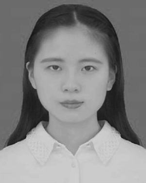 Yang Fang合作论文数1
 Hauke Hennecke合作论文数Institut fur Mikrobiologie, Departement Biologie, Eidgenossische Technische Hochschule Zurich1
 Markus Aebi合作论文数Institute of Microbiology, Eidgenössische Technische Hochschule Zürich;Department of Biology, Eidgenössische Technische Hochschule Zürich1Schmid Andreas合作论文数1
 Nenad Ban合作论文数The Ban Lab, Institute of Molecular Biology and Biophysics, Eidgenössische Technische Hochschule Zürich1Taylor C P合作论文数1
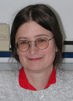 Ursula Kües合作论文数Department of Molecular Wood Biotechnology and Technical Mycology, Faculty of Forest Sciences and Forest Ecology, Georg-August University Goettingen1Bernard Witholt合作论文数1Robert Schwyzer合作论文数1Brigitte Angst合作论文数1
 Andrew J. Petruska合作论文数1
 Thomas Schalch合作论文数Dept Mol Biol, Univ Geneva1
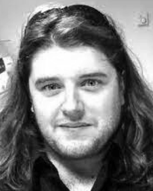 Morgan Madec合作论文数Institut d'Electronique du Solide et des systèmes, rue du Loess, STRASBOURG, France1Hsi-Wen Tung合作论文数1Carl Deluca合作论文数1Christine Deillon合作论文数1Guido Capitani合作论文数1Bernd Gutte合作论文数Biochemisches Institut der Universität Zürich, Winterthurerstrasse 190, CH-8057 Zürich, Switzerland1
 Daniel --- Tanner合作论文数1
 Peter Haebel合作论文数Lehrstuhl für Strukturbiologie, Universität des Saarlandes1




