Idiopathic generalized epilepsies account for about 40% of epilepsy up to age 40 and commonly have a genetic basis. One type is benign familial neonatal convulsions (BFNC), a dominantly inherited disorder of newborns. We have identified a sub-microscopic deletion of chromosome 20q13.3 that co-segregates with seizures in a BFNC family. Characterization of cDNAs spanning the deleted region identified one encoding a novel voltage-gated potassium channel, KCNQ2, which belongs to a new KQT-like class of potassium channels. Five other BFNC probands were shown to have KCNQ2 mutations, including two transmembrane missense mutations, two frameshifts and one splice-site mutation. This finding in BFNC provides additional evidence that defects in potassium channels are involved in the mammalian epilepsy phenotype.
Stargardt disease (STGD, also known as fundus flavimaculatus; FFM) is an autosomal recessive retinal disorder characterized by a juvenile-onset macular dystrophy, alterations of the peripheral retina, and subretinal deposition of lipofuscin-like material. A gene encoding an ATP-binding cassette (ABC) transporter was mapped to the 2-cM (centiMorgan) interval at 1p13-p21 previously shown by linkage analysis to harbour the STGD gene. This gene, ABCR, is expressed exclusively and at high levels in the retina, in rod but not cone photoreceptors, as detected by in situ hybridization. Mutational analysis of ABCR in STGD families revealed a total of 19 different mutations including homozygous mutations in two families with consanguineous parentage. These data indicate that ABCR is the causal gene of STGD/FFM.
A human delayed rectifier K+ channel gene has been localized to the long arm of human chromosome 20q13.2 by fluorescence in situ hybridization of genomic P1 clones from this locus. A polymorphic (GA) microsatellite repeat was identified in one of the P1 clones. The new SSR marker (D20S436) was genotyped in four CEPH pedigrees. Two-point linkage analysis indicated linkage of this marker to a PCR marker, D20S109, with a maximum lod score of 9.32 at θ = 0.001. The assignment of this K+ channel gene to 20q13.2 eliminates it as a candidate for the gene associated with benign familial neonatal convulsions (BFNC), which has been localized to 20q13.3. Genetically, the K+ channel gene maps more than 30 cM proximal to the BFNC locus.
We have genetically mapped the genes encoding four human adrenergic receptors (ARs) of subtypes α1C, α2A, α2B, and β1, which are prototypic G protein coupled receptors that mediate the physiological effects of neurotransmitters, hormones, and drugs. We placed these genes onto the Cooperative Human Linkage Center (CHLC) and Genethon framework maps, within confidence intervals with greater than 1000∶1 odds. With multipoint analysis the α1C gene (locus ADRA1C) mapped to the interval between NEFL and D8S283; α2-C4, the gene encoding the α2C AR (locus ADRA2C), mapped to the interval between D4S126 and D4S62; and the α2-C10 (α2A AR)/β1 haplotype (loci ADRA2A/ ADRB1) mapped to the interval between D10S259 and D10S187. A fifth AR gene, β2, yielded significant LOD scores with markers on the long arm of chromosome 5; however, this locus (ADRB2) could not be mapped to any specific interval with odds of greater than 1000∶1. The two AR genes that are completely linked, α2-C10 and β1, were oriented on their shared 225-kb genomic fragment relative to the direction of transcription, with β1 being 5′ to α2-C10. The positioning of these genes on high-density framework maps allows them to be tested as candidates in a spectrum of diseases that might involve AR dysfunction.
A genetic linkage map for the long arm of human chromosome 13 contains 29 loci derived from 38 probe and enzyme combinations and two protein polymorphisms. Thirteen loci form a continuous linkage map of 106 cM in males and 230 cM in females; each was placed on the map with support of at least 1000:1 against alternative orders. On a sex-combined basis, the mean distance between markers is less than 13 cM. The order of loci on the genetic map agrees with physical localization data that show that together these 13 loci cover 13q13 to 13q34. This map was used to regionally localize the 16 remaining loci. The linkage maps reported here should prove to be useful to investigators mapping disease genes and other genetic markers on human chromosome 13.
We have determined the genetic location of the human gene encoding phenylethanolamine N-methyltransferase (PNMT), the terminal enzyme of the catecholamine pathway catalyzing the synthesis of epinephrine (adrenaline) from norepinephrine. This gene is linked to DNA markers on the long arm of chromosome 17, q21-q22, most closely to the DNA markers MFD15 (D17S250) (Zmax = 15.0, theta = 0.065) and fLB17.1 (Zmax = 14.6, theta = 0.045). Multipoint linkage analysis placed the PNMT locus in the interval fLB17.1-CMM86 (D17S74), at 4 centiMorgans (cM) distal to fLB17.1, and at 17 cM proximal to CMM86. Mapping of the PNMT gene will provide the basis for genetic linkage studies in families with disease which might pathogenetically involve this enzyme. The human chromosomal region 17q21-22 identified here to harbour the PNMT gene may be syntenic to the chromosomal region in the stroke-prone spontaneously hypertensive rat (SHR-SP) recently linked to blood-pressure regulation. As an increase of PNMT activity has been associated with the development of hypertension in SHR-SP, it will be of interest to perform comparative mapping of the PNMT gene.
Despite extensive study since the first report of familial benign hypercalcemia (FBH, or hypocalciuric hypercalcemia) in 1972, there is no evidence of the specific abnormal gene product. FBH is highly suitable for either a candidate gene or a reverse genetics approach to localizing the genetic abnormality, because it is inherited in an autosomal dominant pattern, is highly penetrant, does not affect survival, and can be diagnosed in families with readily available measurements. Importantly, several candidate genes have been cloned and mapped. Therefore, we collected blood samples and extracted leukocyte DNA from 94 members of 4 families with well documented FBH (44 affected, 45 unaffected, and 5 unclassifiable). We digested the DNA samples with various restriction endonucleases, conducted standard Southern blotting, and searched for restriction fragment length polymorphisms for the following candidate genes (probe names in parentheses): multiple endocrine neoplasia (MEN) type 1 (pMCMP.1, pHBI59, p3C7, and pTHH26), MEN 2a (MCK2 and cTB14.34), basic fibroblast growth factor (pHFL1-7), (Ca2+,Mg2+)ATPase isoform 4 (hPMCA4), membrane Na/Ca exchanger (cNC28 M-A), PTH (pPTH-LF), and calbindin-D28K (pSKCalb). In addition, we used the anonymous variable number tandem repeat marker pYNH24 to verify pedigree structures by excluding misinheritances. Data were analyzed using the Linkage program. For none of the genes was there significant linkage with the FBH trait; logarithm of odds scores ranged from -1.3 to -26.0 at a recombination fraction of 0.001, and from 0.6 to -5.6 at a recombination fraction of 0.10. We conclude that FBH is unrelated to the MEN syndromes and is not caused by mutations in any of the calcium-regulating or -binding proteins or growth factors studied thus far.
Journal Article D7S448 detects a Hind III polymorphism located in the centromere region of chromosome 7 Get access M. Dean, M. Dean * * To whom correspondence should be addressed Search for other works by this author on: Oxford Academic PubMed Google Scholar C. Stewart, C. Stewart Search for other works by this author on: Oxford Academic PubMed Google Scholar A. Perry, A. Perry Search for other works by this author on: Oxford Academic PubMed Google Scholar D. Stauffer, D. Stauffer 1Howard Hughes Medical Institute and Department of Human Genetics, University of UtahSalt Lake City, UT 84132, USA Search for other works by this author on: Oxford Academic PubMed Google Scholar B. Otterud, B. Otterud Search for other works by this author on: Oxford Academic PubMed Google Scholar R. White, R. White 1Howard Hughes Medical Institute and Department of Human Genetics, University of UtahSalt Lake City, UT 84132, USA Search for other works by this author on: Oxford Academic PubMed Google Scholar M. Leppert M. Leppert Search for other works by this author on: Oxford Academic PubMed Google Scholar Nucleic Acids Research, Volume 19, Issue 1, 11 January 1991, Page 200, https://doi.org/10.1093/nar/19.1.200-a Published: 11 January 1991
Description of Probe: A cDNA probe of about 3.5 kb which is a PstI-KpnI fragment encoding the majority of the beta chain of cytochrome b-245 transcript in pGEM-4 (1).Polymorphisms: Digestion with NsiI reveals invariant fragments of approximately 7.2, 6.2 and 3.7 kb.In addition to the polymorphic fragments of 1.7 and 1.3 kb described by Battat and Francke (2) a further polymorphism exists with fragment sizes of 2.9 and 2.5 kb.Frequency: In 32 unrelated chromosomes, allele frequencies were 2.9 kb: 19% and 2.5 kb: 81 %.In 29 of the same chromosomes allele frequencies were 1.7 kb: 90% and 1.3 kb: 10%.The observed haplotype distribution does not deviate significantly from that expected on the basis of the above allele frequencies, implying that there is no strong linkage disequilibrium.Not Polymorphic For: BamHI, BglI, EcoRI, HindJH, MspI, PstI, TaqI in 14 chromosomes tested.Chromosomal Localisation: The X-linked CYBB locus has been assigned to Xp21.1 (1).Mendelian Inheritance: X-linked inheritance has been demonstrated in 2 three-generation families.
Usher syndrome is a heterogeneous group of autosomal recessive disorders that combines variably severe congenital neurosensory hearing impairment with progressive night-blindness and visual loss similar to that in retinitis pigmentosa. Usher syndrome type I is distinguished by profound congenital (preverbal) deafness and retinal disease with onset in the first decade of life. Usher syndrome type II is characterized by partial hearing impairment and retinal dystrophy that occurs in late adolescence or early adulthood. The chromosomal assignment and the regional localization of the genetic mutation(s) causing the Usher syndromes are unknown. We analyzed a panel of polymorphic genomic markers for linkage to the disease gene among six families with Usher syndrome type I and 22 families with Usher syndrome type II. Significant linkage was established between Usher syndrome type II and the DNA marker locus THH33 (D1S81), which maps to chromosome 1q. The most likely location of the disease gene is at a map distance of 9 cM from THH33 (lod score 6.5). The same marker failed to show linkage in families segregating an allele for Usher syndrome type I. These data confirm the provisional assignment of the locus for Usher syndrome type II to the distal end of chromosome 1q and demonstrate that the clinical heterogeneity between Usher types I and II is caused by mutational events at different genetic loci. Regional localization has the potential to improve carrier detection and to provide antenatal diagnosis in families at risk for the disease.
Recurrent seizures, commonly known as epilepsies, occur in 1.7% of the general population by age 40. The factors that initiate or underlie seizures are not well understood, but trauma, infectious disease and genetics have been implicated. An understanding of the molecular basis of seizures would shed light on the basic mechanisms of neuronal homeostasis and allow new therapeutic strategies to be explored. Here, we report the mapping of an epilepsy gene to a specific chromosomal region, on the basis of cosegregation of two closely-linked DNA markers with a form of epilepsy known as benign familial neonatal convulsions (BFNC2, 12120 in ref. 3). The linked markers confirm the genetic basis and autosomal dominant inheritance of this trait, and localize the gene causing BFNC in this family to the long arm of chromosome 20. This regional placement is the first step towards the isolation of a gene involved in neuronal activity in the human brain.
The inherited genetic defect in adenomatous polyposis has been localized to a small region on the long arm of chromosome 5. Sixteen DNA marker loci were used to construct a linkage map of the chromosome. When five kindreds segregating a gene for adenomatous polyposis coli were characterized with a number of the markers, significant linkage was found between one marker and the disease gene. Linkage analysis determined the location of the defective gene within a primary genetic map of chromosome 5.
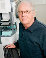 Mark F. Leppert合作论文数Department of Human Genetics, School of Medicine, University of Utah Health16
 B Otterud合作论文数6VE Anderson合作论文数5J.-M. Lalouel合作论文数5Ray White合作论文数5Andy Peiffer合作论文数5
 Michael Dean合作论文数Laboratory of Translational Genomics, Division of Cancer Epidemiology and Genetics, National Cancer Institute;Laboratory of Translational Genomics, NCI3Carole Charlier合作论文数Faculty of Veterinary Medicine, University of Liège (B43)3Peter Oconnell合作论文数3Claudia Stewart合作论文数2
 Richard Alan Lewis合作论文数Department of Molecular and Human Genetics, Baylor College of Medicine2R MELIS合作论文数2William Byerley合作论文数2John Holik合作论文数2
 Margret R. Hoehe合作论文数2
 Elliot S. Gershon合作论文数University of Chicago2Rosemarie Plaetke合作论文数2
 Roberta Melis合作论文数2Nanda Singh合作论文数2Barbara R. DuPont合作论文数1GM Ronen合作论文数1Jt Penniston合作论文数1Philip Smallwood合作论文数1X Zhao合作论文数1Jp Hughes合作论文数1Teodoro O. Rosales合作论文数1WT HSIEH合作论文数1M. Edens合作论文数1
 C David James合作论文数Department of Neurological Surgery, Feinberg School of Medicine, Northwestern University1L Ballard合作论文数1S Woodward合作论文数1M Connolly合作论文数1M Lathrop合作论文数1M Martinez合作论文数Inst Natl Sante & Rech Med1
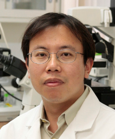 Hui Sun合作论文数University of California, Los Angeles1Dobbs M合作论文数1Singh N合作论文数1
 Peter Scambler合作论文数GOS Institute of Child Health, University College London1
 Rando L. Allikmets合作论文数Department of Pathology and Cell Biology, Columbia University1Don W. Walker合作论文数1Wade H. Berrettini合作论文数Department of Psychiatry,University of Pennsylvania1Raymond F. Gesteland合作论文数1Amy Hutchinson合作论文数1
 James R Lupski合作论文数Department of Molecular and Human Genetics, Baylor College of Medicine1Yixin Li合作论文数1Bernard Gerrard合作论文数1John J Wasmuth合作论文数1Thomas G. Quattlebaum合作论文数1Olaf Pongs合作论文数1




