Abstract Among the different types of systemic therapies used in cancer treatment, differentiation therapy, perhaps the least frequently utilized but typically most tolerable form of treatment, aims to set cancer cells back on the developmental path from which they strayed, leading to cell death through multiple mechanisms. Acute promyelocytic leukemia (APL) is the exemplar for differentiation therapy wherein leukemic blasts arrested in a progenitor-like state are induced to continue their myeloid differentiation after treatment with all-trans retinoic acid (ATRA) and arsenic trioxide, resulting in >95% cure rates. In contrast to APL, other types of acute myeloid leukemia (AML) have only a 5-year survival of ∼33%. However, recent advances in targeted therapies with novel, differentiation-inducing agents for AML with alterations in IDH1/2 (isocitrate dehydrogenase 1 and 2), NPM1 (nucleophosmin 1), and KMT2A (lysine methyltransferase 2A) have demonstrated efficacy in patients who are older, frail, or who have relapsed or refractory disease. However, the underlying mechanism of differentiation induction remains incompletely understood, limiting adoption for other AML subtypes or non-hematologic diseases. Establishing a rational means of discovering regulators of differentiation therapy could accelerate development of this class of drugs. Here, we present a platform for discovering the central regulators of differentiation as a means of identifying targets for downstream therapeutic development. Using single-cell multiomic (RNA- and ATAC-seq - scMultiome) data from clinical samples of IDH1/2-mutated (IDHm) AML as a test case, we utilized a software package called CellOracle to dissect the gene-regulatory networks (GRNs) underpinning the differentiation response throughout the course of IDH inhibition. With these analyses, we recapitulated a known pattern of “BRCAness” in IDHm AML with in silico knockout of BRCA1 (breast cancer gene 1) appearing to support the differentiation effect of IDH inhibition. We also discovered putative as-yet unidentified regulators of the differentiation phenotype including several Ikaros transcription factors. With this new platform, we also sought to identify targets for differentiation therapy in a solid tumor type, wherein examples of differentiation therapy are almost entirely lacking, with the noteworthy exception of ATRA for neuroblastoma. We utilized scMultiome data from H3K27M diffuse midline gliomas of the pons, which are known to have an epigenetically-based inhibition of terminal differentiation. These aggressive and devastating brain cancers affect exclusively school-aged children and have an approximately 95% 2-year mortality. Using the same pipeline we have identified several putative regulators of a malignant-to-normal transition, including several genes known to be important to normal glial development and maintenance of the cancer state. Citation Format: Scott C. Friedland, Tianli Ding, Gary Schweickart, Alice Mims, Katherine Miller, Adithe Rivaldi, Elizabeth Garfinkle, Sunkel Benjamin, Jaye Navarro, Michelle Wedemeyer, Ann-Kathrin Eisfeld, Elaine R. Mardis. Leveraging single-cell multiomics for a generalizable approach to differentiation therapy [abstract]. In: Proceedings of the American Association for Cancer Research Annual Meeting 2026; Part 1 (Regular Abstracts); 2026 Apr 17-22; San Diego, CA. Philadelphia (PA): AACR; Cancer Res 2026;86(7 Suppl):Abstract nr 468.
Quantitative comparison of ChIP-seq profiling between experimental conditions or samples remains technically challenging for the epigenetics field. Here, we report a strategy combining the use of well-defined cellular spike-in ratios of orthologous species' chromatin and a bioinformatic analysis pipeline to facilitate highly quantitative comparisons of 2D chromatin sequencing across experimental conditions. We find that the PerCell methodology results in efficient and consistent levels of spike-in vs. experimental genomic reads. We demonstrate use of the method and pipeline to enable quantitative, internally normalized chromatin sequencing on zebrafish embryos and human cancer cells. Overall, we propose the PerCell method to enable cross-species comparative epigenomics and promote uniformity of data analyses and sharing across labs.
Correct chromosome organization in the cell nucleus is essential for genome function. However, dynamics and regulations of large-scale chromosomal conformations beyond single compartments at larger than ten megabases in vivo in single cells remain largely unknown. Here we use CRISPR-Sirius, a high-resolution and high-sensitivity real-time imaging technique, to directly visualize distinctions in large-scale chromosomal conformations between live osteosarcoma (OS) cells and osteoblasts, suggesting extensive chromatin reorganization during cell transformation. A surprising discovery is that chromosome 19 long arm is primarily extended in osteoblasts and maintained by H3K27me3. Extended chromosome conformation has been reported in fly and mouse but not in human cells yet. However, in OS cells, chromosome 19 primarily folded into collapsed conformations, which reshape in minutes and are regulated by the chromosome architectural proteins CTCF and cohesin in the presence of H3K27ac. Changes in chromosome conformations by knocking down the cohesin subunit RAD21 resulted in altered gene expression, including proto-oncogenes. Transcription inhibition by a small molecule inhibitor did not have detectable effects on large-scale chromosome conformation, suggesting that local transcription events have limited effects on large-scale chromosomal architecture. Our results provide unique insights into the complex regulatory mechanisms of endogenous large-scale chromosome organization in normal and transformed osteogenic tissues. ### Competing Interest Statement The authors have declared no competing interest.
Materials and Methods used for experiments in Supplemental Figures and Supplemental References
Rhabdomyosarcoma (RMS) is a pediatric soft tissue cancer with a lack of precision therapy options for patients. We hypothesized that with a general paucity of known mutations in RMS, chromatin structural driving mechanisms are essential for tumor proliferation. Thus, we carried out high-depth in situ Hi-C in representative cell lines and patient-derived xenografts (PDXs) to define chromatin architecture in each major RMS subtype. We report a comprehensive 3D chromatin structural analysis and characterization of fusion-positive (FP-RMS) and fusion-negative RMS (FN-RMS). We have generated spike-in in situ Hi-C chromatin interaction maps for the most common FP-RMS and FN-RMS cell lines and compared our data with PDX models. In our studies, we uncover common and distinct structural elements in large Mb-scale chromatin compartments, tumor-essential genes within variable topologically associating domains and unique patterns of structural variation. Our high-depth chromatin interactivity maps and comprehensive analyses provide context for gene regulatory events and reveal functional chromatin domains in RMS.
Rhabdomyosarcoma (RMS) is a pediatric malignancy of the muscle with characteristics of cells blocked in differentiation. NOTCH1 is an oncogene that promotes self-renewal and blocks differentiation in the fusion negative-RMS sub-type. However, how NOTCH1 expression is transcriptionally maintained in tumors is unknown. Analyses of SNAI2 and CTCF chromatin binding and HiC analyses revealed a conserved SNAI2/CTCF overlapping peak downstream of the NOTCH1 locus marking a sub-topologically associating domain (TAD) boundary. Deletion of the SNAI2-CTCF peak showed that it is essential for NOTCH1 expression and viability of FN-RMS cells. Reintroducing constitutively activated NOTCH1-ΔE in cells with the SNAI2-CTCF peak deleted restored cell-viability. Ablation of SNAI2 using CRISPR/Cas9 reagents resulted in the loss of majority of RD and SMS-CTR FN-RMS cells. However, the few surviving clones that repopulate cultures have recovered NOTCH1. Cells that re-establish NOTCH1 expression after SNAI2 ablation are unable to differentiate robustly as SNAI2 shRNA knockdown cells; yet, SNAI2-ablated cells continued to be exquisitely sensitive to ionizing radiation. Thus, we have uncovered a novel mechanism by which SNAI2 and CTCF maintenance of a sub-TAD boundary promotes rather than represses NOTCH1 expression. Further, we demonstrate that SNAI2 suppression of apoptosis post-radiation is independent of SNAI2/NOTCH1 effects on self-renewal and differentiation.
Mediator activates RNA polymerase II (Pol II) function during transcription, but it remains unclear whether Mediator is able to travel with Pol II and regulate Pol II transcription beyond the initiation and early elongation steps. By using in vitro and in vivo transcription recycling assays, we find that human Mediator 1 (MED1), when phosphorylated at the mammal-specific threonine 1032 by cyclin-dependent kinase 9 (CDK9), dynamically moves along with Pol II throughout the transcribed genes to drive Pol II recycling after the initial round of transcription. Mechanistically, MED31 mediates the recycling of phosphorylated MED1 and Pol II, enhancing mRNA output during the transcription recycling process. Importantly, MED1 phosphorylation increases during prostate cancer progression to the lethal phase, and pharmacological inhibition of CDK9 decreases prostate tumor growth by decreasing MED1 phosphorylation and Pol II recycling. Our results reveal a novel role of MED1 in Pol II transcription and identify phosphorylated MED1 as a targetable driver of dysregulated Pol II recycling in cancer.
Effective T cell–mediated immune responses require the proper allocation of metabolic resources to sustain growth, proliferation, and cytokine production. Epigenetic control of the genome also governs T cell transcriptome and T cell lineage commitment and maintenance. Cellular metabolic programs interact with epigenetic regulation by providing substrates for covalent modifications of chromatin. By using complementary genetic, epigenetic, and metabolic approaches, we revealed that tricarboxylic acid (TCA) cycle flux fueled biosynthetic processes while controlling the ratio of succinate/α-ketoglutarate (α-KG) to modulate the activities of dioxygenases that are critical for driving T cell inflammation. In contrast to cancer cells, where succinate dehydrogenase (SDH)/complex II inactivation drives cell transformation and growth, SDH/complex II deficiency in T cells caused proliferation and survival defects when the TCA cycle was truncated, blocking carbon flux to support nucleoside biosynthesis. Replenishing the intracellular nucleoside pool partially relieved the dependence of T cells on SDH/complex II for proliferation and survival. SDH deficiency induced a proinflammatory gene signature in T cells and promoted T helper 1 and T helper 17 lineage differentiation. An increasing succinate/α-KG ratio in SDH-deficient T cells promoted inflammation by changing the pattern of the transcriptional and chromatin accessibility signatures and consequentially increasing the expression of the transcription factor, PR domain zinc finger protein 1. Collectively, our studies revealed a role of SDH/complex II in allocating carbon resources for anabolic processes and epigenetic regulation in T cell proliferation and inflammation.
Rhabdomyosarcoma (RMS) is a pediatric soft tissue sarcoma characterized by an impaired myogenic differentiation despite the expression of myogenic master genes MYOD and MYOG. Therefore, the restoration of differentiation is considered an anti-cancer therapy. SKP2 is an oncogenic E3-ubiquitin ligase that promotes cell proliferation by targeting the CDKi p21Cip1 and p27Kip1. Previous works showed that SKP2 overexpression is induced by the fusion oncoprotein PAX3-FOXO1 expressed in fusion positive (FP)-RMS cells, and promotes tumor cell proliferation through p27kip1 degradation. However, the role of SKP2 in fusion negative (FN)-RMS cells, devoid of any fusion gene, remains unclear. We report here that SKP2 transcript and protein levels are up-regulated in RMS patients and cell lines compared to normal tissue. Accordingly, we observed increased acetylation of H3K27 histone mark in RMS patients and cell lines compared to myoblasts and muscle tissue. We then show that in RMS cell lines SKP2 expression is induced by MYOD, which binds two SKP2 regulatory regions, an intronic and a distal enhancers, identified by Hi-C and 3C experiments. SKP2 knockdown in FN-RMS cells leads to p21Cip1 and p27Kip1 protein levels up-regulation coupled with G1/S cell cycle arrest. Rescue experiments showed that SKP2 promotes cell proliferation directly targeting p27Kip1. Moreover, SKP2 binds and promotes degradation of p57Kip2 and its silencing restores myogenic differentiation associated to MYOG and de novo MyHC expression in FN-RMS cells. SKP2 depletion also induces cell senescence and prevents anchorage-independent growth and stemness in vitro, and tumor growth in vivo. In turn, SKP2 forced expression partially rescued the anti-cancer effects preventing the increase of p21Cip1, p27Kip1, p57Kip2 and MYOG, promoting re-entry into cell cycle, inhibiting human myoblasts cell differentiation and restoring the tumorigenic potential in FN-RMS. Since neddylation is an essential step for the activity of SKP2, we used MLN4924, an inhibitor of the Nedd8 Activating Enzyme (NAE), under clinical investigation, to resume SKP2 knockdown features. MLN4924 induces p21Cip1 and p27Kip2 expression, promotes senescence and apoptosis, and hampers cell growth in vitro and in vivo both in FP- and FN-RMS. These results unveil an unprecedented role for SKP2 in governing both proliferation and myogenic differentiation in RMS, suggesting that targeting SKP2 functions through MLN4924 treatment might have clinical relevance in FP- and FN-RMS. The study has been founded by AIRC and 5xmille 2021/Ministero della Salute to RR. Citation Format: Silvia Pomella, Matteo Cassandri, Doris Phelps, Clara Perrone, Michele Pezzella, Marco Wachtel, Benjamin Sunkel, Antonella Cardinale, Zoe Walters, Cristina Cossetti, Sonia Rodriguez, Nadia Carlesso, Janet Shipley, Lucio Miele, Beat Schafer, Enrico Velardi, Peter Houghton, Berkley Gryder, Benjamin Stanton, Concetta Quintarelli, Biagio De Angelis, Franco Locatelli, Rossella Rota. A MYOD-SKP2 axis boosts oncogenic properties of fusion negative rhabdomyosarcoma and is counteracted by neddylation inhibition in vitro and in vivo [abstract]. In: Proceedings of the American Association for Cancer Research Annual Meeting 2022; 2022 Apr 8-13. Philadelphia (PA): AACR; Cancer Res 2022;82(12_Suppl):Abstract nr 668.
In the past decade, we have seen the emergence of sequence-based methods to understand chromosome organization. With the confluence of in situ approaches to capture information on looping, topological domains, and larger chromatin compartments, understanding chromatin-driven disease is becoming feasible. Excitingly, recent advances in single molecule imaging with capacity to reconstruct "bulk-cell" features of chromosome conformation have revealed cell-to-cell chromatin structural variation. The fundamental question motivating our analysis of the literature is, can altered chromatin structure drive tumorigenesis? As our community learns more about rare disease, including low mutational frequency cancers, understanding "chromatin-driven" pathology will illuminate the regulatory structures of the genome. We describe recent insights into altered genome architecture in human cancer, highlighting multiple pathways toward disruptions of chromatin structure, including structural variation, noncoding mutations, metabolism, and de novo mutations to architectural regulators themselves. Our analysis of the literature reveals that deregulation of genome structure is characteristic in distinct classes of chromatin-driven tumors. As we begin to integrate the findings from single cell imaging studies and chromatin structural sequencing, we will be able to understand the diversity of cells within a common diagnosis, and begin to define structure-function relationships of the misfolded genome.
Recent characterizations of pioneer transcription factors provide insights into their structures and patterns of chromatin recognition associated with their roles in cell fate commitment and transformation. Intersecting with these basic science concepts, identification of pioneer factors (PFs) fused together as driver translocations in childhood cancers raises questions of whether these fusions retain the fundamental ability to invade repressed chromatin, consistent with their monomeric PF constituents. This study defines the cellular and chromatin localization of PAX3-FOXO1, an oncogenic driver of childhood rhabdomyosarcoma (RMS), derived from a fusion of PFs. To quantitatively define its chromatin-targeting functions and capacity to drive epigenetic reprogramming, we developed a ChIP-seq workflow with per-cell normalization (pc-ChIP-seq). Our quantitative localization studies address structural variation in RMS genomes and reveal insights into inactive chromatin localization of PAX3-FOXO1. Taken together, our studies are consistent with pioneer function for a driver oncoprotein in RMS, with repressed chromatin binding and nucleosome-motif targeting.
Rhabdomyosarcoma (RMS) is a pediatric malignancy of skeletal muscle lineage. The aggressive alveolar subtype is characterized by t(2;13) or t(1;13) translocations encoding for PAX3- or PAX7-FOXO1 chimeric transcription factors, respectively, and are referred to as fusion positive RMS (FP-RMS). The fusion gene alters the myogenic program and maintains the proliferative state while blocking terminal differentiation. Here, we investigated the contributions of chromatin regulatory complexes to FP-RMS tumor maintenance. We define the mSWI/SNF functional repertoire in FP-RMS. We find that SMARCA4 (encoding BRG1) is overexpressed in this malignancy compared to skeletal muscle and is essential for cell proliferation. Proteomic studies suggest proximity between PAX3-FOXO1 and BAF complexes, which is further supported by genome-wide binding profiles revealing enhancer colocalization of BAF with core regulatory transcription factors. Further, mSWI/SNF complexes localize to sites of de novo histone acetylation. Phenotypically, interference with mSWI/SNF complex function induces transcriptional activation of the skeletal muscle differentiation program associated with MYCN enhancer invasion at myogenic target genes, which is recapitulated by BRG1 targeting compounds. We conclude that inhibition of BRG1 overcomes the differentiation blockade of FP-RMS cells and may provide a therapeutic strategy for this lethal childhood tumor.
SummaryRecent characterizations of pioneer transcription factors have led to new insights into their structures and patterns of chromatin recognition that are instructive for understanding their role in cell fate commitment and transformation. Intersecting with these basic science concepts, the identification of pioneer factors (PFs) fused together as driver translocations in childhood cancers raises questions of whether these fusions retain the fundamental ability to invade repressed chromatin, consistent with their monomeric PF constituents. In this study, we define the cellular and chromatin localization of the translocation, PAX3-FOXO1, an oncogenic driver of childhood rhabdomyosarcoma (RMS), derived from a genetic fusion of PFs. To quantitatively define its chromatin-targeting functions and capacity to drive epigenetic reprogramming, we developed a new method for ChIP-seq with per-cell normalization (pc-ChIP-seq). Our quantitative localization studies address structural variation in RMS genomes and reveal novel insights into heterochromatin localization of PAX3-FOXO1. From these studies, we report novel pioneer function for the major driver oncogene in RMS, with repressed chromatin binding and nucleosome-motif targeting in human cells.
Abstract Ionizing radiation (IR) and chemotherapy are mainstays of treatment for patients with rhabdomyosarcoma, yet the molecular mechanisms that underlie the success or failure of radiotherapy remain unclear. The transcriptional repressor SNAI2 was previously identified as a key regulator of IR sensitivity in normal and malignant stem cells through its repression of the proapoptotic BH3-only gene PUMA/BBC3. Here, we demonstrate a clear correlation between SNAI2 expression levels and radiosensitivity across multiple rhabdomyosarcoma cell lines. Modulating SNAI2 levels in rhabdomyosarcoma cells through its overexpression or knockdown altered radiosensitivity in vitro and in vivo. SNAI2 expression reliably promoted overall cell growth and inhibited mitochondrial apoptosis following exposure to IR, with either variable or minimal effects on differentiation and senescence, respectively. Importantly, SNAI2 knockdown increased expression of the proapoptotic BH3-only gene BIM, and chromatin immunoprecipitation sequencing experiments established that SNAI2 is a direct repressor of BIM/BCL2L11. Because the p53 pathway is nonfunctional in the rhabdomyosarcoma cells used in this study, we have identified a new, p53-independent SNAI2/BIM signaling axis that could potentially predict clinical responses to IR treatment and be exploited to improve rhabdomyosarcoma therapy. Significance: SNAI2 is identified as a major regulator of radiation-induced apoptosis in rhabdomyosarcoma through previously unknown mechanisms independent of p53.
Transcription factors (TFs) are essential mediators of epigenetic regulation and modifiers of penetrance. Studies from the past decades have revealed a sub-class of TF that is capable of remodeling closed chromatin states through targeting nucleosomal motifs. This pioneer factor (PF) class of chromatin remodeler is ATP independent in its roles in epigenetic initiation, with nucleosome-motif recognition and association with repressive chromatin regions. Increasing evidence suggests that the fundamental properties of PFs can be coopted in human cancers. We explore the role of PFs in the larger context of tissue-specific epigenetic regulation. Moreover, we highlight an emerging class of chimeric PF derived from translocation partners in human disease and PFs associated with rare tumors. In the age of site-directed genome editing and targeted protein degradation, increasing our understanding of PFs will provide access to next-generation therapy for human disease driven from altered transcriptional circuitry.
Ewing sarcoma is an aggressive bone cancer of children and young adults defined by the presence of a chromosomal translocation: t(11;22)(q24;q12). The encoded protein, EWS/FLI, fuses the amino-terminal domain of EWS to the carboxyl-terminus of FLI. The EWS portion is an intrinsically disordered transcriptional regulatory domain, while the FLI portion contains an ETS DNA-binding domain and two flanking regions of unknown function. Early studies using non-Ewing sarcoma models provided conflicting information on the roles of each domain of FLI in EWS/FLI oncogenic function. We therefore sought to define the specific contributions of each FLI domain to EWS/FLI activity in a well-validated Ewing sarcoma model and, in doing so, to better understand Ewing sarcoma development mediated by the fusion protein. We analyzed a series of engineered EWS/FLI mutants with alterations in the FLI portion using a variety of assays. Fluorescence anisotropy, CUT&RUN, and ATAC-sequencing experiments revealed that the isolated ETS domain is sufficient to maintain the normal DNA-binding and chromatin accessibility function of EWS/FLI. In contrast, RNA-sequencing and soft agar colony formation assays revealed that the ETS domain alone was insufficient for transcriptional regulatory and oncogenic transformation functions of the fusion protein. We found that an additional alpha-helix immediately downstream of the ETS domain is required for full transcriptional regulation and EWS/FLI-mediated oncogenesis. These data demonstrate a previously unknown role for FLI in transcriptional regulation that is distinct from its DNA-binding activity. This activity is critical for the cancer-causing function of EWS/FLI and may lead to novel therapeutic approaches.
A central component of the epigenome is the pattern of histone post-translational modifications that play a critical role in the formation of specific chromatin states. Following DNA replication, nascent chromatin is a 1:1 mixture of parental and newly synthesized histones and the transfer of modification patterns from parental histones to new histones is a fundamental step in epigenetic inheritance. Here we report that loss of HAT1, which acetylates lysines 5 and 12 of newly synthesized histone H4 during replication-coupled chromatin assembly, results in the loss of accessibility of large domains of heterochromatin, termed HAT1-dependent Accessibility Domains (HADs). HADs are mega base-scale domains that comprise ∼10% of the mouse genome. HAT1 globally represses H3 K9 me3 levels and HADs correspond to the regions of the genome that display HAT1-dependent increases in H3 K9me3 peak density. HADs display a high degree of overlap with a subset of Lamin-Associated Domains (LADs). HAT1 is required to maintain nuclear structure and integrity. These results indicate that HAT1 and the acetylation of newly synthesized histones may be critical regulators of the epigenetic inheritance of heterochromatin and suggest a new mechanism for the epigenetic regulation of nuclear lamina-heterochromatin interactions.
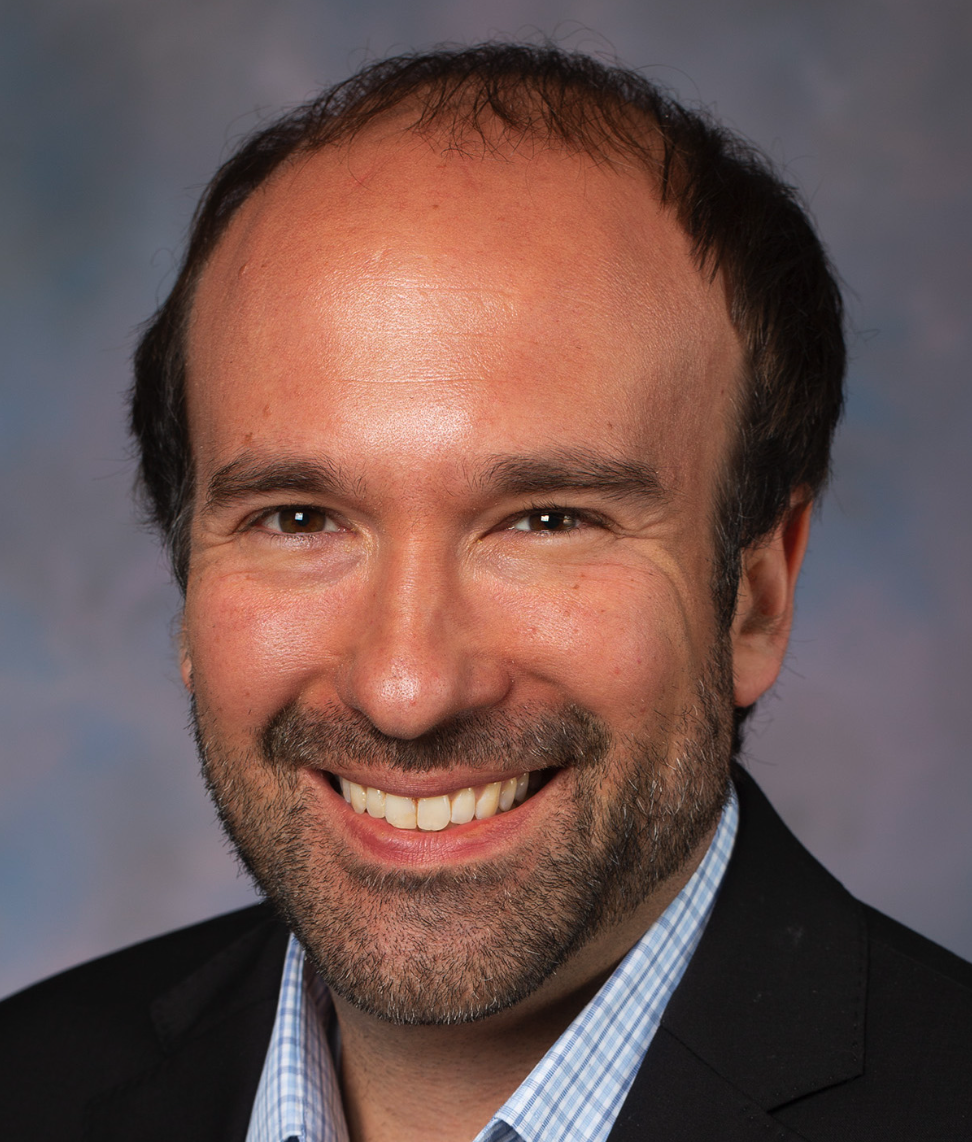 Benjamin Stanton合作论文数Department of Pediatrics, College of Medicine, The Ohio State University;Center for Childhood Cancer & Blood Diseases, Abigail Wexner Research Institute at Nationwide Children’s Hospital18
 Qianben Wang (王前奔)合作论文数Institute of Basic Medical Sciences, Chinese Academy of Medical Sciences16
 Zhong Chen合作论文数Department of Pathology, Duke University School of Medicine12
 Victor X. Jin合作论文数Comprehensive Cancer Center, The Ohio State University12
 Dayong Wu合作论文数10
 Tim Hui-Ming Huang合作论文数Department of Molecular Medicine, Long School of Medicine, The University of Texas Health Science Center at San Antonio9
 Ye Zhenqing合作论文数9
 Steven K. Clinton合作论文数Department of Urology, College of Medicine, The Ohio State University;Division of Medical Oncology, Department of Internal Medicine, Comprehensive Cancer Center, The Ohio State University8
 Berkley Gryder合作论文数Department of Genetics and Genome Sciences, School of Medicine, Case Western Reserve University;Case Comprehensive Cancer Center, School of Medicine, Case Western Reserve University7Meng Wang合作论文数Nationwide Childrens Hosp, Abigail Wexner Res Inst, Ctr Childhood Canc & Blood Dis, Columbus, OH 43210 USA6L. Wang合作论文数6Javed Khan合作论文数6
 Zhihua Liu (刘芝华)合作论文数Cancer Hospital, Chinese Academy of Medical Sciences6
 Jennifer Thomas-Ahner合作论文数Department of Pathology;Ohio State University;Department of Pathology, Ohio State University6
 Jiaoti Huang合作论文数Department of Pathology, School of Medicine, Duke University6
 Myron S Ignatius合作论文数Center for Molecular Neurobiology, The Ohio State University6Prethish Sreenivas合作论文数6
 Rodrigo Moreno-Campos合作论文数Dept Mol Med, Greehey Childrens Canc Res Inst GCCRI5Nicole R Hensch合作论文数The University of Texas at San Antonio, San Antonio, TX 78249, USA5
 Silvia Pomella合作论文数Univ Bologna, Dept Hematol Oncol L&A Seragnoli, Bologna, Italy5Rossella Rota合作论文数Department of Ophthalmology, University of L’Aquila, Aquila, Italy5Hankey William合作论文数5
 Xiang Zhao合作论文数National Oceanography Centre, University of Southampton5
 Peter Houghton合作论文数Department of Molecular Medicine, Long School of Medicine, The University of Texas Health Science Center at San Antonio;Department of Pediatrics, Long School of Medicine, The University of Texas Health Science Center at San Antonio;Greehey Children’s Cancer Research Institute, The University of Texas Health Science Center at San Antonio5
 Xiangtao Liu合作论文数State Key Lab Vet Etiol Biol, Lanzhou Vet Res Inst5Kunal Baxi合作论文数UT Hlth Sci Ctr, Greehey Childrens Canc Res Inst4
 Angelina V. Vaseva合作论文数Department of Pediatrics, Oregon Health Science University;School of Medicine, Oregon Health Science University4
 Kathryn Bondra合作论文数4
 Siyuan Zheng (郑思远)合作论文数jinan university4
 LaHaye Stephanie合作论文数4
 Jiangfei Chen (陈将飞)合作论文数School of Public Health and Management, Wenzhou Medical University4
 Pearlly S Yan合作论文数The James Comprehensive Cancer Center, The Ohio State University;Center for Clinical and Translational Science, The Ohio State University4
 Eleanor Chen合作论文数4Benjamin J. Kelly合作论文数3Connor Geraghty合作论文数3James R Fitch合作论文数3
 Xiaolong Cheng合作论文数Center for Genetic Medicine Research, Children’s National Hospital3Pei Zhao合作论文数3
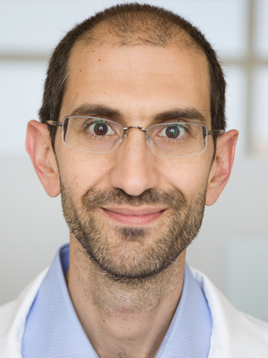 Emmanuel Antonarakis合作论文数Division of Hematology, Oncology, and Transplantation, Medical School, University of Minnesota;Masonic Cancer Center, University of Minnesota3Lu Changxue合作论文数3
 Xun Lan (蓝勋)合作论文数School of Medicine, Tsinghua University;Tsinghua-Peking Center for Life Sciences3
 Qien Wang合作论文数OHIO STATE UNIVERSITY3
 Peter White合作论文数3
 Qingfu Zhang合作论文数Department of Computer Science, College of Engineering, City University of Hong Kong;School of Artificial Intelligence, Xidian University3Cassandra Grenade合作论文数2
 Aaron Marks Horning合作论文数2Chiou-Miin Wang合作论文数Univ Texas Hlth Sci Ctr San Antonio, Dept Mol Med, San Antonio, TX 78229 USA2
 Hsien-Chao Chou合作论文数NCI, Genet Branch, NIH, Bethesda, MD 20892 USA2
 Chun-Liang Chen合作论文数Dept Mol Med, Univ Texas Hlth Sci Ctr San Antonio2




