We present here a quantitative ultrasound tomographic method yielding a sub-mm resolution, quantitative 3D representation of tissue characteristics in the presence of high contrast media. This result is a generalization of previous work where high impedance contrast was not present and may provide a clinically and laboratory relevant, relatively inexpensive, high resolution imaging method for imaging in the presence of bone. This allows tumor, muscle, tendon, ligament or cartilage disease monitoring for therapy and general laboratory or clinical settings. The method has proven useful in breast imaging and is generalized here to high-resolution quantitative imaging in the presence of bone. The laboratory data are acquired in ~ 12 min and the reconstruction in ~ 24 min—approximately 200 times faster than previously reported simulations in the literature. Such fast reconstructions with real data require careful calibration, adequate data redundancy from a 2D array of 2048 elements and a paraxial approximation. The imaging results show that tissue surrounding the high impedance region is artifact free and has correct speed of sound at sub-mm resolution.
Encouraged by progress over the past 10 years in breast imaging, we have generalized 3D ultrasound transmission tomography (UST) to orthopaedic, whole body and clinical applications. We document quantitative breast density measurement, quantitative transmission tomography in the presence of bone and air, and tumor monitoring applications, indicating that 3D UST is a whole body imaging modality with clinical implications.
We describe a novel 3-D ultrasound technology, the quantitative transmission ultrasound system and algorithm to image a pendent breast in a water bath. Quantitative accuracy is verified using phantoms. Morphological accuracy is verified using cadaveric breast and in vivo images, and spatial resolution is estimated. This paper generalizes an earlier 2-D algorithm to a full 3-D inversion algorithm and shows the importance of such a 3-D algorithm for artifact suppression as compared with the 2-D algorithm. The resultant high-resolution ultrasound images, along with quantitative information regarding tissue speed of sound/stiffness, provide a more accurate depiction of the breast anatomy and lesions, contributing to improved breast care.
Quantitative Transmission Ultrasound (QTUS) is a tomographic transmission ultrasound modality that is capable of generating 3D speed-of-sound maps of objects in the field of view. It performs this measurement by propagating a plane wave through the medium from a transmitter on one side of a water tank to a high resolution receiver on the opposite side. This information is then used via inverse scattering to compute a speed map. In addition, the presence of reflection transducers allows the creation of a high resolution, spatially compounded reflection map that is natively coregistered to the speed map. A prototype QTUS system was evaluated for measurement and geometric accuracy as well as for the ability to correctly determine speed of sound.
Water bath breast scanners utilize either ray based or inverse scattering techniques for the quantitative images. The 3D inverse scattering approach we use requires a 3D forward and back-propagation problem to be solved, resulting in ~1.2 mm resolution images. The resulting speed of sound map is then used in a 3D inhomogeneous, eikonal equation based reflection algorithm to account for refraction effects, and yield a 3D co-registered speckle free 360 degree compounded B-scan like volume. There is no harmful ionizing radiation, no compression, and no required contrast agents for proper utilization of our device. The quantitative transmission ultrasound (QTUS) images are independent of operator skill. The patient lies prone on a full length table with the breast pendant in the water bath. The breast is gently immobilized with the use of a breast retention pad that magnetically “attaches” to a magnetic retention rod for imaging of the breast. Scan time for each breast is approximately 10 min. We will show full 3D quantitative images obtained from consented, anonymous patients from several academic collaborating institutions. We will compare our QTUS images with mammographic, MRI, and hand held US images, and correlate biopsy results where appropriate.
Research in quantitative whole breast ultrasound imaging has been developing rapidly. Recently, we published results from 2D transmission inverse scattering algorithms, based on optimization, incorporating diffraction, refraction, and limited multiple scattering effects, using data collected from an early prototype, which showed the feasibility of high resolution quantitative imaging of the breast tissue speed and attenuation, and concomitant refraction corrected reflection imaging. However, artifact problems in speed and attenuation result from the 2D algorithms and the data characteristics. The reflection algorithm uses the speed map to model refractive effects of rays, so these artifacts are unacceptable. The 3D inverse scattering algorithm presented here, using data from a new prototype, overcomes most of these artifacts. We then use a 3D refraction corrected 360° compounded reflection algorithm for high resolution speckle free reflection images. We discuss the transmission and reflection algorithms and the advanced scanner used to collect the data, as well as initial clinical results from the Mayo Clinic, Breast Cancer Imaging Center, Orange County, and the University California, San Diego. We show examples of cysts, fibroadenomas, calcifications, cancers, and DCIS, in dense, fatty, and average breast tissue, and compare these with hand-held ultrasound, MRI, and mammography, where available.
A method was developed to map tissue properties of the entire breast including sound speed and attenuation using fully 3D nonlinear inverse-scattering tomography. Clinical measurements suggest that in breast tissue benign and cancerous lesions may be identified in part by these inherent acoustic parameters. Sound speed accuracy and linearity are very high over a wide range (1325-1700 m/sec) with ~1.5 mm resolution at 2 MHz in transmission mode. Attenuation tomograms provide image contrast over a wide range (0-4 dB/cm/MHz) and assist classification of masses. High resolution 0.6 mm volumetric reflection tomograms are acquired with bandwidth 2-8 MHz, are refraction-corrected with the transmission tissue data and are precisely registered in 3D with the transmission volumes. USCT promises an automated whole-breast scan providing a global view of the entire breast in 3D, facilitating comparison to prior exams in a reproducible geometry. Scanner design, automated operation and results of our trial with over 125 subjects with confirmed breast masses will be presented with detailed comparison to conventional sonography and MRI.
Recent published results in inverse scattering generally show the difficulty in dealing with moderate to high contrast inhomogeneities when employing linearized or iteratively linearized algorithms (e.g., distorted Born iterative method). This paper presents a fully nonlinear algorithm utilizing full wave field data, that results in ultrasound computed tomographic images from a laboratory breast scanner, and shows several such unique images from volunteer subjects. The forward problem, data collection process and inverse scattering algorithm used are discussed. A functional that represents the "best fit" between predicted and measured data is minimized, and therefore requires a very fast forward problem solver, Jacobian calculation, and gradient estimation, all of which are described. The data collection device is described. The algorithm and device yield quantitative estimates of human breast tissue in vivo. Several high resolution images, measuring ∼150 by 150 wavelengths, obtained from the 2D inverse scattering algorithms, using data collected from a first prototype, are shown and discussed. The quantitative values are compared with previous published work.
A full wave nonlinear inverse scattering algorithm is presented and resulting images shown
Acoustic shear waves of low frequency can be detected and measured using a phase contrast based magnetic resonance imaging technique called MR Elastography or phase measurement based ultrasound techniques. Spatio-temporal variations of displacements caused by the propagating waves can be used to estimate local values of the elasticity of the object being imaged. The currently employed technique for estimating the elasticity from the wave displacement maps, the local frequency estimator (LFE), has fundamental resolution limits and also has problems with shadowing and other refraction-related artifacts. These problems can be overcome with an inverse approach using Green's function integrals which directly solve the wave equation problem for the propagating wave. The complete measurements of wave displacements as a function of space and time over the object of interest obtained by the above techniques permit an iterative approach to inversion of the wave equation to obtain elasticity and attenuation maps.
Techniscan has completed a local phase of clinical trials of a unique inverse scattering breast cancer scanner. The scanner is based on transmission of plane waves from 0.3 to 2 MHz through the breast to a receiver on the opposite side of the breast. The receiver consists of a 2-D array of six rows by 160 columns. These elements are multiplexed by a factor of 12 into 80 analog to digital converters. The dual transducer-head design allows the inversion of the transmission data to create two independent quantitative images, one of speed of sound and a second of attenuation. The two heads are mechanically attached to a hollow rotating shaft, to provide 360 deg of mechanical motion. Typically, 180 to 360 steps per 360-deg rotation and 2-mm vertical steps are used to create a data set that is inverted into a 3-D image of 240 by 240 by 70 voxels. A unique set of custom circuit boards was constructed to implement scanner electronics. The next scanner version with anticipated inverse scattering resolution of 1 mm or less and reflection tomography resolution of 0.5 mm or less should improve performance. Clinical images to date and performance will be discussed.
Ultrasound has been proposed as an adjunct diagnostic modality for 25+ years. However, initial studies were hampered by relatively low resolution. Over the past 20 years there has been a tremendous amount of research carried out in inverse scattering, the mathematical foundation behind quantitative determination of the coefficients of a partial differential equation. In particular, the research at TechniScan, Inc., and the Dept. of Bioengineering, Univ. of Utah, has culminated in algorithms that give approximately millimeter resolution, and quantitative accuracy, in 2-D and 3-D problems. We invert the full nonlinear acoustic inverse scattering problem (no Born/Rytov approximation) in 2-D and 3-D. We reconstruct a phantom with known inclusions. The image space is 241×241×135=∼7.8 Megapixels or larger. Each pixel is 0.8×0.8×1.0mm3 in size. The data is collected on a 6 by 160 element array, positioned 180 views surrounding the breast. The transmitted wave is a plane wave chirp. Reconstruction time is ∼2 h on a six-node cluster of Pentium Ms. This paper will discuss theoretical algorithm development, practical issues that result in images, and a device presently being used in a clinical setting. We show several reconstructed images from ∼400 volunteers.
 James Wiskin合作论文数29
 Michael J. Berggren合作论文数11Steven a Johnson合作论文数10s a johnson合作论文数7John Klock合作论文数QT Ultrasound LLC, Novato, CA 94949 USA6
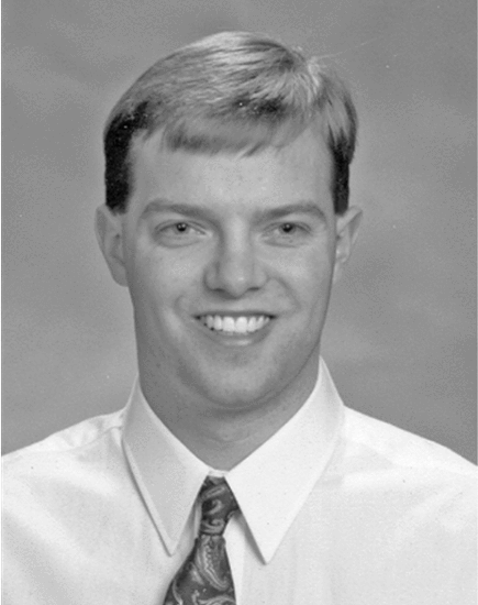 Steven A. Johnson合作论文数6m berggren合作论文数5barry k hanover合作论文数4
 Lenox, M.W.合作论文数CTI PET Syst. Inc.;c;CTI PET Syst. Inc.4
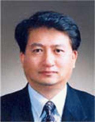 Whan Woo Kim合作论文数4
 Michael P Andre合作论文数3
 Om P. Gandhi合作论文数Department of Electrical Engineering, University of Utah3
 James F. Greenleaf合作论文数Mayo Clinic College of Medicine and Science;Basic Ultrasound Research Laboratory, Department of Physiology and Biomedical Engineering, Mayo Clinic3
 Richard Eidens合作论文数3Elaine Iuanow合作论文数2d a robinson合作论文数2V Dutt合作论文数2
 Raja Muthupillai合作论文数St. Luke's Episcopal Hospital2
 Richard L. Ehman合作论文数Mayo Clinic2
 Armando Manduca合作论文数Department of Physiology and Biomedical Engineering, Mayo Clinic2
 H. C. Frank Natterer合作论文数Fachbereich Mathematik und Informatik, Westfalische Wilhelms-Universitat Munster2Richard S. Eidens合作论文数2Steven Johnson合作论文数1Matthew A. Lewis合作论文数1linda k olson合作论文数1Frank Setinsek合作论文数1
 Bilal Malik合作论文数1T. Abbott合作论文数1
 Haydee Ojeda-Fournier合作论文数Department of Radiology, School of Medicine, University of California, San Diego1Scott Hsieh合作论文数1Nasser Pirshafiey合作论文数1Rajni Natesan合作论文数1Callahan K S合作论文数1
 Michael S. Zhdanov合作论文数Department of Geology & Geophysics, University of Utah1
 Dennis M. Sullivan合作论文数Department of Electrical and Computer Engineering, College of Engineering, University of Idaho1
 Bilal H. Malik合作论文数Dept of Electrical and Computer Engineering;Texas A&M University;Dept of Electrical and Computer Engineering, Texas A&M University1Y. Parisky合作论文数1S.A. Johnson合作论文数1




