Automatically predicting RNA 3D structure from sequence remains an unsolved challenge in biology and biotechnology. Here, we describe a Kaggle code competition engaging over 1700 teams and 43 previously unreleased structures to tackle this challenge. The top three submitted algorithms achieved scores within statistical error of the winners of the recent CASP16 competition. Unexpectedly, the top Kaggle strategy involved a pipeline for discovering 3D templates, without the use of deep learning. We integrated this template-modeling pipeline and other Kaggle strategies to develop a single model RNAPro that retrospectively outperformed individual Kaggle models on the same test set. These results suggest a growing importance of template-based modeling in RNA structure prediction.
The advent of single-particle cryogenic electron microscopy (cryo-EM) has enabled near-atomic resolution imaging of large macromolecules, enhancing functional insights. However, current cryo-EM refinement tools condense all single-particle images into a single structure, which can misrepresent highly flexible molecules like RNAs. Here, we combine molecular dynamics simulations with cryo-EM density maps to better account for the structural dynamics of a complex and biologically relevant RNA macromolecule. Namely, using metainference, a Bayesian method, we reconstruct an ensemble of structures of the group II intron ribozyme, which better matches experimental data, and we reveal inaccuracies of single-structure approaches in modeling flexible regions. An analysis of all RNA-containing structures deposited in the Protein Data Bank reveals that this issue affects most cryo-EM structures in the 2.5-4 Å range. Thus, RNA structures determined by cryo-EM require careful handling, and our method may be broadly applicable to other RNA systems.
Abstract Cryo-EM structure determination of protein-free RNAs has remained difficult with most attempts yielding low to moderate resolution and lacking nucleotide-level detail. These difficulties are compounded for small RNAs as cryo-EM is inherently more difficult for lower molecular weight macromolecules. Here we present a strategy for fusing small RNAs to a group II intron that yields high resolution structures of the appended RNA. We demonstrate this technology by determining the structures of the 86-nucleotide (nt) thiamine pyrophosphate (TPP) riboswitch aptamer domain and the recently described 210-nt raiA bacterial non-coding RNA involved in sporulation and biofilm formation. In the case of the TPP riboswitch aptamer domain, the scaffolding approach allowed visualization of the riboswitch ligand binding pocket at 2.5 Å resolution. We also determined the structure of the ligand-free apo state and observe that the aptamer domain of the riboswitch adopts an open Y-shaped conformation in the absence of ligand. Using this scaffold approach, we determined the structure of raiA at 2.5 Å in the core. Our versatile scaffolding strategy enables efficient RNA structure determination for a broad range of small to moderate-sized RNAs, which were previously intractable for high-resolution cryo-EM studies.
Branching is a critical step in RNA splicing that is essential for 5′ splice site selection. Recent spliceosome structures have led to competing models for the recognition of the invariant adenosine at the branch point. However, there are no structures of any splicing complex with the adenosine nucleophile docked in the active site and positioned to attack the 5′ splice site. Thus we lack a mechanistic understanding of adenosine selection and splice site recognition during RNA splicing. Here we present a cryo-electron microscopy structure of a group II intron that reveals that active site dynamics are coupled to the formation of a base triple within the branch-site helix that positions the 2′-OH of the adenosine for nucleophilic attack on the 5′ scissile phosphate. This structure, complemented with biochemistry and comparative analyses to splicing complexes, supports a base triple model of adenosine recognition for branching within group II introns and the evolutionarily related spliceosome.
Chung et al. recently presented the structure of a primitive group IIC intron with its DNA target, which reveals the structural requirements that this class of intron uses to recognize a transcription terminator stem loop at the DNA level for insertion during retrotransposition.
Recent cryo-EM structures of a group II intron caught in the process of invading DNA have given new insight into the mechanisms of both splicing and retrotransposition. Conformational dynamics involving the branch-site helix domain VI are responsible for substrate exchange between the two steps of splicing. These structural rearrangements have strong parallels with the movement of the branch-site helix in the spliceosome during catalysis. This is strong evidence for the spliceosome evolving from a group II intron ancestor. We observe other topological changes in the overall structure of the catalytic domain V that may occur in the spliceosome as well. Therefore, studying group II introns not only provides us with insight into the evolutionary origins of the spliceosome, but also may inform the design of experiments to further probe structure-function relationships in this eukaryotic splicing apparatus. This article is categorized under: RNA Processing > Splicing Mechanisms RNA Structure and Dynamics > RNA Structure, Dynamics, and Chemistry RNA Structure and Dynamics > Influence of RNA Structure in Biological Systems RNA Evolution and Genomics > RNA and Ribonucleoprotein Evolution
Group II introns are a class of retroelements that invade DNA through a copy-and-paste mechanism known as retrotransposition. Their coordinated activities occur within a complex that includes a maturase protein, which promotes splicing through an unknown mechanism. The mechanism of splice site exchange within the RNA active site during catalysis also remains unclear. We determined two cryo-EM structures at 3.6-Å resolution of a group II intron reverse splicing into DNA. These structures reveal that the branch-site domain VI helix swings 90°, enabling substrate exchange during DNA integration. The maturase assists catalysis through a transient RNA-protein contact with domain VI that positions the branch-site adenosine for lariat formation during forward splicing. These findings provide the first direct evidence of the role the maturase plays during group II intron catalysis. The domain VI dynamics closely parallel spliceosomal branch-site helix movement and provide strong evidence for a retroelement origin of the spliceosome.
 Navtej Toor合作论文数Department of Chemistry and Biochemistry, Division of Physical Sciences, University of California, San Diego7
 Dmitry Lyumkis合作论文数2Xiaodong Yan合作论文数1Anna Lewicka合作论文数1P JANOS合作论文数1Maggie Demkin合作论文数1
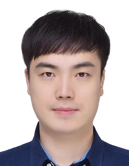 Youhan Lee合作论文数Kakao Brain Corp1
 Chaitanya K. Joshi合作论文数Department of Computer Science & Technology, Clare Hall College, University of Cambridge1
 Shanshan Li (李珊珊)合作论文数School of Life Sciences, Division of Life Sciences and Medicine, University of Science and Technology of China1Shujun He合作论文数1
 Kai Ming Zhang (张开明)合作论文数1Jian Zou (邹剑)合作论文数1
 Zhaoming Su (苏昭铭)合作论文数West China Hospital of Sichuan University1
 Rhiju Das合作论文数Biochemistry Department, Stanford University1
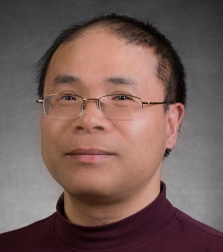 Shi-Jie Chen合作论文数Department of Physics and Astronomy, College of Arts and Science, University of Missouri1
 Koirala Deepak合作论文数Department of Chemistry &Biochemistry, Kent State University;Department of Chemistry & Biochemistry, Kent State University1
 Joseph Piccirilli合作论文数Department of Chemistry, The University of Chicago1Miao Zhichao合作论文数Institut de biologie moléculaire et cellulaire du CNRS, Université de Strasbourg1
 Giovanni Bussi合作论文数Molecular and Statistical and Biophysics group, Scuola Internazionale Superiore di Studi Avanzati1
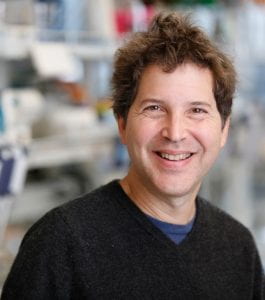 David Baker合作论文数Institute for Protein Design, University of Washington;Department of Biochemistry, University of Washington1Baker Timothy S合作论文数1
 Massimiliano Bonomi合作论文数Dept Chem, Univ Cambridge1
 Cheng Zhang合作论文数1
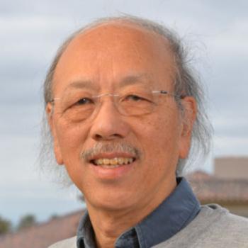 Wah Chiu合作论文数Departments of Bioengineering, School of Engineering, Stanford University;Department of Microbiology and Immunology, School of Medicine, Stanford University;SLAC National Accelerator Laboratory1
 John Moult合作论文数Institute for Bioscience and Biotechnology Research, University of Maryland1
 Kevin Weeks合作论文数Department of Chemistry, College of Arts and Sciences, The University of North Carolina at Chapel Hill;Ribometrix1
 Alessandra Magistrato合作论文数SISSA, CNR IOM DEMOCRITOS Natl Simulat Ctr1
 Marco Marcia合作论文数Dept Mol Membrane Biol, Max Planck Inst Biophys1
 Eric Westhof合作论文数Institut de Biologie Moleculaire et Cellulaire, CNRS;University of Strasbourg;Université Louis Pasteur1
 Walter Reade合作论文数Advanced Combustion Engineering Research Center (ACERC), Brigham Young University1
 Andriy Kryshtafovych合作论文数Protein Structure Prediction Center, University of California, Davis1




