CASE:A patient presented with midline back pain radiating into the left lower extremity. Magnetic Resonance images revealed an abnormal midline dorsal structure described as a facet cyst. Rupture was attempted by spinal needle under fluoroscopic guidance. His pain returned in less than 2 weeks. Further study of the images showed the structure was more medial and cephalad than typical facet cysts, leading to a revised diagnosis of Baastrup disease. The cyst was surgically removed by minimally invasive techniques; an uninstrumented spinal arthrodesis was performed. CONCLUSION:Baastrup disease is commonly misdiagnosed. Despite the delay in treatment, surgical intervention was effective.
STUDY DESIGN:Retrospective matched cohort study.SUMMARY OF BACKGROUND DATA:With a growing interest in minimally invasive spine surgery (MIS), the question of which technique is the most advantageous for patients with low-grade degenerative lumbar spondylolisthesis (DLS) still remains unclear.OBJECTIVE:To compare patient-reported outcomes, perioperative morbidity, and rates of reoperation between MIS decompression with either unilateral noninstrumented facet fusion (MIS-F) or with transforaminal interbody fusion (MIS-T) for grade 1 DLS.METHODS:Twenty patients who underwent MIS-T and 20 patients with MIS-F were matched based on age, sex, and preoperative ODI, VAS back, and VAS leg. All patients had DLS with at least 4 millimeters of translation on standing radiographs. Exclusion criteria included prior level surgery, multilevel instability, disk impinging on the exiting nerve root, spondylolisthesis from significant facet arthropathy, or foraminal compromise from disk collapse. ODI, VAS back, VAS leg, and patient satisfaction measured by the North American Spine Society questionnaire were tracked at 3, 6, 12, and 24 months postoperatively. Minimum clinically important differences and substantial clinical benefits were calculated.RESULTS:MIS-F and MIS-T resulted in decreased ODI at 3, 6, and 12 months following the index procedure. Sixty percent of MIS-F and 83% of MIS-T patients reached minimum clinically important difference at 1 year postoperatively; however, using the threshold of 30% ODI reduction from baseline, 67% of MIS-F and 83% MIS-T ( P = 0.25) achieved this goal. Forty-three percent of MIS-F and 59% of MIS-T patients met substantial clinical benefits. Satisfaction at 1 year, measured by a score of 1 or 2 on the North American Spine Society questionnaire, was 64% for MIS-F and 83% for MIS-T.CONCLUSIONS:MIS-F and MIS-T are effective treatment options for spinal stenosis associated with low-grade DLS. Both techniques result in comparable patient-reported outcomes and satisfaction up to 2 years and have similar long-term reoperation rates. More evidence is required to delineate optimal selection characteristics for MIS-F versus MIS-T.
Pediatric olecranon osteochondral flap fractures are shear injuries of the humeroulnar joint with elevation of an articular cartilaginous flap from the subchondral bone of the olecranon articular surface. All previously reported cases included an osteochondral flap containing the coronoid, with varying imaging and fixation methods used. We treated 2 pediatric patients with this injury. One of our patients had a large, displaced osteochondral fracture of the medial semilunar notch including the coronoid, in addition to avulsions of the medial flexor mass and distal ulnar collateral ligament. The other patient sustained a displaced, rotated osteochondral fracture including the coronoid with subsequent humeroulnar subluxation. Both fractures were anatomically reduced and fixed with absorbable suture, leading to excellent results at 1 year. A high degree of suspicion, thorough work-up, and anatomical reduction of all injured structures are paramount in treatment of this rare, difficult-to-diagnose injury. Copyright (C) 2021 by the American Society for Surgery of the Hand. All rights reserved.
Case: Lumbar paraspinal compartment syndrome (LPCS) is a rare but potentially catastrophic pathology caused by increased intracompartmental pressures leading to potential ischemic necrosis of muscle, local denervation, and complications of rhabdomyolysis. A combination of unique clinical history and examination findings, laboratory findings, and radiologic imaging help in diagnosis. Conclusion: Early detection and intervention with minimally invasive percutaneous fasciotomy can minimize local soft tissue trauma while providing extensive fascial release with good clinical outcomes. Radiographic follow-up in this case suggests that the muscle at risk in LPCS can be spared with early surgical intervention.
Hip fractures are one of the most common major orthopaedic injuries in the United States, with an incidence that is growing with the aging population. These fractures vary significantly in anatomy, pathogenesis, and treatment. Orthopaedists must be able to carefully distinguish between femoral neck, intertrochanteric, and subtrochanteric femoral fractures because diagnosis is essential in guiding treatment. The extent of complex decision-making and management is often underappreciated in this diverse group of fractures. In this review, the relevant background, anatomy, and current treatment considerations essential to optimize management of these common and significant injuries are discussed.
Surgery still offers a cure to the majority of intramedullary tumours. The challenge of the surgery is taking out the tumour while preserving the function. Very often the patient has a huge tumour with very minimal symptoms, such as mild numbness. The slow growth of the tumour nature provides enough time for the cord fibres to adapt accordingly. Usually, the motor function is preserved and most of the pathologies are benign. For this reasons, the majority of cases have good long-term tumour control. The functional outcome is depending on the preoperative functional state, especially for motor function. The patient should be educated prior to surgery, especially to anticipate the post-surgical rehabilitation period. The surgical technique should preserve the motor function, but the fine movement usually gets worst for several months after surgery and slowly recover within 6 months. We share our experience of 45 surgical cases with intramedullary tumour (14F;31M), the pathologies distributions are 20 ependymomas, 8 astrocytomas (1/8 anaplastic astrocytoma), 7 cavernomas, 8 hemangioblastomas, 1 glioblastoma multiforme, and 1 tuberculoma. The location distribution varied from 27 at the cervical cord, 11 thoracal, 4 thoracolumbal, and 3 MO-upper cervical. The surgical outcome for all cases experienced sensory changes and recovered over 6 months. Almost all cases experienced some degree of spasticity and fine movement difficulty and they are improving over 6 months. Motor strength is usually preserved. One case of GBM, improve gradually for the first two months then followed by the disease course regardless of the treatment. The tuberculoma case required one year to recover her neurological function with adequate treatment.
 David G. Kline合作论文数School of Medicine, Louisiana State University25
 Murovic Judith A合作论文数13Max C. Lee合作论文数13Ung-Kyu Chang合作论文数12
 Salahadin Abdi合作论文数Miller School of Medicine, University of Miami8
 Ho-Yeol Zhang合作论文数7Robert L. Tiel合作论文数6Midha Rajiv合作论文数5
 Hoang Le合作论文数5Shane V. Abdunnur合作论文数4Sang-Kook Lee合作论文数4
 Richard G. Fessler合作论文数Department of Neurosurgery, RUSH Medical College, Rush University4Alan R. Hudson合作论文数4R. Fessler合作论文数3
 Vincent C. Traynelis合作论文数Department of Neurosurgery, Medical College, Rush University3
 Dosang Cho合作论文数3Fahim Daniel K合作论文数3Saetia Kriangsak合作论文数3
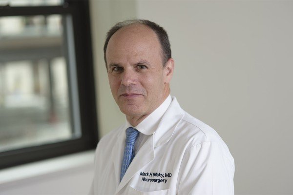 Mark H. Bilsky合作论文数Multidisciplinary Spine Tumor Center, Memorial Sloan Kettering Cancer Center;Weill Medical College, Cornell University3
 Se-Hoon Kim合作论文数Department of Neurosurgery, Ansan Hospital, Korea University Medical Center3
 Paul C Mccormick合作论文数Irving Medical Center, Columbia University;NewYork-Presbyterian Allen Hospital;College of Physicians and Surgeons, Columbia University;The Och Spine Hospital, The Neurological Institute of New York3Gun Choi合作论文数3Eric L. Zager合作论文数3T. Jesse Lim合作论文数3Adriane Nelson合作论文数3Kurtus Dafford合作论文数3
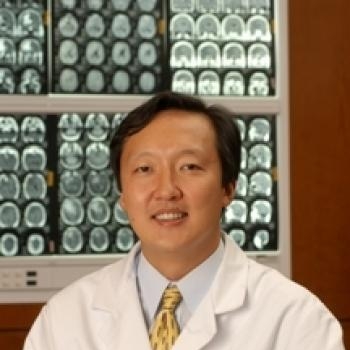 Jon Park合作论文数Neurosurgery Department, Stanford University3Issada Thongtrangan合作论文数3
 Raju Balabhadra合作论文数3Ashwin Viswanathan合作论文数Department of Neurosurgery, M.D. Anderson Cancer Center3F. Cammisa合作论文数2Sang Hyun Park合作论文数2K. Hussey合作论文数2
 Kedar Padhye合作论文数Division of Orthopaedic Surgery, IWK Health Centre2Kim Keung Nyun合作论文数2Paterno Josemaria合作论文数2
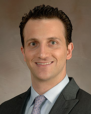 Yoshua Esquenazi合作论文数Department Of Neurosurgery, University of Texas Health Science Center at Houston2Daniel K. Resnick合作论文数2R.K Sinha合作论文数2
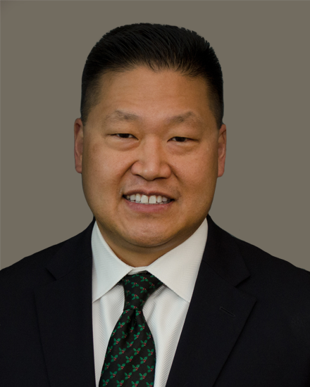 Michael Yung-Shun Wang合作论文数Department of Neurological Surgery, Miller School of Medicine, University of Miami;University of Miami Hospital2
 Edward C Benzel合作论文数Department of Neurosurgery, Neurological Institute, Cleveland Clinic;Cleveland Clinic Lerner College of Medicine, Case Western Reserve University2
 Volker K. H. Sonntag合作论文数Barrow Neurological Institute2Seong Yi合作论文数2John Gachiani合作论文数2Stephen M. Russell合作论文数2Hyun Chul Shin合作论文数2Dong Yeob Lee合作论文数2
 Sang-Ho Lee合作论文数Wooridul Spine Hospital2




