OBJECTIVE:Flexible approach to treating Zenker's diverticulum (ZD) has gained traction in recent years. However, objective measures comparing pre- and post-surgical outcomes have not been reported. The purpose of this investigation is to describe the changes in quantitative measurements in videofluoroscopic swallowing studies (VFSS) after flexible Zenker's diverticulotomy using modern per oral endoscopic myotomy (Z-POEM) technique. METHODS:A retrospective review of all patients undergoing the Z-POEM technique for treatment of ZD was conducted from July 2022 to December 2024. The primary fluoroscopic outcome measures were the size of the diverticulum, pharyngoesophageal segment opening (PESo) in lateral and anterior-posterior (AP) views, pharyngeal constriction ratio (PCR), and penetration aspiration scale (PAS) score. The secondary outcome measure was the Eating Assessment Tool-10 (EAT-10) score. RESULTS:Twenty-nine patients had pre- and post-surgical VFSS performed. The mean age was 74.1 years (±9.5) and 55% were male. The mean diverticulum size decreased from 1.076 cm (±0.702) to 0.469 cm (±0.573), p < 0.001. The mean PESo in lateral improved from 0.392 cm (±0.201) to 0.709 (±0.303), p < 0.001. The mean PCR increased from 0.0746 (±0.116) to 0.0583 (±0.102), p = 0.03. The mean EAT-10 score decreased from 16.59 (±8.7) to 6.35 (±7.03) (p = 0.001). There was no significant difference in postoperative PAS score or PESo in the AP view. CONCLUSION:Our data indicate that flexible surgery for ZD using the Z-POEM technique is both fluoroscopically and clinically successful in improving swallowing dysfunction in patients suffering from Zenker's diverticulum. LEVEL OF EVIDENCE: 4:
Objective Examine the association between age and treatment outcomes in conditions causing single- versus multilevel airway restriction. Study Design Multi-institutional retrospective cohort study. Setting Tertiary laryngology centers. Methods Participants included younger (18-64 years) and geriatric (>= 65 years) adults with posterior glottic stenosis (PGS), multilevel airway stenosis (MLAS), and bilateral vocal fold paralysis (BVFP). Subgroup demographics, comorbidities, type, and etiology of airway compromise were described. Associations between age and primary outcome variables (i.e., tracheostomy and decannulation rates, number of surgeries performed, time between surgeries, and change in quality-of-life patient-reported outcome measures [PROMs]) were evaluated. Statistical analyses included independent t tests, chi(2), Fisher's exact, or Mann-Whitney tests. Results In 158 patients [96 younger (30 PGS, 29 MLAS, 37 BVFP) and 62 geriatric (24 PGS, 9 MLAS, 29 BVFP)], age differences were not significant for gender (P = .990), tracheostomy placement (70% vs 66%, P = .629), or decannulation success (40% vs 24%, P = .091) in younger versus geriatric groups, respectively. In younger patients, MLAS was more common (30.2% vs 14.5%, P = .024), and BVFP patients were more likely to decannulate (50% vs 12%, P = .017). Geriatric patients were more likely to have a history of prior radiation (26% vs 10%, P = .016), stenosis due to malignancy (23% vs 9%, P = .022), and fewer overall surgeries (median 1 vs 3, P = .003). Median PROMs were comparable between age subgroups (P > .05). Conclusion Younger adults underwent more surgeries, but overall comorbidities, tracheostomy decannulation rates, and PROMs were comparable between groups. Age does not negatively impact treatment outcomes and should not be a deterrent in treatment decision-making.
BACKGROUND:Malnutrition is a major consequence of head and neck cancer (HNC), often leading to decreased skeletal muscle mass and impacting survival. The goal of this study is to determine the effect of sarcopenia, as defined by skeletal muscle index (SMI), on survival in patients with HNC. METHODS:This is a retrospective review of patients with HNC treated with surgery and/or radiation at a single tertiary care institute. All had pre-treatment imaging available for skeletal muscle index (SMI) measurements, and SMI was calculated at the level of the 3rd cervical vertebra. Sarcopenia was defined as an SMI < 41 cm2/m2 in females and as <43 cm2/m2 in underweight or healthy weight males. Sarcopenia was defined as <53 cm2/m2 in overweight or obese males. Chi-square analysis was performed to compare recurrence and survival rates, and survival analysis was performed via Kaplan-Meir curve. RESULTS:Hundred and twelve patients with HNC were evaluated, 84 men and 28 women with an average age of 60.9 years. Tumors were primarily located in the oral cavity (24.1%) and oropharynx (42%). The majority (69.6%) underwent surgery. Mean body mass index prior to treatment was 28. Sixty-nine patients (61.6%) in our cohort had low SMI. Mean follow-up was 3.9 ± 2.2 years. Recurrence rate was 26% in those with low SMI versus 2% in those without. Patients with low SMI were more likely to have a recurrence (p = 0.02). Overall survival was 72.5% in those with low SMI and 81% in those with normal SMI (p = 0.09). CONCLUSIONS:Defining sarcopenia as a low skeletal muscle index at the third cervical vertebra is clinically relevant. This study demonstrates that low SMI at this level, and thus sarcopenia, was strongly associated with higher rates of recurrence.
Importance:Late effects of head and neck cancer (HNC) treatment include profound dysphagia, chronic aspiration, and death. Functional laryngectomy (FL) can improve patient survival and quality of life (QoL); however, removing a failing larynx for a noncancer reason is a difficult decision. Data regarding the ability of FL to improve self-perceptions of voice, swallowing, and QOL in survivors of HNC with intractable aspiration are inconclusive. Objective:To investigate the association of FL with changes in self-reported perceptions of voice, swallowing, oral intake, QoL, and mood in survivors of HNC experiencing profound dysphagia and intractable aspiration. Design, Settings, and Participants:This cohort study was conducted at a single academic institution and included survivors of HNC with profound swallowing dysfunction and intractable aspiration who underwent FL from July 2016 through March 2022. Of the initial 22 patients enrolled, 2 patients (15%) died of aspiration pneumonia before receiving FL. Data analyses were performed from July 2016 through March 2023. Main Outcomes and Measures:Self-reported measures of voice using the VHI (30-item Voice Handicap Index), swallowing using the EAT-10 (10-item Eating Assessment Tool), functional oral intake scale using the FOIS (Functional Oral Intake Scale), and quality of life using the FACT-H&N (Functional Assessment of Cancer Therapy-Head & Neck) were assessed before FL and at 1, 3, and 6 months after FL. Mood states were evaluated using the POMS (Profile of Mood States, second edition), before FL and at 6 months after FL. Results:The study analyses included 20 patients (mean [SD] age, 72.4 (7.0) years; 19 [95%] males and 1 [5%] female) who underwent FL and had complete data across all time points. Among these, 12 patients (60%) had received chemoradiation for oropharyngeal, 7 (35%) for laryngeal, or 1 (5%) for nasopharyngeal cancer. The mean (SD) time from completion of oncologic treatment to FL was 15.5 (5.5) years. Mean (SD) score on the EAT-10 improved from 33.2 (7.4) to 23.1 (10.8) at 1 month; 12.1 (9.1) at 3 months; and 8.3 (7.4) at 6 months, with a large effect size (η2 = 0.72; 95% CI, 0.54-0.80). Mean (SD) score on the FOIS improved from 2.0 (1.5) to 2.9 (1.7) at 1 month; 4.8 (2.5) at 3 months; and 5.2 (1.7) at 6 months, with a large effect size (η2 = 0.6; 95% CI, 0.38-0.71). Improvement in oral intake was achieved in 19 patients (95%), and feeding tubes were removed in 10 of 16 patients (63%) who were feeding tube-dependent; 6 patients (27%) continued to require supplemental tube feedings. Mean (SD) score on the VHI improved from 63.6 (34.0) to 86.9 (33.7) at 1 month; 71.3 (36.1) at 3 months; and 39.7 (26.9) at 6 months, with a large effect size (η2 = 0.42; 95% CI, 0.19-0.56). Seventeen patients (85%) were able to use a tracheoesophageal voice prosthesis for alaryngeal communication. Mean (SD) score on the FACT-H&N improved from 86.2 (17.8) to 93.6 (18.4) at 1 month; 109.0 (18.4) at 3 months; and 121.0 (16.8) at 6 months, with a large effect size (η2 = 0.64; 95% CI, 0.42-0.74). Mean (SD) score on the POMS improved from 58.9 (13.2) at baseline to 44.5 (9.9) at 6 months, with a large effect size (Cohen d = 1.04; 95% CI, 0.48-1.57). None of the patients experienced major complications of FL; 1 patient (5%) had a postoperative pharyngocutaneous fistula. Conclusions and Relevance:The findings of this cohort study indicate that FL was associated with marked improvements in self-perception of voice and swallowing, functional oral intake, QoL, and mood state among survivors of HNC. These findings can serve as a framework for FL counseling among HNC survivors experiencing profound dysphagia and intractable aspiration.
OBJECTIVE:Sarcopenia, characterized by decreased skeletal muscle mass, is associated with poorer oncologic outcomes in head and neck cancer (HNC) patients. The effect of sarcopenia on swallowing following HNC treatment is unknown. This study aims to investigate the association of sarcopenia and swallowing dysfunction in patients treated for HNC. STUDY DESIGN:Retrospective cohort study. SETTING:Academic medical center. METHODS:Pretreatment sarcopenia was assessed using the skeletal muscle index calculated from cross-sectional imaging at the third cervical vertebra. Feeding tube dependence, patient-reported dysphagia, and swallowing safety were assessed before and after treatment with the Functional Oral Intake Scale, Eating Assessment Tool-10, and Penetration Aspiration Scale, respectively. The association between sarcopenia and swallowing dysfunction was evaluated. RESULTS:A total of 112 patients were included, 84 males (75%) and 28 females (25%). A total of 69 (61.6%) had sarcopenia prior to initiating HNC therapy. Sarcopenia was significantly associated with an elevated risk of patient-reported dysphagia (odds ratio [OR] = 2.71 [95% confidence interval, CI, 1.12-6.79]; P < .05). Multivariate logistic regression demonstrated that sarcopenia (OR = 15.18 [95% CI, 1.50-453.53]; P < .05) is an independent predictor for aspiration following treatment for HNC. CONCLUSION:Patients with pretreatment sarcopenia had higher rates of dysphagia before treatment and were more likely to develop aspiration after completion of HNC therapy. Sarcopenia is readily measured using cross-sectional imaging and may be useful for identifying patients at risk of swallowing dysfunction and those most likely to benefit from prehabilitation efforts.
ObjectiveWe sought to validate a laryngeal simulation model and subsequently demonstrate its efficacy in improving surgical technique.Study DesignPre-post interventional study.SettingOtolaryngology Program at a Tertiary Care Center.MethodsA low-cost, high-fidelity laryngeal model was created using a 3-dimensional-printed cast and multilayered silicone to mimic vocal fold lesions. Participants (attendings and trainees) were first given a series of tasks including mucosal vocal fold lesion resection and microflap excision of a submucosal lesion. Trainees were then provided with an instructional video from a laryngologist and asked to repeat the same tasks on the model. Performance data was then assessed using validated surveys and blinded expert reviewers.ResultsEighteen participants completed the simulation. All subjects agreed that the "simulation experience was useful" and 93% agreed "the simulator helped improve my ability to do microsurgical tasks." In the postinstruction self-evaluation, trainees reported a significant decrease in mental demand (95% confidence interval [CI]: 0.37-0.91; P = .038) and significant increase in subjective performance (95% CI: 1.51-51.89; P = .016) compared to the preinstruction self-evaluation. On the postinstruction attempt, there was a significant improvement in all domains of the adapted objective structured assessment of technical skills as measured by 3 blinded, expert reviewers.DiscussionThis study demonstrates the usefulness of a silicone larynx model and the value of instructional video in developing laryngeal microsurgical skills. Participants positively reviewed the laryngeal model and trainees saw both a subjective and objective improvement indicating tangible operative benefits from the use of this laryngeal simulation.
Endoscopic Zenker's diverticulotomy (EZD) is typically performed via stapling (endoscopic staple diverticulotomy; ESD) or CO2 laser (endoscopic laser diverticulotomy; ELD). Conflicting reports exist on which approach provides optimal outcomes. This investigation compared objective fluoroscopic data between ESD and ELD.
OBJECTIVE:To compare rates of sialadenectomy in a veteran population before and after introduction of sialendoscopy.MATERIALS AND METHODS:All Veterans Affair (VA) Northern California Healthcare System (NCHS) encounters from 2006 to 2021 for benign obstructive salivary etiologies were identified using International Classification of Disease 9/10 codes. This cohort was then cross referenced with Current Procedural Terminology codes to identify patients who underwent a procedure for their salivary pathologies. The rates of sialadenectomy and minimally invasive procedures were measured before and after sialendoscopy was introduced to the VA NCHCS in 2016. Data was obtained via chart review and demographic information, diagnosis, and procedure type were extracted. Rates of sialadenectomy, minimally invasive procedures, and other patient and procedural characteristics were compared between the Pre-Sialendoscopy Era (PSE) and Sialendoscopy Era (SE). Statistical analysis was performed using Microsoft Excel (Microsoft, version 16.66).RESULTS:An increasing number of patients per year sought care for benign obstructive salivary pathology in the SE when compared to the PSE cohort and a higher rate of female patients were treated in the SE cohort. A simultaneous reduction in sialadenectomy rates and increase in minimally invasive procedure rates was observed in the SE. Sialendoscopy represented most of the increase in minimally invasive techniques, but there was a slight increase in other procedure types. Submandibular gland obstructive pathologies required the most procedures and resulted in the most gland excisions.CONCLUSION:This retrospective review strongly suggests introducing sialendoscopy reduced the incidence of sialadenectomy for benign non-tumor obstructive pathology in this VA NCHS population. Further prospective studies are needed to evaluate this in a more generalized setting.
INTRODUCTION:Dysphagia can result in malnutrition, dehydration, social isolation, depression, pneumonia, pulmonary abscess, and death. The effect of dysphagia on the health and quality of life (QOL) of the life partners of persons with dysphagia is uncertain. We hypothesize that the partners of individuals with significant dysphagia will experience a significant reduction in quality of life. PURPOSE:To evaluate the QOL of the significant others of persons with swallowing dysfunction. METHODOLOGY:Persons with significant swallowing dysfunction (defined as EAT10® > 10) and their significant others were prospectively administered the 10-item Eating Assessment Tool (EAT10®) patient-reported outcome measure and the 12-item SF quality of life instrument (SF12). Summary data from the 8 mean health domains were compared between patients and their significant others. Mean scores for each domain are calibrated at 50, and a score below 47 implies significantly diminished QOL for a particular domain. RESULTS:Twenty-three couples were evaluated. The mean ± SD EAT10 score for persons with significant dysphagia was 21 ± 7; mean EAT10 for their significant others or for couples in the control group was 0.3 ± 0.8. The mean physical health composite score (PCS) was significantly lower for patients with dysphagia compared to their significant others (39.1 ± 10 and 46.2 ± 11, respectively) (p < 0.05). Both patients and their significant others had comparable mean mental health composite scores (MCS) of 46.6 ± 10 and 46.4 ± 10, respectively (p > 0.05). CONCLUSION:Although significant others of persons with swallowing dysfunction have higher physical well-being than their partners, they exhibit the same reduction in mental well-being, which is significantly lower than the general population. The data suggest that clinicians should address the mental well-being of the partners of persons with severe swallowing dysfunction.
OBJECTIVE:To evaluate the safety and biocompatibility of bone wax as an implant material for medialisation laryngoplasty in a large animal model.METHODS:Three Dorper-cross ewes underwent type I thyroplasty of the right vocal fold with bone wax. The animals were monitored for four weeks for general wellbeing. The animals were euthanised and the larynges harvested. Histological evaluation was performed to assess for adverse tissue reaction and biocompatibility.RESULTS:The mean (± standard deviation) amount of bone wax implanted was 0.49 g (± 0.12 g). No adverse events were reported. Ex vivo vibration was present on high-speed imaging for all medialised vocal folds. Histology demonstrated implanted paraffin embedded within the thyroarytenoid muscle with no evidence of resorption, a minimal inflammatory infiltrate, and a thin fibrotic capsule.CONCLUSION:The results of this investigation suggest that bone wax may be a safe and efficacious implant material for medialisation laryngoplasty. Further studies are necessary to assess its long-term safety and efficacy.
Abstract Objectives To evaluate the feasibility of using Calcium Hydroxylapatite (CaHA) to augment the tongue base for patients with swallowing impairment due to tongue base atrophy. Methods A fresh human cadaver was obtained through the institution's body donation program and baseline lateral fluoroscopic images were obtained. A total of 2 mL of CaHA (Prolaryn Plus) were injected into three sites of the base of tongue under flexible endoscopic guidance with a 22G, 1.5‐inch needle (Monoject, Cardinal Health). Post‐lateral fluoroscopic images were obtained and pharyngeal area (cm2) and tongue base to pharyngeal wall distance (cm) was measured pre‐ and postinjection using SwallowTail fluoroscopic measurement software (Belldev Medical). Results The procedure was easily performed and the CaHA flowed easily into the cadaveric tongue without evidence of extrusion. The pre‐procedural pharyngeal area decreased from 24.36 to 23.14 cm after augmentation. The base of tongue to pharyngeal wall distance decreased from 2.21 to 1.32 cm after augmentation. Conclusion Tongue base augmentation with CaHA may be a feasible adjuvant therapy for the management of swallowing impairment secondary to tongue base atrophy. Further investigation is necessary to evaluate the clinical safety and efficacy. Level of Evidence 4
Abstract Objectives Vocal fold medialization is commonly performed for glottic insufficiency and vocal fold immobility. Currently available materials are temporary injectables or synthetic implants. Acellular scaffolds may allow vocal fold augmentation with autologous tissue via host cell migration. The purpose of this investigation was to evaluate the use of a novel carbohydrate scaffold as a medialization implant. Study Design Animal model. Setting Academic medical center. Methods Unilateral type I medialization thyroplasty was performed in 3 Dorper cross ewes using a hypercrosslinked carbohydrate polymer (HCCP) scaffold. Animals were monitored for 4 weeks for general well‐being, dyspnea, and weight loss. The animals were euthanized at 4 weeks and the larynges harvested. Histologic evaluation was performed to assess for adverse tissue reaction, migration, degradation, and biocompatibility. Results No adverse events were reported. No animals lost weight or displayed evidence of dyspnea. Histology demonstrated ingrowth of host cells and neovascularization with minimal peri‐implant inflammatory reaction. Cellular ingrowth into the scaffold was predominately made up of fibroblasts and early inflammatory cells. Scaffold shape was grossly maintained as it underwent degradation and replacement with host tissue. Migration of the implant material was not observed. Conclusion Vocal fold medialization in an ovine model with an HCCP scaffold resulted in the ingrowth of host cells with minimal peri‐implant inflammation. Scaffold shape was maintained without evidence of migration as it underwent replacement with host tissue. Further research is required to assess long‐term efficacy in comparison to currently available implants.
OBJECTIVES:Chemoradiation therapy (CRT) for head and neck cancer can have profound effects on swallowing function. Identification of risk factors for development of swallowing dysfunction after CRT may improve allocation of preventive strategies. The purpose of this study is to determine the association between pre-CRT dysphagia and post-CRT swallowing dysfunction.STUDY DESIGN:Retrospective cohort study.SETTING:Academic medical center.METHODS:Patients with head and neck cancer were evaluated before and 3 months after CRT. Dysphagia was assessed with the Eating Assessment Tool (EAT-10). Swallowing dysfunction was evaluated by a videofluoroscopic swallow study and the Penetration-Aspiration Scale and swallowing safety was evaluated by the Functional Oral Intake Scale. The association between pre-CRT EAT-10 scores and post-CRT swallowing dysfunction was evaluated.RESULTS:The mean ± SD post-CRT Functional Oral Intake Scale score was 6.5 ± 1.2 for individuals with a pre-CRT EAT-10 score <3 and 5.3 ± 1.9 for those with a pre-CRT EAT-10 score ≥3 (P < .001). The mean post-CRT Penetration-Aspiration Scale score was 2.2 ± 1.5 for individuals with a pre-CRT EAT-10 score <3 and 3.8 ± 2.6 for those with a pre-CRT EAT-10 score ≥3 (P < .001). After multivariate analysis adjusting for age, tumor subsite, T classification, and primary surgery, pre-CRT dysphagia (EAT-10 ≥3) was an independent predictor of post-CRT aspiration (odds ratio, 4.19 [95% CI, 1.08-16.20]; P = .04) and feeding tube dependence (odds ratio, 3.54 [95% CI, 1.01-12.33]; P = .04).CONCLUSION:The presence of dysphagia before chemoradiation for head and neck cancer is an independent predictor of swallowing dysfunction after completion of therapy. These results may help target intervention toward preventing swallowing dysfunction in high-risk individuals.
ObjectivesTo develop an expert consensus statement on the clinical use of swallowing fluoroscopy in adults that reduces practice variation and identifies opportunities for quality improvement in the care of patients suffering from swallowing impairment.MethodologyA search strategist reviewed data sources (PubMed, Embase, Cochrane, Web of Science, Scopus) to use as evidence for an expert development group to compose statements focusing on areas of controversy regarding swallowing fluoroscopy. Candidate statements underwent two iterations of a modified Delphi protocol to reach consensus.ResultsA total of 2184 publications were identified for title and abstract review with 211 publications meeting the criteria for full text review. Of these, 148 articles were included for review. An additional 116 publications were also included after reviewing the references of the full text publications from the initial search. These 264 references guided the authors to develop 41 candidate statements in various categories. Forty statements encompassing patient selection, fluoroscopic study choice, radiation safety, clinical team dynamics, training requirements, videofluoroscopic swallow study and esophagram techniques, and interpretation of swallowing fluoroscopy met criteria for consensus. One statement on esophagram technique reached near‐consensus.ConclusionsThese 40 statements pertaining to the comprehensive use of swallowing fluoroscopy in adults can guide the development of best practices, improve quality and safety of care, and influence policy in both the outpatient and inpatient settings. The lack of consensus on some aspects of esophagram technique likely reflects gaps in knowledge and clinical practice variation and should be a target for future research. Laryngoscope, 133:255–268, 2023
Objective Airway stenosis—particularly multi‐level—presents complex management challenges. This study assessed rates of tracheostomy, decannulation, and the number of surgeries required in patients with posterior glottic stenosis (PGS), multi‐level airway stenosis (MLAS), and bilateral vocal fold paralysis (BVFP). Methods Airway stenosis patients treated between 2016 and 2021 at three tertiary medical centers were identified. Demographics, etiology of stenosis, medical comorbidities, and patient‐reported outcome measures (PROMs) were collected. Results 158 patients (84 women, mean age 56.98 ± 15.5 years) were identified (54 PGS, 38 MLAS, and 66 BVFP). 72.3% required tracheostomy, including 72.2%, 86.8%, and 63.6% in these groups, respectively. Decannulation rates were 43.6%, 21.2%, and 32.5% in these groups, respectively. Patients with MLAS had higher rates of tracheostomy than BVFP ( p < 0.05). However, decannulation rates were not different between groups ( p > 0.05). MLAS required more surgeries (mean 4.0 ± 3.9) than PGS (2.4 ± 2.2, p = 0.02) or BVFP (1.0 ± 1.8, p < 0.0001). Mean PROMs scores at the latest follow‐up were abnormal: 15.4 ± 12.2 (Dyspnea Index), 19.9 ± 12.2 (Voice Handicap Index‐10), and 9.67 ± 11.1 (Eating Assessment Tool‐10). Co‐morbidities present included body mass index >30 (41.4%), diabetes (31.8%), pulmonary disease (50.7%), gastroesophageal reflux disease (39.4%), autoimmune disease (22.9%), and tobacco use history (55.2%). Conclusions Airway stenosis is a challenging clinical problem that negatively impacts patients' quality of life and often requires numerous surgeries. PGS more frequently requires tracheostomy compared to BVFP, but patients can often decannulate successfully. Patients with multi‐level stenosis have lower decannulation rates and require more surgeries than glottic stenosis alone; these patients may benefit from earlier and/or more aggressive intervention. Level of Evidence 4 Laryngoscope , 133:528–534, 2023
Objectives Dysfunction of the pharyngoesophageal segment (PES) is a common cause of oropharyngeal dysphagia. Surgical dilation of the PES uses cylindrical dilators that expand radially in a circular shape. Animal studies, however, suggest that the PES is kidney‐shaped. The purpose of this investigation was to evaluate the 3D shape of the human PES with a novel casting method. Methods A platinum‐cured liquid silicone polymer was infused under pressure into the upper aerodigestive tract of nine fresh human cadavers to construct 3D casts of the maximally distended PES. Cross‐sectional and volumetric analysis were performed using computed tomography and serial sectioning of the models. Canonical variate analysis was used to identify the shape features that best distinguishes the PES from the cervical esophagus. Results The mean age of the cadavers was 77.9 (SD ± 10.6) years, with 67% women. Analysis of the casts confirms that the human PES possesses a kidney‐shaped structure at maximal distention, which is discretely different from the adjacent esophagus ( P < 0.001). The posterior body of the cricoid cartilage formed the anterior wall of the PES, which provided a rigid structure responsible for preservation of the kidney shape during distention. The diameter of the maximally distended PES at the cricopharyngeus was highly variable (range = 0.86–4.68 cm 2 ; SD = 1.33 cm 2 ). Conclusion The data suggest that the human PES is not round and that targeted expansion at the level of the cricopharyngeus with an eccentrically shaped dilator may provide improved distention. Level of Evidence 4 Laryngoscope , 2019
Renal cell carcinoma (RCC) is the most common malignant kidney tumor and originates from the renal cortex. Metastatic disease may affect up to 18% of patients on presentation and commonly involves the lungs, bone, liver, and brain. Two prior case reports have documented metastatic RCC to the trachea resulting in respiratory distress. In the modern era of immunotherapy, patients with metastatic RCC are being treated with a class of immune checkpoint inhibitors known as anti-programmed death receptor-1 (anti-PD-1) axis inhibitors. These medications disinhibit T-cell antitumor function, which can subsequently lead to a constellation of systemic side effects. A small subset of patients on these medications may have atypical treatment responses including ‘‘hyperprogression’’—a rapid increase in tumor growth compared to the expected growth rate—and ‘‘pseudoprogression,’’ which represents a tumor flare characterized by transient tumor growth followed by decreased total burden. When located in the airway, tumor hyperprogression can quickly become life-threatening. We present a case of endoscopic management of metastatic RCC to the trachea resulting in respiratory distress due to an atypical treatment response to anti-PD-1 therapy. A 68-year-old Filipino male with long-standing widely metastatic RCC presented as a referral to Otolaryngology Clinic due to the incidental finding of tracheal lesion on surveillance computed tomography (CT) imaging (Figure 1A). He had been undergoing treatment with anti-PD1 inhibitor pembrolizumab 3 months prior to evaluation. In-office flexible fiber-optic bronchoscopy demonstrated an exophytic hypervascular endotracheal mass causing approximately 40% airway obstruction. Over the course of the next few weeks, the patient became increasingly symptomatic presenting to the emergency department with hoarseness, cough, hemoptysis, and stridor. Repeat CT (interval 3 weeks) demonstrated tumor growth (Figure 1B). He was admitted for definitive airway management. The patient was taken to the operating room (OR) for definitive airway management. Initially, spontaneous ventilation was attempted, but airway obstruction quickly ensued. After suspension laryngoscopy with supraglottic jet ventilation failed to adequately ventilate the patient, a rigid ventilating bronchoscope was inserted which revealed a friable multilobulated mass nearly filling the tracheal lumen (Figure 2A). A flexible fiber-optic Nd:YAG laser bronchoscope was passed through the rigid bronchoscope to quickly coagulate the proximal surface of the tumor mass followed by debulking with optical forceps (Figure 2B). To minimize further bleeding from the friable tumor, the remaining volume of disease was treated in a piecemeal fashion using cryoablation, resulting in substantial improvement in airway patency (Figure 2C). The patient was reintubated, transferred to the intensive care unit, and returned
 Peter C. Belafsky合作论文数Department of Otolaryngology Head and Neck Surgery, University of California, Davis18
 Kuhn Maggie A合作论文数7
 Lisa Evangelista合作论文数6
 Nogah Nativ-Zeltzer合作论文数5
 Derrick R Randall合作论文数5Clark A. Rosen合作论文数5
 Emily Plowman合作论文数3
 Marianne Abouyared合作论文数3
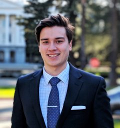 E. Brandon Strong合作论文数Department of Biological Sciences, California Polytechnic State University-San Luis Obispo3Shumon I Dhar合作论文数3
 Yuval Nachalon合作论文数3Libby J. Smith合作论文数3Angela A. Colback合作论文数2Kaicheng Yen合作论文数2Daniel V. Arkfeld合作论文数2Alireza Paydar合作论文数2Omid B Mehdizadeh合作论文数2Osama Raslan合作论文数2Shambavi J Rao合作论文数2Young VyVy N合作论文数2Siddiqui M Tausif合作论文数Department of Otolaryngology-Head &amp;Neck Surgery, University of California, Davis2
 Madden Lyndsay L合作论文数University of Pittsburgh, University of Pittsburgh Medical Center2Naren N. Venkatesan合作论文数2Gregory N. Postma合作论文数2Jui-Te Lin合作论文数1Steven D Stockton合作论文数1Inderpreet Kaur Khalsa合作论文数1
 Amanda Domer合作论文数1Chanticha Laohakittikul合作论文数1Brian O Hernandez合作论文数1Kychelle L Del Rosario合作论文数1Mayuri Srikanth合作论文数1
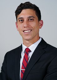 Benjamin T Ostrander合作论文数Dept Otolaryngol Head & Neck Surg, Univ Calif San Diego1Anastasiya Stasyuk合作论文数1
 Grant E. Gochman合作论文数1Andrew Yousef合作论文数1
 Philip Weissbrod合作论文数Department of Surgery, School of Medicine, University of California, San Diego1
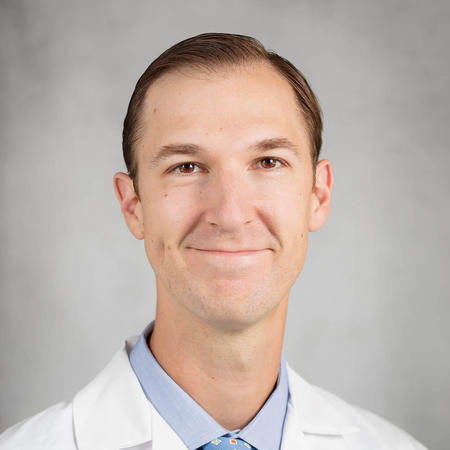 Andrew Vahabzadeh-Hagh合作论文数Department of Surgery, School of Medicine, University of California, San Diego1
 Tania Morimoto合作论文数Department of Mechanical and Aerospace Engineering, Jacobs School of Engineering, University of California, San Diego1Ashli O'Rourke合作论文数1
 Mark A Fritz合作论文数1Steven G Hoshal合作论文数1Amanda L. Johnson合作论文数1Ariadne A Nichol合作论文数1Apoorva T. Ramaswamy合作论文数1Halum Stacey L合作论文数Dept Otolaryngol Head & Neck Surg, Indiana Univ Sch Med1
 Khadijeh Bijangi-Vishehsaraei合作论文数1Jacqueline Allen合作论文数1




