The forms and locations of centrosomes in mouse oocytes and in sea urchin eggs were followed through the whole course of fertilization and first cleavage by immu- nofluorescence microscopy. Centrosomes were identified with an autoimmune antiserum to centrosomal material. Staining of the same preparations with tubulin antibody and with the DNA dye Hoechst 33258 allowed the correlation of the forms of the centrosomes with the microtubule structures that they generate and with the stages of meiosis, syngamy, and mitosis. The results with sea urchin eggs conform to Boveri's view on the paternal origin of the functional centrosomes. Centrosomes are seen in spermatozoa and enter the egg at fertilization. Initially, the centrosomes are compact, but as the eggs enter the mitotic cycle the forms of the centrosomes go through a cycle in which they spread during interphase, apparently divide, and con- dense into two compact poles by metaphase. In anaphase, they spread to form flat poles. In telophase and during reconstitu- tion of the daughter nuclei, the centrosomal material is disposed as hemispherical caps around the poleward surfaces of the nuclei. Mouse sperm lack centrosomal antigen. In the unfertilized mouse oocyte, the meiotic spindle poles are dis- played as broad-beaded centrosomes. In addition, centrosomal material is detected in the cytoplasm as particles, about 16 in number, which are foci of small aster-like arrays of microtu- bules. The length and number of astral microtubules correlate with the size of the centrosomal foci. After sperm incorpo- ration, as the pronuclei develop and more cytoplasmic micro- tubules assemble, a few of the foci associate with the peripheries of the nuclei. The number of foci multiplies during the first cell cycle. At the end of interphase, all of the centrosomal foci have concentrated on the nuclear peripheries and the cytoplasmic microtubules have disappeared. At prophase, the centrosomes are seen as two irregular clusters, marking the poles which, at metaphase and anaphase, appear as rough bands with foci, and the spindle is typically barrel-shaped. At telophase, the centrosomes are seen as arcs that lie on the nuclear peripheries after cleavage. The ordering of microtubules in all the stages reflects the shapes of the centrosomes. The findings on the sea urchin confirm the classical theory of the paternal origin of centrosomes and contrast with observations tracing the mitotic poles of the mouse egg to maternal centrosomal material. This evidence strengthens the conclusion that mouse centrosomes derive from the oocyte.
Monopolar mitotic apparatus can be produced in sea urchin eggs by a manoeuvre that distributes the four poles of the second mitosis into four separate blastomeres. The pole of the monopolar mitotic apparatus generates a half-spindle that is similar in structural details to the half-spindle of a normal bipolar mitotic apparatus, although the chromosomes are not as well aligned as in a normal metaphase plate. The chromosomes are oriented; one kinetochore faces the pole while its sister kinetochore faces away from the pole. The poleward kinetochore is connected to the pole by bundles of microtubules. No microtubules are seen on the sister kinetochore that faces away from the pole. Therefore, a single pole can direct most of the events in the establishment of a mitotic apparatus. Our interpretation examines the cooperation of kinetochores and poles in the formation of microtubules between them, stressing the half-spindle as the medium of cooperation and leaving open the question whether the kinetochores are origins or terminations of microtubules.
The fertilization reaction of sea urchin ( Lytechinus pictus ) and sand dollar ( Dendraster excentricus ) eggs was followed by simultaneous measurements of membrane potential and membrane resistance. In both species, the fertilization action potential (phase I) was associated with a considerable decrease in resistance, consistent with the interpretation that the first events of activation were associated with an increase in Na + permeability. As the membrane repolarized and the potential paused at a level more negative than that of the unfertilized egg (phase II), the membrane resistance increased to values higher than those of the unfertilized egg. In Dendraster the development of phase III (further repolarization associated with the development of K + permeability to its equilibrium value) was paralleled by a decrease in membrane resistance which reflected the increase in K + conductance. In Lytechinus , the decrease in resistance lagged behind the increase in negative potential as the latter developed to its equilibrium value. If dinitrophenol was added during phase I or early in phase II, the high membrane resistance associated with phase II was sustained and the further development of the membrane potential was blocked. If the DNP was added at the end of phase II, a partial development of the phase III potential was observed. The effects of dinitrophenol were reversible. It is suggested that the development of K + conductance in phase III involves an energy requirement.
In normally developing embryos of the sand dollar Dendraster excentricus the blastomeres are not tightly bound to each other at the end of cytokinesis. However, within 6 to 10 min the apposing cell surfaces appear flat against each other. Electron micrographs reveal that direct connection of the cell membranes exists. This process is called post-division adhesion.
Fast, low-cost, accurate detection of time-resolved dynamic nucleic acid conformational variation in response to small molecule binding: 1) enriches our understanding of nucleic acid structure, and regulation of structure by small molecules as ...Nucleic acids can undergo conformational changes upon binding small molecules. These conformational changes can be exploited to develop new therapeutic strategies through control of gene expression or triggering of cellular responses and can also be used ...
This chapter discusses the mitotic apparatus as a whole. As the cell enters mitosis, and proceeds through mitosis, several structures interplay in an orderly way and their collective expression is the mitotic apparatus. The chapter discusses the problems of the mitotic apparatus at two levels: (1) its assembly, organization, and disassembly as a large body, which is not seen in cells that are not dividing and represents a considerable volume of the cell when they are dividing, and (2) its very specific operations in engaging and moving the chromosomes with astounding precision. Conventional representations of typical mitosis in animal cells show a double system of polarized fibers: continuous fibers runningfrom centriole t o centriole and chromosomal fibers running from kinetochores t o centrioles. During anaphase, the chromosomal fibers are represented as shortening as sister chromosomes separate, leaving at the equator an interzonal region. T h e pole-to-pole fibers are represented as elongating in conjunction with the elongation of the spindle, which takes place either during or after chromosome-to-pole movement .
The reproduction of biological systems may be referred to the reproduction of cells. Subcellular entities such as viruses cannot maintain themselves indefinitely without parallel reproduction of the cells in which they live. The asexual reproduction of organisms such as flatworms is limited by the production of new cells, and all cases of sexual reproduction imply the generation of new cells after the mixing of genetic material from the parent cells. An evolutionary consequence of cellularity is implicit in the designation of multicellular organisms as higher organisms. Single cells can develop a high level of variety and complexity of structure and function. In the development of a multicellular organism, cell division, beginning with the egg, is the essential step toward differentiation. If mitosis is a device for the distribution of sister genes to sister cells, the chromosomes may be viewed as a system in which the numerous genes are packaged into a small number of units. Mitosis is accompanied by some changes in the physical state or texture of the cytoplasm, which have been assessed as viscosity changes.
Annals of the New York Academy of SciencesVolume 90, Issue 2 p. 455-469 THE ANALYSIS OF CELL REPRODUCTION* Daniel Mazia, Daniel Mazia Department of Zoology, University of California, Berkeley, Calif.Search for more papers by this author Daniel Mazia, Daniel Mazia Department of Zoology, University of California, Berkeley, Calif.Search for more papers by this author First published: October 1960 https://doi.org/10.1111/j.1749-6632.1960.tb23264.xCitations: 28 † Work done in the author's laboratory described in this paper has been supported in part by the American Cancer Society, New York, N. Y.; the Office of Naval Research, Washington, D. C., the University of California Cancer Coordinating Committee, Berkeley, Calif.; the Miller Institute for Basic Research in Science, Berkeley, Calif.; and the National Institutes of Health, Public Health Service, Bethesda, Md. AboutPDF ToolsRequest permissionExport citationAdd to favoritesTrack citation ShareShare Give accessShare full text accessShare full-text accessPlease review our Terms and Conditions of Use and check box below to share full-text version of article.I have read and accept the Wiley Online Library Terms and Conditions of UseShareable LinkUse the link below to share a full-text version of this article with your friends and colleagues. Learn more.Copy URL Citing Literature Volume90, Issue2Second Conference on the Mechanisms of Cell DivisionOctober 1960Pages 455-469 RelatedInformation
 Neidhard Paweletz合作论文数4G Sluder合作论文数2Jean M. Clark合作论文数2Maurice H. Bernstein合作论文数1G Blumenthal合作论文数1Ag Szentgyorgyi合作论文数1Victor D Vacquier合作论文数Scripps Institution of Oceanography, University of California San Diego1Ron Balczon合作论文数1NOVELLI G D合作论文数1
 Heide Schatten合作论文数Department of Veterinary Pathobiology, College of Veterinary Medicine, University of Missouri1E. L. Tatum合作论文数1David M Prescott合作论文数1
 Gerald P. Schatten合作论文数University of Pittsburgh Medical Center;McGowan Institute for Regenerative Medicine, University of Pittsburgh1John D. Roslansky合作论文数1Rollin D. Hotchkiss合作论文数1C. Fred Fox合作论文数1
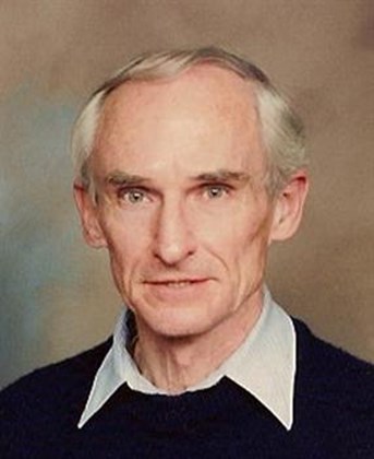 David M. Prescott合作论文数University of Colorado Boulder1
 Calvin R Simerly合作论文数1Ellen R. Dirksen合作论文数1Sheldon Shen合作论文数1Richard A. Steinhardt合作论文数1
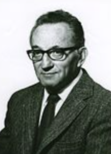 Sol Spiegelman合作论文数College of Physicians and Surgeons, Columbia University1Linda J. Mullins合作论文数1Eva-Maria Finze合作论文数1W. Eugene Knox合作论文数1Henry I Hirshfield合作论文数1




