Abstract Tryptophan (Trp) metabolism is recognised to have immunomodulatory effects in cancer, with downstream metabolites such as kynurenine suppressing T cell responses. However, the role of Trp catabolism in cancer cell biosynthesis and tumor growth is poorly understood. Herein, we investigated for the first time the role of Trp as a source of one-carbon metabolism through formate production in triple-negative breast cancer (TNBC) and its effect on serine synthesis via phosphoglycerate dehydrogenase (PHGDH), a rate-limiting enzyme for de novo serine synthesis. CRISPR-Cas9 system was used to generate tryptophan 2,3-dioxygenase (TDO2) and/or indoleamine 2,3-dioxygenase (IDO1) knockout TNBC cell lines. Multi-omics approach using LCMS Q-TOF system and RNA-seq was employed to integrate targeted and untargeted metabolomics, 13C fluxomics, and transcriptomics in TNBC cell lines. For fluxomics studies, cells were cultured with 13C-labeled tryptophan to trace the metabolic flux of carbon atoms through Trp metabolism into kynurenine pathway. While serine is recognised as a major source of one-carbon metabolism, our fluxomics data revealed that one-carbon units are released during the conversion of N-formylkynurenine to kynurenine. Labelled metabolites derived from tryptophan that give rise to tetrahydrofolate and methionine cycles were identified and these were subsequently incorporated to generate de novo purines. Integrated transcriptome and metabolomic analysis of tryptophan dioxygenase KO versus WT, as well as formylkynurenine and kynurenine treated cells, revealed a link between Trp catabolism and serine anabolism via alteration in PHGDH expression. Moreover, KO cells deprived of one-carbon units derived from Trp metabolism showed upregulation of PHGDH gene expression. Interestingly, KO cells showed a significantly lower growth rate in serine-glycine free media and also when PHGDH was inhibited with 2.5 μM NCT-503. This study provides evidence that Trp metabolism contributes to one-carbon metabolism in TNBC beyond its immunosuppressive functions. We also show that disruption of Trp metabolism upregulates PHGDH as a compensatory mechanism. This study identifies simultaneous inhibition of both pathways as a promising dual-targeting therapeutic strategy for TNBC treatment. Citation Format: Jamshid Motalebzadeh, Hossein Anani, Chloe Thompson-Peach, Theresa Hickey, Lisa Butler, Nirmal Robinson, Daniel Thomas. Tryptophan and serine metabolic axis reveals novel therapeutic vulnerabilities in triple-negative breast cancer [abstract]. In: Proceedings of the American Association for Cancer Research Annual Meeting 2026; Part 1 (Regular Abstracts); 2026 Apr 17-22; San Diego, CA. Philadelphia (PA): AACR; Cancer Res 2026;86(7 Suppl):Abstract nr 2023.
CD47 is an innate immune checkpoint that inhibits phagocytosis by myeloid cells, contributing to immune evasion by cancer cells. CD47-blocking antibodies have limited efficacy in glioblastoma (GBM), and the cell-intrinsic role of CD47 is poorly understood. In this study, we show that CD47 is highly expressed at the invasive edge of GBM tumors, and its elevated expression correlates with poor patient survival. We demonstrate that CD47 loss impairs GBM cell proliferation, migration, and invasion, independent of immune activity, and leads to reduced tumor burden and prolonged survival in vivo. Our study identifies ROBO2 signaling as a key downstream effector of CD47 and demonstrates that loss of ROBO2 similarly reduces GBM cell proliferation and migration. Importantly, we have uncovered that CD47 stabilizes ROBO2 by sequestering the E3 ubiquitin ligase ITCH, thereby blocking ubiquitination and proteasomal degradation of ROBO2. These findings establish CD47 as a key regulator of GBM cell plasticity and highlight the therapeutic potential of targeting CD47-ROBO2 signaling in GBM.
Mitochondrial DNA (mtDNA) mutations are frequently observed in cancer, but their clinical and functional significance in chronic myeloid leukemia (CML) remains incompletely defined. Here, we show that a distinct mtDNA mutational landscape is associated with mitochondrial metabolic programs and response to imatinib therapy in CML. We performed comprehensive profiling of somatic mtDNA mutations in 120 patients with chronic-phase CML. At diagnosis, 241 somatic mtDNA mutations were identified in 92 patients, including 29 homoplasmic mutations. In a clinically annotated cohort of 79 imatinib-treated patients, a higher number of mtDNA mutations (≥3 mutations) and higher variant allele frequency were associated with superior molecular responses, and remained significant in multivariable analyses. mtDNA mutational patterns were associated with distinct metabolic phenotypes in CD34+ leukemic stem/progenitor cells. Suboptimal responders exhibited increased mitochondrial respiration, spare respiratory capacity, mitochondrial content, and enrichment of mitochondrial biogenesis and lipid metabolic programs, consistent with enhanced oxidative phosphorylation dependence. In contrast, favorable responders displayed higher mtDNA mutational burden together with reduced respiratory reserve and increased mitophagy-related programs. Pharmacologic Complex I inhibition reduced clonogenic potential and enhanced imatinib sensitivity. Collectively, these findings identify mtDNA mutational states as a biomarker of metabolic fitness and therapeutic response in CML, while supporting further investigation of mitochondrial metabolism as a potential therapeutic vulnerability in CML.
Abstract Introduction: Tryptophan (Trp) metabolites are increased in various cancer types due to elevated expression of Trp dioxygenases, TDO2, IDO1, and IDO2. While recent studies have highlighted the immunosuppressive functions of these metabolites, their intrinsic significance to cancer cell survival remains poorly understood. This study explores the role of formylkynurenine and kynurenine, the first two metabolites in Trp catabolism on triple-negative breast cancer (TNBC) resistance to ferroptosis. Methods: We performed comprehensive gene expression analysis using 46 ER+, 103 TNBC tumours, and 21 adjacent normal tissue samples from the SRA database. BRCA-TCGA RNA-seq and DNA-seq data were analysed to identify alterations in three Trp dioxygenases in TNBC patients. RNA-seq was conducted on BT549 cell line cultured with 50 μM formylkynurenine and kynurenine. Gene expression changes were validated using western blotting and real-time qPCR. Intracellular glutathione (GSH) and oxidized glutathione (GSSG) levels were quantified by LC-MS and MS/MS approach. Cell sensitivity to ferroptosis was evaluated by treating cells with 300 nM RSL3. Results: Our analysis revealed increased expression of all three Trp dioxygenases, TDO2, IDO1, and IDO2, in TNBC tumours compared to luminal and adjacent normal tissues. DNA amplification was detected in IDO1 and IDO2, and reduced IDO1 promoter methylation in TNBC samples compared to luminal subtypes. Formylkynurenine and kynurenine both at concentrations of 50-100 μM significantly enhanced BT549 cell line proliferation. Transcriptomic analysis indicated that these metabolites regulate genes involved in ROS metabolism and iron transport. Metabolomics analysis further demonstrated alterations in the intracellular GSH/GSSG ratio. Notably, we report for the first time that kynurenine upregulates GPX4 protein expression, while formylkynurenine does not. However, both metabolites increased cellular resistance to RSL3-induced ferroptosis. Conclusion: Formylkynurenine and kynurenine both increase GSH/GSSG ratio. While kynurenine upregulates GPX4 expression, formylkynurenine does not alter its levels. These findings establish a novel metabolic axis linking Trp catabolism to ferroptosis resistance in TNBC, suggesting that targeting this pathway could represent a promising therapeutic strategy for this aggressive subtype of breast cancer. Citation Format: Jamshid Motalebzadeh, Hossein Anani, Chloe Thompson-Peach, Theresa Hickey, Lisa Butler, Nirmal Robinson, Daniel Thomas. Tryptophan metabolites drive ferroptosis resistance in triple-negative breast cancer cells [abstract]. In: Proceedings of the American Association for Cancer Research Annual Meeting 2026; Part 1 (Regular Abstracts); 2026 Apr 17-22; San Diego, CA. Philadelphia (PA): AACR; Cancer Res 2026;86(7 Suppl):Abstract nr 2022.
Human mononuclear cells derived from peripheral blood and bone marrow are valuable resources for the study of hematological malignancies, including acute myeloid leukemia (AML) and chronic myelomonocytic leukemia (CMML). Cryopreservation enables long-term storage of patient samples for downstream assays; while thawing protocols have been described, subsequent recovery of viable cells after thawing can be challenging, particularly for fragile blast and monocyte populations. Here, we describe a reliable protocol for thawing cryopreserved AML and CMML mononuclear cells designed to preserve post-thaw viability, recovery, and functional integrity. The method incorporates controlled dilution of cells out of cryoprotectant with anticoagulant-supplemented thaw buffer, DNase I treatment, and gentle resuspension steps. Using this approach, post-thaw viability consistently exceeded 80% with a mean recovery of 55.6% across samples. Recovered cells retained functional capacity, as demonstrated by colony-forming assays, and maintained immunophenotypic characteristics by flow cytometry. This protocol provides a robust and reproducible method for the recovery of cryopreserved AML and CMML mononuclear cells and may be broadly applicable to other fragile or monocyte-rich patient-derived hematopoietic samples.
T-cell acute lymphoblastic leukemia (T-ALL) is a biologically heterogeneous and clinically aggressive malignancy, where outcomes remain poor for patients with early relapse or refractory disease. Although tyrosine kinase alterations represent potentially actionable lesions, understanding of their functional and therapeutic relevance in T-ALL is limited. Here, we report the first case of an IRF2BP2::JAK2 fusion in a pediatric patient with high-risk T-ALL, identified through whole-genome and transcriptome sequencing. Using CRISPR-Cas9 genome engineering, we modeled the Irf2bp2::Jak2 fusion in Ba/F3 cells and showed that it confers cytokine-independent growth, localizes to the cytoplasm, and drives constitutive JAK-STAT signaling. Importantly, both type I (ruxolitinib) and type II (CHZ868) JAK inhibitors potently inhibited the fusion. These findings establish IRF2BP2::JAK2 as a novel oncogenic driver and druggable vulnerability in T-ALL and represent the first reported instance of an IRF2BP2 fusion directly activating a non-receptor tyrosine kinase. More broadly, this work underscores the critical role of functional modeling in defining the biological and therapeutic significance of rare genomic alterations to enable the translation of precision medicine to the clinic.
Somatic frameshift mutations in the gene encoding calreticulin (CALR) give rise to myelofibrosis and are classified as Type 1 (del52) or Type 2 (ins5) according to the degree of wildtype sequence retained adjacent to the neopeptide, with each type conferring different clinical outcomes. Targeting strategies specific for Type 1 vs Type 2 mutations would have enormous clinical utility in the treatment and prevention of myelofibrosis as responses to tyrosine kinase inhibitors are not durable nor mutation-specific. Here we show that dual targeting of Type 1 (del52) mutant CALR with two monoclonal antibodies directed against distinct epitopes in CALR have significant advantages compared to single agent treatment in the eradication of primary megakaryocyte progenitors in vitro and in a humanized ossicle microenvironment leading to improved survival in xenograft models. Dual targeting was superior in blocking constitutive STAT5 and ERK phosphorylation induced by del52 and prevented accumulation of JAK2 phosphorylation, overcoming ruxolitinib resistance. In contrast, Type 2 mutations showed increased CALR dimerization and were partially resistant to antibody targeting but could be impacted by ruxolitinib triple combination. Together, our data demonstrate an ultra-precision medicine approach tailored to either Type 1 OR Type 2 mutation classes will be required for maximal efficacy and complete blockade of JAK/STAT signalling, with far-reaching implications for patient management.
Summary of the key interactions in the IL-3R ternary complex in the IL-3R ternary complex crystal structure.
Enrichment of the IL-3R hexamer versus dodecamer gene signature in primitive normal and leukemic stem cells.
In a recent study published in Cell,Xu et al.have uncovered a critical role for the endoplasmic reticulum(ER)stress sensor inositol-requiring enzyme 1 alpha(IRE1α)in modulating the immunogenic effects of taxane chemotherapy in triple-negative breast cancer(TNBC).They reveal how IRE1α acts as a defense mechanism in cancer cells,preventing the accumulation of danger signals and subsequent immunogenic cell death(ICD).
Increasing IL3Rα/βc ratios lead to hexameric receptor assembly and augmented quiescence.
Key interactions between distinct residues in the IL-3R ternary complex crystal structure.
The World Health Organization (WHO-5) and International Consensus Classification (ICC) acknowledge the poor prognosis of TP53-mutated (TP53mut) myeloid neoplasm (MN). However, there are substantial differences between the two classifications that may lead to under- or overestimation of the prognostic risk. We retrospectively applied WHO-5 and ICC to 603 MN cases harboring TP53mut (variant allele frequency, VAF ≥ 2%). WHO-5 and ICC would not classify 64% and 20% of these cases as TP53mut MN, respectively. Moreover, of those classified, 67.5% would be classified discrepantly. Primary drivers of discrepancies included: (i) prognostic importance of TP53mut acute myeloid leukemia (AML), (ii) interaction of the blast percentage and allelic status, (iii) 17p.13.1 deletion detected by cytogenetics, (iv) complex karyotype (CK) as multi-hit equivalent, and (v) TP53mut VAF threshold, we analyzed survival outcomes of each of these groups with an aim to provide clarity. TP53mut AML was associated with significantly poor survival compared to TP53-wild type TP53wt AML, myelodysplasia-related (AML, MR 4.7 vs. 18.3 months; P < 0.0001), supporting its inclusion within TP53mut MN as a distinct subentity. Secondly, the survival of TP53mut with blast 10–19% was poor regardless of the allelic status. Thirdly, for cases with a single TP53mut with VAF < 50%, 17p13.1 del or CK serve as practical surrogates of biallelic inactivation, obviating the need for an additional copy number analysis. Finally, TP53mut AML, MDS multi-hit/multi-hit equivalent with VAF < 10% had significantly poorer survival compared to TP53mut MDS VAF < 10% without CK and 17p del, and were comparable to those with VAF ≥ 10% (14.1 vs. 48.8 vs.7.8 months, P < 0.0001). Collectively, these findings address key areas of contention and provide valuable insights that will guide future revisions of the WHO and ICC classifications.
Somatic mutations in mitochondrial DNA (mtDNA) are not typically considered key oncogenic drivers of cancer, primarily because of a high synonymous to non-synonymous variant ratio. Here, we surveyed 248 matched diagnosis and remission samples from patients with chronic myeloid leukemia (CML) and found a 75% had mitochondrial mutations with a median number of 2 mutations per patient. mtDNA mutations were predominantly non-synonymous, enriched in the D-loop control region, and likely originated from replication and transcriptional errors. Functionally, mtDNA mutations were associated with reduced oxidative phosphorylation (OXPHOS), as measured by Seahorse analyser. This metabolic vulnerability could be phenocopied by treatment with the complex I inhibitor IACS-10759 in combination with the targeted tyrosine kinase inhibitor (TKI) imatinib, which significantly reduced the colony-forming potential of TKI resistant leukemic stem/progenitor cells (LSPCs). Strikingly, we show that mtDNA mutations were associated with increased sensitivity to imatinib therapy in the clinic. Patients with ≥3 mutations and patients with mutations in the D-loop showed significantly higher cumulative incidence of major molecular response at 24 months (90% vs. 68%, p = 0.004, and 89% vs 68%, p = 0.004 respectively). Single-cell RNA sequencing further revealed enrichment in non-synonymous mtDNA variants in LSPCs from TKI-sensitive patients, while TKI-resistant cells exhibited upregulated gene signatures related to glycerolipid and phospholipid metabolism and mitochondrial biogenesis. Together, our findings demonstrate that mtDNA mutations are key determinants of sensitivity to targeted therapy, rather than oncogenic drivers of leukemogenesis. Mechanistically, non-synonymous mtDNA mutations appear to restrict mitochondrial metabolic plasticity, with widespread implications for precision oncology. ### Competing Interest Statement The authors have declared no competing interest. We declare that data supporting the findings of this study are available within this manuscript and its supplementary information files. Supplementary information accompanies the manuscript on the Signal Transduction and Targeted Therapy website SAHMRI Mid-Career Seed Funding Grant, Adelaide Cancer Council SA National Health and Medical Research Council, 2007908 Contributing Haematologists Committee Research Grant, Adelaide National Medical Research Council Singapore, CIRG/1468/2017, MOH-000602, MOH-000059, CIRG16nov032 Leukemia & Lymphoma Synergistic Team Award with support from the Mike & Sofia Segal Foundation NHMRC Ideas Grants The Medical Research Future Fund
Acute myeloid leukemia (AML) exhibits a hierarchical cellular organisation, with a minor fraction of self-renewing leukemic stem cells (LSCs) at the apex of this hierarchy. LSCs confer resistance to chemotherapy and are implicated in AML relapse. Compared to normal haematopoietic stem cells, upregulated CD123 or IL-3Rα was one of the first cell surface markers identified on LSCs and is particularly highly expressed in FLT3-ITD and RUNX1 mutated AML. Thus, identifying the molecular mechanisms responsible for upregulating CD123 could offer a promising avenue for the eradication of LSCs. Here we illustrate a novel role for TGF-β in upregulating CD123 and promoting quiescence and chemotherapy resistance. Method We performed flow cytometry, qPCR and western blot analysis of cytokine stimulations and inhibitor studies with ligand-trap, TGF-β receptor 1 (TβRI) inhibitor, gilteritinib and midostaurin in AML cell lines and primary samples, and CD34⁺ cord blood (CB) stem cells. We did SMAD2/4 co-immunoprecipitation, chromatin immunoprecipitation, immunofluorescence and shRNA knockdown studies in the TF-1 and MOLM13 cells. We performed CFU assays for TGF-β-treated CD34⁺, TβRI inhibitor–treated primary AML samples and cytarabine/TGF-β-treated TF-1 cells. We did propidium iodide cell cycle and cytarabine-induced apoptosis assessment via AnnexinV/7AAD. ResultWe first identified TGF-β1 mediates upregulation of IL-3Rα by 4 to 5-fold increase on the cell surface and total protein, as well as corresponding IL3RA RNA (6-fold increase) in the TF-1 cells. Similarly, healthy common myeloid progenitors (CMPs) (CD34+CD38+CD45RA-CD123low) from CB exhibited a 2-fold increase in IL-3Rα expression following TGF-β1 treatment. Focusing on TGF-β family members, we found that only TGF-β1 and TGF-β2, but not BMP4, through TβRI/TβRII activation could upregulate IL-3Rα expression. TGF-β1 binding to TβRI/TβRII results in consistent SMAD2 phosphorylation at its C-terminal region, forming a complex with SMAD4. Using chromatin immunoprecipitation, we detected SMAD2 and SMAD4 directly bound to the IL3RA proximal enhancer region upon TGF-β1 stimulation. We further confirmed that SMAD4 knockdown blocked the ability of TGF-β1 to upregulate IL-3Rα.Mechanistically, we found that IL-3 exerted a positive feedback effect on the TGF-β1 signalling pathway by upregulating the expression of TβRI, leading to enhanced phosphorylation of SMAD2. This, in turn, resulted in a further increase in expression of the IL-3Rα, suggesting a reinforcing loop between IL-3 and TGF-β1 signalling components. Both TGF-β1 alone and in combination with IL-3 induced cellular quiescence, as evidenced by a ~30% increase in the proportion of cells in the G0/G1 phase and a corresponding decrease in the S and G2/M phases of the cell cycle. This quiescent state was accompanied by elevated expression of the cyclin-dependent kinase inhibitor CDKN1A (p21), further supporting the role of TGF-β1 in cell cycle arrest. Functionally, TGF-β1 conferred a survival advantage following cytarabine treatment. While cytarabine alone induced significant apoptosis, detected by AnnexinV and 7AAD, TGF-β1 co-treatment reduced cell death by approximately 50%. This protective effect was corroborated by colony-forming unit (CFU) assays, which showed a 20-fold increase in colony numbers in cells treated with both TGF-β1 and cytarabine compared to cytarabine treatment alone.We then investigated leukemias harboring FLT3-ITD and RUNX1 mutations, which have high CD123 expression. We found FLT3-ITD mutations hijack the TGF-β1 pathway by inducing constitutive phosphorylation of SMAD2 at its linker region, sensitive to gilteritinib and midostaurin. This aberrant signalling is associated with increased expression of TGF-β1-responsive genes, including TGFB1, SMAD2, CDKN1A, and IL3RA. Primary RUNX1 mutated patient sample with high CD123 expression LSCs population (CD34+CD38- CD45low) were sensitive to TβRI inhibition (≅2-fold decrease) and their colony formation ability decreased by 50% when they were pretreated with TβRI inhibitor. ConclusionGiven the well-established role of LSCs with high expression of CD123 in post-chemotherapy relapse and the frequent co-occurrence of FLT3-ITD and RUNX1 mutations in AML patients, our study underscores the clinical relevance of targeting the TGF-β1 pathway. These findings highlight TGF-β1 signalling as a key modulator of LSC persistence and a potential therapeutic vulnerability in AML.
 Angel F Lopez合作论文数SA Pathology47
 Ravi Majeti合作论文数Institute for Stem Cell Biology and Regenerative Medicine, Cancer Institute, Division of Hematology, Department of Medicine, Stanford University40
 Devendra K. Hiwase合作论文数37
 David M Ross合作论文数27
 Yeung David T合作论文数27Kelly Lim合作论文数26
 Richard J D'Andrea合作论文数24Denis Tvorogov合作论文数22
 Paul G Ekert合作论文数20
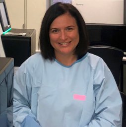 Anna L. Brown合作论文数Hanson Institute;Institute of Medical and Veterinary Science;Institute of Medical and Veterinary Science, Hanson Institute20Chung H Kok合作论文数19
 Mark Guthridge合作论文数19
 Timothy Hughes合作论文数Adelaide Medical School, Faculty of Health and Medical Sciences, The University of Adelaide;South Australian Health and Medical Research Institute19
 Andreas Reinisch合作论文数Med Univ Graz19Jason A. Powell合作论文数Receptor Laboratory;Department of Human Immunology;Hanson Institute;Receptor Laboratory, Hanson Institute19
 Emma F. Barry合作论文数The Division of Human Immunology, Centre for Cancer Biology19Chloe Thompson-Peach合作论文数18
 Hamish S Scott合作论文数17
 Nirmal Robinson合作论文数17
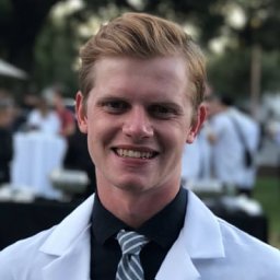 Brooks Benard合作论文数Stanford Canc Inst, Stanford Univ16
 Kaylene Simpson合作论文数16
 Saumya E Samaraweera合作论文数Discipline of Genetics, School of Molecular &Biomedical Science and ARC Special Research Centre for the Molecular Genetics of Development, The University of Adelaide;School of Molecular & Biomedical Science and ARC Special Research Centre for the Molecular Genetics of Development, The University of Adelaide16Toubia John合作论文数Centre for Cancer Biology, University of South Australia15Mara Dottore合作论文数15
 Yusuke Nakauchihiroshi合作论文数Department of Hematology, Tokyo Medical and Dental University15
 Christopher N Hahn合作论文数15
 Timothy R Hercus合作论文数15
 Greg Goodall合作论文数Centre for Cancer Biology, University of South Australia14Deepak Singhal合作论文数Adelaide Medical School, University of Adelaide14Kan Winnie L合作论文数The Centre for Cancer Biology, the University of South Australia14
 Mrinal Patnaik合作论文数College of Medicine, Mayo Clinic-Rochester14
 Mithun Vinod Shah合作论文数14Monika M Kutyna合作论文数13Rakchha Chhetri合作论文数13
 Michael Parker合作论文数Department of Biochemistry and Pharmacology, The University of Melbourne;St Vincent’s Institute of Medical Research13
 Thomas Kohnke合作论文数Department of Medicine III, University Hospital Grosshadern13
 Aref Al-Kali合作论文数12Tracy L. Nero合作论文数Department of Medicine, Clinical Pharmacology and Therapeutics Unit, The University of Melbourne12
 Li-Qing Jin合作论文数11
 Stuart M Pitson合作论文数Centre for Cancer Biology11
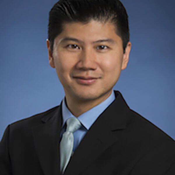 Steven Chan合作论文数Department of Medical Biophysics, Temerty Faculty of Medicine, University of Toronto11
 John Dick合作论文数Department of Molecular Genetics, Temerty Faculty of Medicine, University of Toronto11
 Ayalew Tefferi合作论文数Comprehensive Cancer Center, Mayo Clinic11
 Colin Glenn Begley合作论文数BridGene Biosciences, Inc.11Naseema Gangat合作论文数11
 Sophie E Broughton合作论文数School of Biomedical Sciences, Monash University11
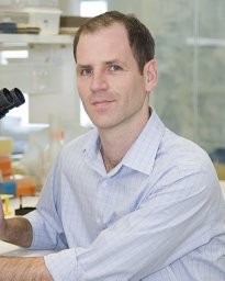 Steven W. Lane合作论文数Department of Haematology, Princess Alexandra Hospital11
 David Dill合作论文数Department of Computer Science, School of Engineering, Stanford University;Facebook;Meta11




