OBJECTIVES:The goal of this task force was to examine the 1992 definition of the intensivist, identify gaps, and initiate a path forward to define a concise and practical definition that could be applied globally. DESIGN:A modified Delphi technique was used to develop a revised definition and roles of the intensivist. We determined a priori that 75% or greater participant agreement for the definition and essential roles of the intensivist was required. SETTING:A task force consisting of physicians, a respiratory therapist, advanced practice providers, and a pharmacist that practice in critical/intensive care medicine, in various settings, was established with the goal of evaluating and revising the previous definition considering evolving healthcare. SUBJECTS:The task force participated in online questionnaires related to the definition and roles of the intensivist. INTERVENTIONS:None. MEASUREMENTS AND MAIN RESULTS:The task force agreed on the following definition of an intensivist: "A physician who has successfully completed an accredited program or equivalent critical care/intensive care medicine training and maintains advanced certification (if available); and shows dedication to the area of critical/intensive care medicine in the way of professional work." Additionally, the task force determined a list of essential roles of the intensivist categorized into Direct Clinical Care, Unit Management/Unit Involvement, Responsibility to the Community, and Administration and Leadership. CONCLUSIONS:The revised definition of the intensivist seeks to integrate the intensivist in the current realm of team-based healthcare. The intensivist is a physician who provides care to critically ill patients in collaboration with an interprofessional team. Establishment of a single, revised definition is intended to render clarity of an intensivist's role and responsibilities for patients, families, and the interprofessional team.
Introduction Diagnostic radiology interpretive errors in trauma patients can lead to missed diagnoses, compromising patient care. Due to this, our level II trauma center implemented a reread protocol of all radiographic imaging within 24 hours on our highest trauma activation level (Code T). We sought to determine the efficacy of this reread protocol in identifying missed diagnoses in Code T patients. We hypothesized that a few, but clinically relevant errors, would be identified upon reread. Methods All radiographic study findings (initial read and reread) performed for Code T admissions from July 2015 to May 2016 were queried. The reviewed radiological imaging was given one of four designations: agree with interpretation, minor (non-life threatening) nonclinically relevant error(s)—addendum/correction required or clinically relevant error(s) (major [life threatening] and minor)—addendum/correction required, and trauma surgeon notified. The results were compiled, and the number of each type of error was calculated. Results Of the 752 radiological imaging studies reviewed on the 121 Code T patients during this period, 3 (0.40%) contained minor clinically relevant errors, 11 (1.46%) contained errors that were not clinically relevant, and 738 (98.1%) agreed with the original interpretation. The three clinically relevant errors included a right mandibular fracture found on X-ray and a temporal bone fracture that crossed the clivus and bilateral rib fractures found on computerized tomography. Discussion Clinically relevant errors, although minimal, were discovered during rereads for Code T patients. Although the clinical errors were significant, none affected patient outcomes. We propose that the implementation of reread protocols should be based upon institution-specific practices.
Learning Objectives: The current protocol at our hospital for a patient suffering mild or minor traumatic head injury (GCS 13-15) requires a preliminary CT scan, admission to a step down unit or a monitored surgery floor for at least 24 hours and a repeat CT scans in 6-8 hours. Our objective was to determine the rate of delayed intracerebral hemorrhage in patients who sustained a mild head injury that had an initial head CT that was negative for bleeding. Methods: We conducted a retrospective chart review study involving anticoagulated (warfarin) patients who had sustained minor head injury and a negative initial head CT scan at a level 2 regional trauma cente from October 2013 to December 2017. Our inclusion criteria included patients above 18 years of age; only blunt mechanism; Minor head injury with a GCS on admission 13-15;ISS <15;Injury sustained within the previous 48 hours of admission; Patients with no focal neurodeficit and/or no evidence of cranial fracture; All patients admitted for at least 24 hours observation with a repeat CT scan performed before discharge; Preinjury use of anticoagulation(only warfarin and use for at least 1 week)Initial CT scan on admission were negative for any intracranial bleed. Results: All 61 patients were on Coumadin and (9/61) took aspirin as an additional anticoagulant. The first CT scan was negative in all 61 patients. The second CT scan, performed at ≥6 hours, was positive in 3/61 of the patients. Ninety-eight percent of the patients (60/61) had no neurological deterioration during their hospital stay. The one patient (1.6%) who had neurological deterioration later died of cardiac arrest. 92% of patients did not have a CT scan at time 3 or time 4 and 8% of patients (5/61) did have a scan during this period. Of the 5 patients undergoing a CT scan, 3 patients had a positive scan, showing some abnormality. Fifty-four percent of the patients (33/61) had a hospital stay of greater than 3 days; 19.7% of patients (12/61) had a hospital stay of 2-3 days and 26.2% of patients (16/61) had a hospital stay between 0 – 1 days. Conclusions: A mean 4 day hospital stay as was evident from our study comprising of elderly patient population would amount to $16,304USD. This excludes the anciliary costs of labs and transport etc. Patients with a mild head injury, a therapeutic INR, and negative initial CT brain can be safely discharged home with close family monitoring, proper education and detailed instructions on when to return.
Compassion fatigue (CF), a state of physical/emotional distress caused by repeatedly caring for those experiencing traumatic episodes, is a prevalent issue for today's healthcare provider. We sought to characterize levels of CF within a surgeon population, particularly comparing trauma surgery with other surgical specialties. A survey containing the Professional Quality of Life Scale (ProQOL), a validated tool assessing compassion satisfaction (CS), CF, and burnout (BO) was distributed via electronic newsletter to members of the American College of Surgeons. Demographic data and Professional Quality of Life Scale scores for CS, BO, and CF were collected and compared within specialty and gender subgroups. A total of 178 surgeons completed surveys. Respondents were predominantly male, general surgeons, >55 years old. Trauma surgeons composed the second largest subgroup. Levels of CS were significantly lower in the trauma surgeon subgroup compared to other surgical specialties (trauma: 37.1 ± 5.28, other: 39.5 ± 6.30; P = 0.044). Female surgeons from all specialties exhibited significantly higher levels of BO (female: 26.7 ± 6.10, male: 24.6 ± 6.79; P = 0.035) and CF (female: 24.2 ± 6.29, male: 21.9 ± 6.11; P = 0.021) compared with male surgeons. Subanalyses comparing female trauma surgeons to female surgeons in other specialties found female trauma surgeons exhibited significantly lower levels of CS (trauma: 34.8 ± 4.63, other: 38.8 ± 5.99; P = 0.038) and higher levels of BO (trauma: 29.1 ± 3.14, other: 25.3 ± 6.41; P = 0.049). Trauma surgeons, particularly female trauma surgeons, may be at a heightened risk for developing a poorer overall professional quality of life compared with surgeons of other specialties. In addition, female surgeons may be at greater risk for developing CF compared with male counterparts.
BACKGROUND:The appropriate managing center for adolescent trauma patients is debated. We sought to determine whether outcome differences existed for adolescent severe traumatic brain injury (sTBI) patients treated at pediatric versus adult trauma centers. We hypothesized that no difference in mortality, functional status at discharge (FSD), or overall complication rate would be observed between center types. METHODS:All adolescent trauma patients (aged 15-17 years) presenting with isolated sTBI (head Abbreviated Injury Scale [AIS] score ≥3; all other AIS body region scores ≤2) to accredited Levels I to II trauma centers in Pennsylvania from 2003 to 2015 were extracted from the Pennsylvania Trauma Outcome Study database. Dead on arrival, transfer, and penetrating trauma patients were excluded from analysis. Adult trauma centers were defined as non-pediatirc (PED) (n = 24), whereas standalone pediatric hospitals and adult centers with pediatric affiliation were considered Pediatric (n = 9). Multilevel mixed effects logistic regression models and a generalized linear mixed models assessed the adjusted impact of center type on mortality, overall complications, and FSD. Significance was defined as a p value less than 0.05. RESULTS:A total of 1,109 isolated sTBI patients aged 15 to 17 years presented over the 13-year study period (non-PED, 685; PED, 424). In adjusted analysis controlling for age, shock index, head AIS, Glasgow Coma Scale motor, trauma center level of managing facility, case volume of managing facility, and injury year, no significant difference in mortality (adjusted odds ratio, 0.82; 95% confidence interval [CI], 0.23-2.86; p = 0.754), FSD (coefficient, -0.85; 95% CI, -2.03 to 0.28; p = 0.136), or total complication rate (adjusted odds ratio, 1.21; 95% CI, 0.43-3.39; p = 0.714) was observed between center types. CONCLUSION:Although the optimal treatment facility for adolescent patients is frequently debated, patients aged 15 to 17 years presenting with isolated sTBI may experience similar outcomes when managed at pediatric and adult trauma centers. LEVEL OF EVIDENCE:Epidemiologic study, level III; therapeutic study, level IV.
Wu, Daniel; Gross, Brian; Rinehart, Cole; Lynch, Caitlin; Morrison, Chet; Bradburn, Eric; Rogers, Frederick
Copyright © 2016 by the Society of Critical Care Medicine and Wolters Kluwer Health, Inc. All Rights Reserved.
The checklist concept has received much attention as a result of its ability to improve patient care by minimizing complications. We hypothesized daily sign-out rounds using a checklist, by improving team communication and consistency of clinical care, could lead to expedited throughput for patients at a major trauma center. A retrospective study examined patients admitted to a mature trauma center. Two time periods, PRE (September 2008 to January 2009) and POST (September 2009 to January 2010), were selected to match for seasonal variation in admission diagnosis. An organ system-based checklist was used during daily sign-out for all admitted trauma patients in the POST period. We examined discharge status, complications and rates, and intensive care unit (ICU) and overall hospital length of stay for differences. There were similar numbers of patients (824 PRE vs 798 POST) admitted in these two cohorts. We found no statistical differences in the incidence of complications or mortality rate. We did discover statistically significant differences in the median ICU days (2 PRE vs 1 POST, P = 0.007) as well as median hospital length of stay (2 days, interquartile differences Q1 to Q3 PRE [1 to 5] and POST [1 to 4] P = 0.000). These trends remained valid even among the severely injured (Injury Severity Score 16 or greater) with a hospital length of stay of 5 (PRE) versus 3 days (POST; P = 0.021). A simple, organ system-based checklist can be successfully adopted for daily sign-out round on a busy, multiprovider trauma service. We were able to expedite trauma patient throughput in both ICU and overall hospital stays with a trend toward decreasing mortality. This improved throughput may potentially translate into a cost saving for the hospital.
INTRODUCTION:Approximately one in three older adults fall each year, resulting in a significant proportion of geriatric traumatic injuries. In a hospital with a focus on geriatric fall prevention, we sought to characterize this population to develop targeted interventions. As mild hyponatremia, defined as a serum sodium <135meq/L, has been reported to be associated with falls, unsteadiness and attention deficits, we hypothesized that hyponatremia is associated with falls in our geriatric trauma population. METHODS:Gender, age, pre-existing conditions (cardiac disease, diabetes, hematologic disorder, liver disease, malignancy, musculoskeletal disorder, neurological disorder, obesity, psychiatric disorder, pulmonary disease, renal disease, thyroid disease), mechanism of injury and admitting serum sodium level were queried for all geriatric trauma admissions from 2008 to 2011. Mechanism of injury was coded as falls admissions and non-falls admissions. Admitting serum sodium levels were coded as hyponatremic (<135mmol/L) and not hyponatremic (≥135mmol/L). RESULTS:Of the 2370 geriatric trauma admissions during the study period, there were 1841 (77.7%) falls admissions and 293 (12.4%) patients who were hyponatremic. Gender, age, neurological disorder, hematologic disorder, and hyponatremia were found to be significant predictors of falls in both univariate and multivariable analyses. CONCLUSION:Hyponatremic patients are significantly more likely to be admitted for a fall than non-hyponatremic patients, when adjusting for age, neurological disorder, and hematologic disorder. Consequently, hyponatremia identification and management should be an integral part of any geriatric trauma fall prevention programme. Additionally, if hyponatremia is found during a geriatric fall workup, it should be corrected prior to discharge and closely monitored by a primary care physician to prevent recurrent episodes of falls.
BACKGROUND As we enter the brave new world of the Patient Protection and Affordable Care Act of 2010, it is imperative that trauma centers provide not only excellent but also cost-effective trauma care. To that end, we sought to determine those factors that contribute significantly to barrier days (BDs), when a patient is medically cleared for discharge but unable to leave the hospital. We hypothesized that there would be significant demographic and payor factors associated with BDs. METHODS All trauma admissions to a Level II trauma center discharged alive from 2010 to 2012 were queried from the trauma registry. BDs were identified and recorded at daily sign-out. Patients with a hospital length of stay of 24 hours or less or transferred to another hospital were excluded. Univariate logistic regression was used to analyze which factors were significant (p ⩽ 0.05) for BDs. Significant variables were then included in a multivariate logistic regression model. RESULTS A total of 3,056 patients were included in the study, 105 (3.44%) of whom had at least one BD. Multivariate analysis revealed that patients awaiting nursing home placement and rehabilitation placement were at 6.39 and 2.79 times higher odds of having significant barriers to discharge, respectively, compared with patients who were discharged home. The multivariate model also showed that Medicaid coverage, one or more comorbidities, Injury Severity Score of 9 or greater, and one or more ventilation days had a significant correlation with the incidence of BDs. CONCLUSION This study suggests that discharge destination is a significant factor associated with BDs. Understanding what type of patient is prone to develop barriers to discharge will allow case managers and social workers to intervene with discharge planning early in that patient’s hospital course to secure placement and possibly reduce health care costs and improve functional outcome. LEVEL OF EVIDENCE Prognostic/epidemiologic study, level III.
The American College of Surgeons Committee on Trauma (ACS-COT) has determined that a 5 % pre-hospital undertriage [UT; defined as Injury Severity Score (ISS) > 15 and not sent to a trauma center] is an acceptable rate for pre-hospital transfer to a non-trauma center. We sought to determine if this level of undertriage is acceptable within a mature Level II trauma center as a measure of the adequacy of its trauma activation system.
BACKGROUND: This study aimed to determine the relative "weight" of risk factors known to be associated with venous thromboembolism (VTE) for patients with trauma based on injuries and comorbidities.METHODS: A retrospective review of 16,608 consecutive admissions to a trauma center was performed. Patients were separated into those who developed VTE (n = 141) versus those who did not (16,467). Univariate analysis was performed for each risk factor reported in the trauma literature. Risk factors that were shown to be significant (p < 0.05) by univariate analysis underwent multivariate analysis to develop odds ratios for VTE. The Trauma Embolic Scoring System (TESS) was derived from the multivariate coefficients. The resulting TESS was compared with a data set from the National Trauma Data Bank (2002-2006) to determine its ability to predict VTE.RESULTS: The multivariate analysis demonstrated that age, Injury Severity Score, obesity, ventilator use for more than 3 days, and lower-extremity trauma were significant predictors of VTE in our patient population. The TESS was from 0 to 14, with the best prediction for those patients with a score of more than 6 (sensitivity, 81.6%; specificity, 84%). Overall, the model had excellent discrimination in predicting VTE with a receiver operating characteristic curve of 0.89. The VTE rates for TESS in the National Trauma Data Bank data set were similar for all integers except for 3 and 4, in which the VTE rates were significantly higher (3, 0.2% vs. 0.6%; 4, 0.4% vs. 1.0%).CONCLUSION: The TESS provides an objective measure of classifying VTE risk for patients with trauma. The TESS could allow informed decision making regarding prophylaxis strategies in patients with trauma. (J Trauma Acute Care Surg. 2012; 73: 511-515. Copyright (C) 2012 by Lippincott Williams & Wilkins)
BACKGROUND:Temporary inferior vena cava filters (IVCF) are uniquely suited for trauma patients in whom the high risk of venous thromboembolism is transient. Currently, few "retrievable filters" are actually retrieved, with most published series documenting a retrieval rate between 20% and 50%. We sought to determine whether we could achieve a higher rate of retrieval with an improved process of care.METHODS:All permanent and temporary filters were entered prospectively into a dedicated filter registry. Within 60 days of filter placement, all temporary filter patients were contacted by a trauma case manager to evaluate ongoing venous thromboembolism risk. Low-risk patients were then evaluated by radiology for removal of the IVCF. If appropriate, removal of the IVCF was scheduled. Initial contacts with patients were made by telephone. If unsuccessful with phone contact, family members, rehabilitation facility, and social work were all contacted to obtain the most recent phone number and address. A follow-up letter was sent to the patient with follow-up visit instructions. Finally, if prior contact measures did not work, a certified letter was sent to the last known address.RESULTS:Between 2006 and 2009, of 7,949 trauma admissions, 420 (5.2%) met indications for filter placement. Of those, 160 were available for removal and 94 were successfully removed (59%).CONCLUSIONS:A retrieval rate of 59% can be achieved with an explicit process of care emphasizing disciplined follow-up.LEVEL OF EVIDENCE:III.
Patient satisfaction surveys are increasingly being used as a measure of physician performance in a hospital setting. We sought to determine what role the clinical condition the physician is treating has on overall patient satisfaction scores. Patient satisfaction scores were calculated for elective and emergent general surgery and trauma patients for eight surgeons taking care of all three types of patients. Both physician satisfaction (PP) and hospital satisfaction (GP) scores were calculated. Mean scores (± standard deviation) between groups were compared with P < 0.05 significance. Of 1521 trauma patients and 3779 general surgery patients, there was 14.8 and 15.1 per cent response rate, respectively, to the survey. Trauma patients had a significantly lower PP than general surgery patients (81.0 ± 19.4 vs 85.7 ± 16.4; P < 0.001). However, the GP between trauma and general surgery was not significant (84.0 ± 13 vs 84.0 ± 12.3; nonsignificant) When general surgery patients were divided into emergent versus elective, the PP was significantly higher for elective than emergent (87.9 ± 14.6 vs 82.7 ± 18; P < 0.001). A patient's underlying clinical condition may influence response to patient satisfaction surveys. Further research needs to be performed before patient satisfaction surveys can be adopted as a overall measure of physician competency.
The geriatric trauma patient poses unique challenges to the trauma surgeon due to occult injuries and occult hypoperfusion. We hypothesized that those elderly patients with significant injuries, who were not initially evaluated via trauma activation, would suffer worse outcomes. All cases of elderly (age ≥ 65) admitted to the trauma service from the years 2000 to 2010 were included. Our trauma activation system encompasses anatomic, physiologic, and mechanism of injury criteria. We defined patients as undertriaged (UT) if they had an ISS > 15 and did not undergo a trauma team activation, but had a regular workup by an emergency department physician and trauma team consultation. Factors that contributed to being UT in the emergency department were investigated by univariate and multivariate analysis. A total of 4534 elderly patients constitute this analysis, of which 15.1 per cent were UT. The UT patients were more likely to die, when adjusted for Revised Trauma Score, Glasgow Coma score, the occurrence of $1 complication, and whether the patient was on Coumadin. UT has a high risk of death in elderly patients. Trauma triage guidelines need to be better tailored to identify the high-risk geriatric trauma patient.
Background: The state of Pennsylvania (PA) has one of the oldest, most well-established trauma systems in the country. The requirements for verification for Level I versus Level II trauma centers within PA differ minimally (only in the requirement for patient volume, residency, and research). We hypothesized that there would be no difference in outcome at Level I versus Level II trauma centers.Methods: Odds of mortality for 16 Level I and 11 Level II hospitals in PA over a 5-year period (2004-2008) was computed using a random effects logistic regression model. Overall adjusted mortality rates at Level I versus Level II hospitals were compared using the nonparametric Wilcoxon's rank sum test. The crude mortality rates for 140,691 patients over the 5-year period were similar (5.07% Level II vs. 5.48% Level I), but statistically significant (odds ratio mortality at Level I = 1.084, p = 0.002 Fisher's exact test).Results: Although Level I centers had on average crude mortality rates that were higher than those of Level II centers, median adjusted mortality rates were not different for the two types of centers (Wilcoxon's rank sum test). Performance of Level I versus Level II shows considerable variability among centers (basic random effects model, age, blunt/penetrating, and Injury Severity Score [ISS]). However, Level II centers seem no different from Level I.Conclusion: As trauma systems mature, the distinction between Level I and Level II trauma centers blurs. The hierarchal descriptors "Level I" or "Level II" in a mature trauma system is pejorative and implies in those hospitals labeled "Level II" as inferior, and as such should be replaced with nonhierarchal descriptors.
 Frederick Rogers合作论文数Department of Surgery, Perelman School of Medicine, University of Pennsylvania;Department of Surgery, Larner College of Medicine, University of Vermont16
 Edavettal Mathew合作论文数8
 Eric H Bradburn合作论文数8Amelia Rogers合作论文数7John C. Lee合作论文数7
 Michael A Horst合作论文数6Tracy Evans合作论文数5Jo Ann Miller合作论文数4
 Gross Brian合作论文数4Cole D Rinehart合作论文数3Caitlin Lynch合作论文数Trauma Dept, Penn Med Lancaster Gen Hlth3
 Rittenhouse Katelyn合作论文数Trauma Services, Lancaster General Health,3Krasne Margaret D合作论文数Department of Medicine, Memorial Sloan Kettering Cancer Center3
 Tawnya Vernon合作论文数2
 Madison Morgan合作论文数2Sakorafas Lois U合作论文数2
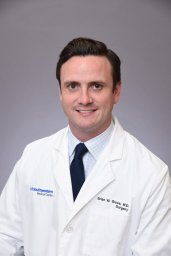 Brian Gross合作论文数Lancaster General Health2Steven R. Shackford合作论文数Department of Surgery, University of Vermont College of Medicine2
 Turner M. Osler合作论文数University of Rochester Medical Center, The University of Vermont2James Alzate合作论文数1Carissa Harnish合作论文数1Roxanne Chandler合作论文数1Shreya Jammula合作论文数1Catherine T Brown合作论文数1Micheal Horst合作论文数1Autumn Vogel合作论文数1Maria Gillio合作论文数1
 Taha Ahmed合作论文数1
 Enyo Ablordeppey合作论文数1
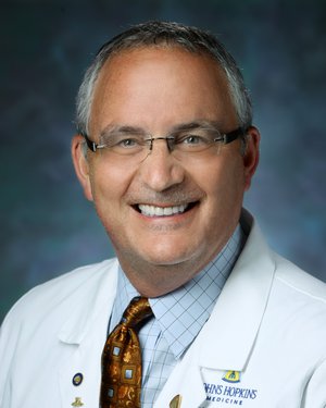 Todd Dorman合作论文数Department of Anesthesiology, The Johns Hopkins University School of Medicine/Department of Surgery, The Johns Hopkins University School of Medicine/Department of Internal Medicine, The Johns Hopkins University School of Medicine/Johns Hopkins University School of Nursing1
 Samuel A. Tisherman合作论文数Department of Surgery, School of Medicine, University of Maryland;R Adams Cowley Shock Trauma Center, University of Maryland Medical Center;Surgical Intermediate Care Unit, University of Maryland Medical Center1
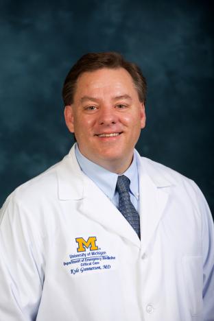 Kyle Gunnerson合作论文数Department of Emergency Medicine, University of Michigan Medical School;Department of Anesthesiology, University of Michigan Medical School1Livesay Sarah合作论文数1Miller Jo Ann合作论文数1Dzierba Amy L合作论文数1
 Piyush Mathur合作论文数Surgical Intensive Care Unit, Anesthesiology Institute &amp;Outcomes Research*, Cleveland Clinic Foundation;Anesthesiology Institute &amp; Outcomes Research*, Cleveland Clinic Foundation1
 Cook Alan合作论文数Division of Trauma, Baylor University Medical Center1Edavettal Matthew合作论文数1
 Claire Mooney合作论文数1Clark Elizabeth Hoffer合作论文数1Gambler Angela合作论文数1To Tuc合作论文数1
 C. William Schwab合作论文数The University of Pennsylvania School of Medicine1
 Bin Du (杜斌)合作论文数Peking Union Medical College, Chinese Academy of Medical Sciences1
 Tamas Szakmany合作论文数Whiston Hospital, St Helens and Knowsley Hospital NHS Trust1
 Sheila Nainan Myatra合作论文数Tata Memorial Hospital1
 Chet Morrison合作论文数1




