Introduction Usher syndrome (USH) represents a group of genetically heterogenous autosomal recessive disorders, characterized by combined vision and sensorineural hearing loss, and in some cases vestibular dysfunction.1–3 It primarily affects the light-sensitive photoreceptor cells in the retina and the auditory hair cells in the cochlea. Vision loss in all cases is progressive, and manifests as retinitis pigmentosa (RP), characterized by the gradual degeneration of the photoreceptor cells.4 Peripheral rod function is lost first, leading to night blindness and constricted visual fields, followed by the death of cones and severe visual impairment. There are currently no drugs or biological therapies proven to be effective in treating USH syndrome. Cochlear implantation, which bypasses the damaged hair cells and stimulates the primary auditory neurons directly, is an effective approach to alleviate the hearing impairment in patients. However, there is no remedy for the progressive loss of the photoreceptor neurons in the retina, and thus a critical unmet need exists to develop therapeutic strategies to prevent blindness in USH syndrome. The generation of animal models that faithfully mimic the human USH disorder, the timing of gene therapy interventions, the identification of correct target cells and patient selection are key factors in successfully reaching this goal. USH syndrome was named after Dr Charles Howard Usher, a Scottish ophthalmologist who described 69 patients with the disease in 1914.5 As is commonly the case in medicine, the disease was first described much earlier (1858) in brothers suffering from blindness and deafness by von Gräfe6 and was further characterized by his student, Liebreich,7 3 years later in a larger population of patients. From its discovery until recently, USH syndrome and its subgroups were delineated by clinical characteristics.8 Now the disease is recognized by symptomology and classified by genotyping. To date, there are at least 10 causative genes that are associated with USH syndrome, summarized in previous reviews.1,2,9 In general, based on its diverse clinical symptoms, particularly the onset and severity of the sensorineural hearing impairment and the presence of vestibular dysfunction, USH syndrome is grouped into 3 clinical subtypes. Type I disease is associated with severe-to-profound prelingual hearing loss, vestibular abnormalities and prepubescent onset of RP symptoms.10,11 USH1 results from mutations in MYO7A (USH1B), HARMONIN (USH1C), CDH23 (USH1D), PCDH15 (USH1F), SANS (USH1G), and CIB2 (USH1J). Patients with type II disease display moderate-to-severe congenital hearing loss without vestibular dysfunction, and account for >50% of all USH cases. Their RP symptoms generally begin in the second decade.12 USH2 is caused by mutations in USH2A (USH2A), ADGRV1 (USH2C), and WHRN (USH2D). Patients with type III disease caused by mutations in CLRN1 (USH3A) gene have postlingual progressive hearing loss, variable vestibular dysfunction, and variable onset of RP that rapidly progresses to legal blindness by the fourth decade of life.13–17 Mutations in the HARS gene (Histidyl-tRNA synthetase) are associated with the ultra-rare form USH3B.18,19 Other genes have been described in cases of atypical USH, including: CEP250 and CEP78, encoding members of the CEP family of centrosome-associated proteins; ESPN, encoding the F-actin cross-linker espin; ARSG, encoding the arylsulfatase G enzyme.20 Gene Therapy for USH Syndrome: Current Challenges The success of gene therapy for Leber Congenital Amaurosis (LCA2) due to defects in the RPE65 gene and subsequent FDA and EMA approval of Luxturna (voretigene neparvovec) have sparked hope that many other autosomal recessive retinal (and neurological) diseases may also be treated using a vector-based gene delivery approach.21,22 USH syndrome was no exception and several laboratories embarked on USH gene transfer studies in animal models. Conventional recombinant adeno-associated viral vectors (AAVs) contain a single-stranded DNA expression cassette, which includes a cell-specific or ubiquitous promoter, the transgene and polyadenylation signal, flanked by palindromic inverted terminal repeats at both ends. In contrast to other gene therapy systems, AAV vectors have low immunogenicity (especially when given subretinally), display long-lasting expression in postmitotic cells due to their ability to primarily persist as episomes, and efficiently transduce both photoreceptors and the retinal pigmented epithelium (RPE), where most RP-associated genes are expressed.23 Overall, AAV vectors hold tremendous potential to prevent retinal degeneration in USH syndrome. However, there are numerous challenges in the field, which become apparent by acknowledging the critical disease-specific parameters that account for the success of AAV-based gene augmentation therapy in RPE65 LCA. These include the availability of small and large animal models of RPE65-deficiency displaying a robust phenotype, a slow degenerative component of the disease accompanying a marked retinal dysfunction, a large window for treatment, and the lack of detrimental effects following long-term AAV-mediated transgene overexpression in RPE.24 In contrast to ocular diseases in USH syndrome, the molecular basis of retinal dysfunction in LCA2 is well understood. Moreover, the lack of RPE65 causes a substantial loss of visual function in murine and canine animal models, as demonstrated by the significantly reduced or nonrecordable electroretinograms.25–27 The marked loss of vision in animal models, which contrasts sharply with the remarkable preservation of retinal architecture, has allowed testing of long-term gene therapy effects.28–30 The RPE65 protein is a 65 kDa retinoid isomerohydrolase, which plays an essential role in the retinoid visual cycle by converting all-trans-retinyl ester to 11-cis-retinol in RPE cells.31–33 The 11-cis retinal chromophore deficiency in LCA2 photoreceptor outer segments can be corrected by restoring RPE65 enzyme to RPE, either by using a cell-specific human RPE65 promoter or a modified version of the ubiquitous chicken-beta actin promoter. Furthermore, one RPE cells interdigitates with 20 to 40 photoreceptor cells at the apical interface, amplifying the effects of gene therapy while minimizing the potential downside of nonuniform AAV-mediated transgene expression in neighboring RPE cells. Subretinal injection of an AAV2 vector delivering the canine wild-type RPE65 cDNA in dog models has produced long-term beneficial effects on visual function and protection from degeneration especially when treatment was initiated early.29,30,34,35 Ultimately, the successful restoration of vision in a large animal model paved the way towards an AAV-based gene therapy for LCA2 patients.36–40 Unlike the case of RPE65-associated disease, the majority of the genetically engineered mouse models of USH syndrome do not exhibit a visual phenotype, although they all display hearing loss.41,42 Some of these models were reported to display only a progressive retinal dysfunction with aging.42 It has been suggested that the lack of a retinal phenotype in mouse models could result from a distinct species-specific cell pattern of gene expression or to morphologic differences between human and mouse photoreceptors, for example, mice lack the well-developed, actin-filled calyceal processes, the subcellular structures extending from the inner segments in frogs, pigs, zebrafish, and primates.43,44 The absence of a robust ocular phenotype in animal models has severely limited the ability to perform therapeutic, preclinical proof-of-concept testing for vision loss in USH syndrome. However, gene replacement, gene editing, and small molecule therapy have been used to preserve hearing in many USH animal models with positive results.9,45–53 There are several recent, in-depth reviews of these therapies.9,54,55 These data, combined with the progressive nature of the vision loss due to USH syndrome, have motivated a few companies to empirically study treatments on human subjects. One example is the lentiviral-mediated MYO7A gene delivery in USH1B.56,57 This approach was shown to correct the melanosome mislocalization and opsin accumulation at the photoreceptor connecting cilium in the Myo7a-deficient shaker1 mouse model of USH1B.57 Conventional AAV has a small packaging capacity compared with lentiviral vector (4.7 vs. 8.5 kb). MYO7A cDNA, which is expressed in photoreceptors and the RPE cells, is too large (~6.7 kb) to be packaged in AAV vectors.58 Therefore, an equine infectious anemia virus-based lentiviral vector (equine infectious anemia virus-CMV-MYO7A, UshStat) was subretinally delivered to USH1B patients with the goal to prevent or slow down the RP progression (ClinicalTrials.gov identifier: NCT01505062). Long-term safety evaluation of this treatment in patients who received UshStat is currently being performed in a clinical trial (NCT02065011, results pending). Another clinical trial (UshTher) is currently underway, which uses a dual AAV vector approach to express MYO7A in the retina of patients following subretinal delivery (https://cordis.europa.eu/project/id/754848/it). Dual AAV vectors take advantage of the ability of the 2 distinct AAV genomes containing the 5′ and 3′ half of MYO7A transgene cDNA to undergo intermolecular concatamerization, followed by homologous recombination to reconstitute the whole large gene expression cassette.59,60 To date, there has been no clinically positive data reported from either one of these trials. The disease associated with much larger genes, for example, USH2A (cDNA~15.6 kb), may benefit from genome editing approaches, such as CRISPR-Cas9-sgRNAs, and those that promote exon skipping to restore functional protein restoration.61–66 A splicing-modulating antisense oligonucleotide approach designed to cause in-frame skipping of the mutated exon 13 of the USH2A disease-associated transcripts results in a shortened but functional usherin protein, which localizes correctly in photoreceptors and hair cells.66–68 Interestingly, recent studies have shown that zebrafish models of USH2A disease display early-onset retinal dysfunction, indicating they may represent useful tools to investigate the pathophysiology underlying USH syndrome and test therapeutic strategies.69,70 Morpholino antisense oligonucleotides were shown to mediate ush2a exon 13 skipping and rescue the visual dysfunction in a zebrafish USH2A model.66 This mutation-specific therapeutic strategy is being evaluated in a clinical trial using an antisense oligonucleotide (QR-421a) administered through intravitreal injections, with early evidence of efficacy reported at the 3-month interim analysis (phase 1/2 Stellar trial of QR-421a, ProQR Therapeutics, Clinicaltrials.gov ID: NCT03780257). Critical for the success of future therapeutic approaches is the generation and characterization of novel animal models that mimic the human ocular USH disease. These models will not only serve as preclinical platforms to test potential therapies, including viral-based gene delivery strategies, but can also be used to gain insight into the molecular mechanisms underlying retinal dysfunction and degeneration in USH syndrome. Interestingly, a pig model of USH1C lacking harmonin, USH1C (R31X), was recently reported to mimic the human disease, displaying combined deafness, changes in photoreceptor architecture, and significantly reduced visual function.71 USH syndrome has an estimated prevalence ranging from 4 to 17 cases per 100,000 individuals worldwide.72,73 Approximately 98% of USH syndrome patients belong to either type 1 or type 2 subgroups of disease. In contrast, USH3 is relatively rare, accounting for ~2% of all cases.74 However, it is the most prevalent form of USH disease in the Finnish population, where it represents an estimated 40% of all USH cases, and it also segregates to a high degree in patients of Ashkenazi-Jewish descent.16,17,74 Through funding from the Usher III initiative, a team of researchers was brought together in 2007 to uncover the roles of CLRN1 in sensory organs and the pathology that ensues when the gene is defective. Several therapeutic approaches were developed as a result of this sustained effort, with the ultimate goal of treating USH3A patients. An offshoot of this effort also resulted in several NIH-funded grants specifically directed towards understanding the complex pathophysiology of USH3 in the retina and cochlea. The rest of this review will highlight important findings of this research, and discuss strategies for developing a potentially effective gene therapy approach for USH syndrome. The USH Protein Network The lack of therapeutic approaches to prevent vision loss in USH patients has motivated a considerable amount of research on the pathophysiology of disease. Fluorescence colocalization studies on tissue sections from different species and reciprocal pull-down assays suggest that all proteins encoded by USH genes interact with each other and organize into large complexes within specific subcellular regions in photoreceptors and hair cells.75 They form highly dynamic networks, which consist of proteins from different classes with critical roles in transport, adhesion, and signaling processes in both sensory cell types. USH proteins include the actin-based motor protein myosin VIIa (MYO7A, USH1B); the cell-adhesion transmembrane proteins cadherin 23 (CDH23, USH1D), protocadherin 15 (PCDH15, USH1F), and usherin (USH2A); the very-large G protein-coupled receptor-1 VLGR1 (ADGRV1, GPR98, USH2C); the calcium and integrin binding protein 2 (CIB2, USH1J); the PDZ-domain scaffolding proteins harmonin (USH1C) and whirlin (WHRN, USH2D); the ankyrin and SAM-domain containing scaffolding protein SANS (USH1G); the tetraspanin-like protein clarin-1 (CLRN1, USH3A). In the retina, USH proteins were found to concentrate in specialized regions of photoreceptor cells, the connecting cilium, calyceal processes, and periciliary membrane.2 Some of the USH proteins were also detected at ribbon synapses in both photoreceptors and hair cells, which enable a rapid and sustained release of neurotransmitters necessary for the high frequency transmission of signals in sensory cells.76 The auditory system has often served as a mirror to gain insight into potential functions of USH proteins in the retina. In the cochlea, USH proteins were detected in the hair cell bundle, a mechanosensory antenna containing numerous F-actin rich stereocilia projecting from the apical surface of these auditory cells. As an elegant example, PCDH15 and CDH23 form both transient and permanent extracellular filamentous links between neighboring stereocilia, while harmonin, Sans, and myosin VIIa anchor these links to the intracellular actin core.77–80 In adult stages, PCDH15 and CDH23 form tip links, suggested to act similar to gating springs and open the sensory mechanoelectrical transduction ion channels in response to mechanical stimuli provided by sound waves.81,82 In the retina, USH1 proteins were detected at the fragile actin-rich calyceal processes in macaque, zebrafish, and humans, and may play a role in their development and maintenance.43,83 Similar to the interstereociliary links found in the hair bundle, USH1 proteins may form and stabilize membrane-membrane connection sites linking the calyceal processes to photoreceptor outer segments, allowing them to withstand daily mechanical stress.43 Consistent with this possibility, USH proteins organize and stabilize interactions between microvilli of absorptive intestinal cells.84,85 In contrast to USH1, the USH2 proteins usherin, VLGR1, and whirlin are mainly located in the periciliary ridge membrane complex region surrounding the photoreceptor connecting cilium, where they provide structural support and may also participate in vesicle docking with the plasma membrane during protein translocation to the outer segment.63,86 Pathogenic mutations leading to dysfunction or absence of a particular USH protein were found to alter the localization of other members in the network, a property which has been exploited to evaluate the efficacy of gene therapy experiments in animal models.87 Considering their subcellular localization to specific regions critical for transport and structural maintenance, careful AAV vector dosing and choice of regulatable promoters in gene therapy studies will be needed to ensure that long-term vector-mediated USH proteins expression mimics endogenous levels and does not lead to detrimental effects on photoreceptors structure and function.88 The use of novel penetrating AAV capsids with higher transduction efficiency may enable photoreceptor targeting following a surgically safer intravitreal approach, reducing the concentration of vector reaching the outer retina, and avoiding potential overexpression effects.89,90 CLRN1: an “Invisible” Tetraspanin Protein of Unknown Biological Function A major challenge in the USH field lies in the difficulty of reliably detecting the USH proteins in the retina, due to limited epitope availability, a consequence of USH protein complexes and their low levels of expression. This aspect, combined with the lack of thoroughly validated antibody reagents and proper negative controls in certain cases, has generated conflicting information regarding the cellular expression pattern of some of the USH proteins in the retina. This is especially true for CLRN1, a tetraspanin-like glycoprotein whose biological function is currently unknown.91 The main CLRN1 isoform encodes a 232 amino acid protein that contains a single glycosylation site at asparagine 48 (N48) in the first extracellular loop and a potential PDZ binding motif within its C-terminal end.91,92 In vitro experiments using a hemagglutinin (HA)-epitope tagged CLRN1 have shown that following its overexpression in HEK cells, the protein concentrates at specific regions within plasma membrane and forms CLRN1-enriched microdomains, recruiting diverse proteins involved in cell-adhesion, focal adhesion, and tight junctions, with an essential role in organizing the actin cytoskeleton.93 In the cochlea, CLRN1 has been suggested to interact with USH1 proteins harmonin and PCDH15.47,94Clrn1 knockout and the N48K knock-in mouse models of USH3 do not develop a retinal degeneration phenotype, but display an early-onset hearing loss and disorganized F-actin-rich stereocilia, indicating that one function of CLRN1 in the cochlea is to maintain the structural integrity of the hair bundle.95–97 A zebrafish model further established that CLRN1 is as an essential hair bundle protein.98 Other studies reported that CLRN1 absence also causes synaptic defects in cochlea hair cells.47,94 Three recent studies have shown that gene therapy can successfully prevent hearing loss in different mouse models of USH3 following early postnatal AAV-mediated Clrn1 cochlear delivery, when the sensory hair cell structure is preserved.47–49 These proof of principle studies raise hope that AAV-mediated CLRN1 expression to the correct retinal target cells will also be successful, providing that therapy is initiated before significant structural damage to photoreceptor cells occurs. Studies aimed to define the cellular expression of endogenous CLRN1 in the eye detected it either in the inner or outer retinal cells, or both, depending on the method used (in situ hybridization for mRNA or immunostaining for protein).97,99–101 Localization was further confounded by differences in RNA hybridization probes or antibodies used, variability in sample processing conditions, as well as the particular species analyzed. In one study, Clrn1 transcripts were transiently detected during postnatal development in the inner retina by in situ hybridization (ISH), in a pattern consistent with progenitor cells and Müller glia expression.97 By using rodless aged rd1 mice and laser capture microdissection, that study further showed that in adult mouse retinas Clrn1 mRNA was confined to the inner nuclear layer, detectable only by RT-PCR. In the same year, another group reported that CLRN1 protein was expressed in photoreceptor synapses and connecting cilium regions following immunostaining of the mouse retina with a custom made anti-CLRN1 antibody.101 In zebrafish, CLRN1 was detected in both the inner and outer retina.99 However, several studies concluded that endogenous CLRN1 protein remains below the levels of detection of immunostaining.47,49,97,100 Recently, we have generated and characterized a novel N-terminus epitope-tagged CLRN1 knock-in mouse to facilitate the detection of endogenous CLRN1 protein expression in retinal sections with a high affinity anti-HA antibody.100 We noted a similar background labeling pattern in both HA-tagged CLRN1 knock-in and the C57BL/6J controls. Importantly, the HA-tagged CLRN1 in this mouse was only detectable by western blot analysis of retinal homogenates, and was expressed continuously during postnatal development and adulthood.100 The HA-tagged CLRN1 protein in retinal sections could only be successfully detected following its viral-mediated overexpression using a subretinal or intravitreal delivery approach.100,102 The availability of single-cell RNA sequencing (scRNAseq) technology has provided an unprecedented opportunity to firmly map the localization of CLRN1 and other USH gene transcripts in the retina in multiple species, providing a complementary tool to immunodetection.103,104 In our recently published study, by using scRNAseq analysis of large data sets, we bypassed the challenges of immunostaining experiments, and convincingly demonstrated that CLRN1 transcripts in adult retina are present in Müller glia in human, mouse and non-human primates.100 Müller glia enrichment of CLRN1 transcripts in the retina has been corroborated in human samples by other scRNAseq studies.105–108 Interestingly, scRNAseq data sets have provided evidence that HARMONIN (USH1C), CDH23 (USH1D), and SANS (USH1G) transcripts are also present Müller glia, supporting the need to consider the involvement of multiple cell types in USH disease.108 Furthermore, by using the highly sensitive RNAscope ISH assay we detected CLRN1 transcripts in the inner retina and not photoreceptors, both in the adult mouse and human tissue, a result consistent with the scRNAseq data.100 No signal was detected when the ISH Clrn1 target probes were used on Clrn1 knockout mouse sections, confirming the high specificity of the assay.100 We have recently examined CLRN1 mRNA expression in the pig retina using a similar RNAscope ISH chromogenic assay. We determined that CLRN1 transcripts are abundantly expressed early in postnatal development, before the photoreceptor outer segments are fully developed, concentrating within the middle part of the inner nuclear layer of piglet retinas (Fig. 1). Taken together, these experiments provide solid evidence that CLRN1 transcripts are expressed in the inner retina in multiple species, and highlight the complexities inherent in understanding the molecular pathophysiology of vision loss in USH syndrome and potentially, a novel biology associated with retinal degeneration caused by Müller glia dysfunction. However, these studies do not completely rule out the possibility that CLRN1 might also be expressed in photoreceptor cells at very low levels, undetectable with our current technologies.Figure 1: Detection of CLRN1 transcripts in piglet retina at postnatal day P2 using the RNAscope ISH assay (red punctate staining). GCL indicates ganglion cell layer; INL, inner nuclear layer; ISH, in situ hybridization; ONL, outer nuclear layer; RPE, retinal pigment epithelium. The paraffin-embedded retina tissue sections were counterstained with Gill hematoxylin to visualize nuclei (blue). Scale bar: 20 µm.The potential roles of CLRN1 in protein scaffolding and signaling within the retina, as well as its specific molecular impact on Müller glia and photoreceptor neurons are currently unknown. Müller glia are involved in many different aspects of retinal biology, including neuronal development, modulation of neuronal synaptic activity and structural support of photoreceptor cells, and there are species-specific differences between the density and architecture of these cells in the retina.109,110 Previous studies have also suggested a possible function for Müller glia as radial tension sensors, showing that these extraordinary cells are sensitive to mechanically enforced changes in retinal structure.111,112 They have been compared with taut springs, providing mechanical resilience to the retina, and protecting the retina from ripping apart.111 Under normal physiological conditions, the retina is subjected to daily mechanical stress, including during its postnatal expansion. As a tetraspanin, CLRN1 may be involved in maintaining adhesion links between Müller glia and retinal neurons, protecting photoreceptors from mechanical stress, thus enabling their proper function throughout development and with aging. USH3 patients often display a severe impairment in retinal function at very early stages of the disease, with electroretinogram amplitudes being notably decreased in children as young as 3 to 5 years of age.16 Novel animal models of USH3 that mimic human retinal disease could be used to answer several critical questions with regard to gene therapy approaches. First, is presymptomatic transgene expression of CLRN1 required within photoreceptors, Müller glia, or both cell types to prevent blindness. Second, does ectopic CLRN1 expression in either photoreceptors or Müller glia actually promote degeneration. This could happen if exogenous CLRN1 protein prevents normal USH complexes formation in those cells. Finally, does CLRN1 delivery in adult stages, following symptomatic onset, restore vision loss and slow down the disease progression in USH3 patients. A variety of AAV-based gene therapy vector capsids and promoters targeting either photoreceptors or Müller glia are already available, and more are currently being generated and continuously optimized.23,89,113–116 The development and characterization of large animal models of USH syndrome is urgently needed in the field, as it will contribute greatly to the advancement of our understanding of the pathophysiology of this disease, and critically, provide an opportunity to study the cellular autonomy of retinal degeneration in USH patients. Conclusions On the surface, gene replacement appears to be a logical path to treat autosomal recessive retinal pathologies. This has led to the many laboratories and companies moving directly to gene therapeutic strategies to treat certain USH subtypes. The success of this path is yet to be determined. This review has highlighted several hurdles that should be overcome to optimize development of gene therapy towards specific inherited diseases. These fall into 2 general areas, (1) understanding the molecular pathology of the specific disease, and (2) developing gene therapy vectors that safely reach the target cells and express in a manner that effectively corrects biochemical defects without causing collateral damage. In LCA2, the detailed understanding of RPE65 role in vision, the simplicity of the gene structure and availability of both engineered and naturally occurring small (mouse) and large (dog) animal models enabled many researchers to successfully treat these animals, leading to the regulatory approvals of Luxturna. This review and others painfully demonstrate the complexities inherent in understanding the molecular pathophysiology of vision loss secondary to USH disease. Both the lack of solid understanding of the normal function of the associated proteins, and how the omission of these proteins specifically impacts the retina, limit the ability to develop USH therapies. This is true for all forms of USH, but perhaps is most evident for USH3, caused by mutations in an “invisible” CLRN1 protein. A key tool necessary to reveal this information is the generation of suitable animal models displaying a robust visual dysfunction and retinal degeneration phenotype. These models will allow basic scientists to better understand the molecular basis of disease and translational laboratories to develop effective therapies.
Usher syndrome type III (USH3) characterized by progressive loss of vision and hearing is caused by mutations in the clarin-1 gene (CLRN1). Clrn1 knockout (KO) mice develop hair cell defects by postnatal day 2 (P2) and are deaf by P21-P25. Early onset profound hearing loss in KO mice and lack of information about the cochlear cell type that requires Clrn1 expression pose challenges to therapeutic investigation. We generated KO mice harboring a transgene, TgAC1, consisting of Clrn1-UTR (Clrn1 cDNA including its 5′ and 3′ UTR) under the control of regulatory elements (Atoh1 3′ enhancer/β-globin basal promoter) to direct expression of Clrn1 in hair cells during development and down regulate it postnatally. The KO-TgAC1 mice displayed delayed onset progressive hearing loss associated with deterioration of the hair bundle structure, leading to the hypothesis that hair cell expression of Clrn1 is essential for postnatal preservation of hair cell structure and hearing. Consistent with that hypothesis, perinatal transfection of hair cells in KO-TgAC1 mice with a single injection of AAV-Clrn1-UTR vector showed correlative preservation of the hair bundle structure and hearing through adult life. Further, the efficacy of AAV-Clrn1 vector was significantly attenuated, revealing the potential importance of UTR in gene therapy.
UNLABELLED:Restoring vision in inherited retinal degenerations remains an unmet medical need. In mice exhibiting a genetically engineered block of the visual cycle, vision was recently successfully restored by oral administration of 9-cis-retinyl acetate (QLT091001). Safety and visual outcomes of a once-daily oral dose of 40 mg/m2/day QLT091001 for 7 consecutive days was investigated in an international, multi-center, open-label, proof-of-concept study in 18 patients with RPE65- or LRAT-related retinitis pigmentosa. Eight of 18 patients (44%) showed a ≥20% increase and 4 of 18 (22%) showed a ≥40% increase in functional retinal area determined from Goldmann visual fields; 12 (67%) and 5 (28%) of 18 patients showed a ≥5 and ≥10 ETDRS letter score increase of visual acuity, respectively, in one or both eyes at two or more visits within 2 months of treatment. In two patients who underwent fMRI, a significant positive response was measured to stimuli of medium contrast, moving, pattern targets in both left and right hemispheres of the occipital cortex. There were no serious adverse events. Treatment-related adverse events were transient and the most common included headache, photophobia, nausea, vomiting, and minor biochemical abnormalities. Measuring the outer segment length of the photoreceptor layer with high-definition optical coherence tomography was highly predictive of treatment responses with responders having a significantly larger baseline outer segment thickness (11.7 ± 4.8 μm, mean ± 95% CI) than non-responders (3.5 ± 1.2 μm). This structure-function relationship suggests that treatment with QLT091001 is more likely to be efficacious if there is sufficient photoreceptor integrity.TRIAL REGISTRATION:ClinicalTrials.gov NCT01014052.
Background Leber congenital amaurosis, caused by mutations in RPE65 and LRAT, is a severe form of inherited retinal degeneration leading to blindness. We aimed to assess replacement of the missing chromophore 11-cis retinal with oral QLT091001 (synthetic 9-cis-retinyl acetate) in these patients.Methods In our open-label, prospective, phase 1b trial, we enrolled patients (aged >= 6 years) with Leber congenital amaurosis and RPE65 or LRAT mutations at McGill University's Montreal Children's Hospital. Patients received 7 days of oral QLT091001 (10-40 mg/m(2) per day). We assessed patients at baseline and days 7, 9, 14, and 30, and then 2 months and every 2 months thereafter for up to 2.2 years for safety outcomes and visual function endpoints including Goldmann visual fields (GVF), visual acuity, and functional MRI assessment. We regarded patients as having an improvement in vision if we noted at least a 20% improvement in retinal area on GVF compared with baseline or a visual acuity improvement of five or more letters compared with baseline in two consecutive study visits (or any improvement from no vision at baseline). This study is registered with ClinicalTrials.gov, numberNCT01014052.Findings Between December, 2009, and June, 2011, we enrolled and treated 14 patients aged 6-38 years who were followed up until March, 2012. Ten (71%) of 14 patients had an improvement in GVF areas (mean increase in retinal area of 28-683%). Six (43%) patients had an improvement in visual acuity (mean increase of 2-30 letters). Self-reported or parent-reported improvements in activities of daily living supported these findings. After 2 years, 11 (79%) patients had returned to their baseline GVF retinal area and ten (71%) had returned to baseline visual acuity letter values. Thus, three (21%) patients had a sustained GVF response and four (30%) had a sustained visual acuity response. Four patients had functional MRI scans, which correlated with visual response or absence of response to treatment. No serious adverse events occurred, although we noted transient headaches (11 patients), photophobia (11 patients), reduction in serum HDL concentrations (four patients), and increases in serum triglycerides (eight patients) and aspartate aminotransferase concentrations (two patients).Interpretation Non-invasive oral QLT091001 therapy is well tolerated, and can rapidly improve visual function in some patients with Leber congenital amaurosis and RPE65 and LRAT mutations.
Following vitreoretinal surgery, patients may develop elevated intraocular pressure (IOP), which may be due to multiple different mechanisms. Preexisting glaucoma may be a potential cause of any postoperative IOP elevation in patients undergoing treatment for retinal disorders. Neovascularization of the anterior segment from underlying ischemic retinopathy may lead to neovascular glaucoma during the perioperative and postoperative periods. Also, prolonged treatment with topical or intravitreal steroids following retinal surgery can cause a steroid-induced glaucoma in susceptible patients.
OBJECTIVE To study the phenotypic characteristics of patients with a novel p.E292K mutation in BEST1. METHODS Affected individuals underwent ophthalmic examination and testing that included photography, autofluorescence, optical coherence tomography, and electrophysiological testing. Their DNA was analyzed for BEST1 mutations. RESULTS Five patients aged 5 to 59 years who expressed the p.E292K mutation in BEST1 were identified in 3 families. Electro-oculographic light-rise was subnormal in all probands and carriers. Carriers had normal findings from fundus examination, multifocal electroretinography, and visual acuity, and were emmetropic or myopic. Only probands had hyperopia and fundus findings typical of Best macular dystrophy. Optical coherence tomography of vitelliform lesions demonstrated retinal pigment epithelium elevation without subretinal fluid; atrophic lesions exhibited disruption of the hyperreflective outer retina-retinal pigment epithelium complex. Intense hyperautofluorescence correlated with the vitelliform lesion. CONCLUSIONS Patients with the Glu292Lys variation in BEST1 exhibit intrafamilial and interfamilial phenotypic variability. A disproportionate fraction (26%) of Best disease-causing mutations occurs in exon 8, suggesting that the portion of protein encoded by this exon (amino acids 290-316) may be especially important to bestrophin's function. Relatively good visual acuity with vitelliform lesions can be explained by preservation of the outer retina, demonstrated by optical coherence tomography. Clinical Relevance A novel mutation in this region of BEST1 carries implications for disease pathogenesis.
PURPOSE Long-term effects of treatment with 9-cis-retinyl acetate (9-cis-R-Ac), an artificial retinoid prodrug, were tested on changes in rod and cone visual functions in mice. METHODS The acetyl ester of the functional geometric chromophore 9-cis-retinal was delivered by oral gavage to C57BL/6 female mice. In initial experiments, 10-month-old mice were used for the single treatment with 9-cis-R-Ac or the control vehicle. In long-term experiments, 4-month-old mice were treated with 9-cis-R-Ac monthly for 6 and 10 months. Photoreceptor status was evaluated by various electroretinographic (ERG) techniques, retinoid analyses, and retinal morphology. Opsin, the predicted target of oxidized 9-cis-R-Ac, was purified and its chromophore was characterized. RESULTS Age-related changes observed in vehicle-treated mice at 10 months of age, compared with those in 4-month-old mice, included a progressive decline in ERG responses, such as a decreased rate of dark adaptation and a lowered rhodopsin/opsin ratio. Administration of 9-cis-R-Ac increased the rhodopsin regeneration ratio, and improved ERG responses and dark adaptation. Compared with vehicle-treated control animals, 10- and 14-month-old mice treated monthly with 9-cis-R-Ac for 6 or 10 months exhibited improved dark adaptation. In 14-month-old mice treated monthly, changes in the expression of retina-specific genes in the eye were detected by mRNA expression profiling, but no significant effects in gene expression were detected in the liver and kidney. CONCLUSIONS Deteriorating photoreceptor function documented in mice at 10 and 14 versus 4 months of age was improved significantly by long-term, monthly administration of 9-cis-R-Ac. These findings suggest a potential therapeutic approach to prevent age-related retinal dysfunction.
Purpose: To evaluate combination treatment with reduced-fluence photodynamic therapy (PDT) and intravitreal triamcinolone acetonide (IVT) injection for choroidal neovascularization (CNV) in age-related macular degeneration (AMD).Methods: This is a retrospective consecutive case series of 23 previously untreated eyes of 22 patients with subfoveal CNV secondary to AMD. Six eyes were treated with 50 J/cm(2); 8, with 40 J/cm(2); and 9, with 25 J/cm(2). PDT was immediately followed by a 4-mg IVT injection. Patients were observed for 6 months at 6-week intervals.Results: The 50 J/cm(2) subset lost a mean of 2.2 lines of Snellen visual acuity at the 6-month follow-up compared with a 1-line lost in the 40 J/cm(2) subset and a 0.9-line gain in the 25 J/cm(2) subset. In the 50 J/cm(2) subset, 60% lost <= 3 lines,of Snellen visual acuity, 33% gained >= 0 line, and 33% gained >= 3 lines. In the 40 J/cm(2) subset, 75% lost <= 3 lines of Snellen visual acuity, 50% gained >= 0 line, and 25% gained >= 3 lines. In the 25 J/cm(2) subset, 89% lost <= 3 lines of Snellen visual acuity, 56% gained >= 0 line, and 33% gained >= 3 lines. Fifty percent of the 50 J/cm(2) subset, 50% of the 40 J/cm(2) subset, and 33% of the 25 J/cm(2) subset required retreatment by 6 months.Conclusion: Although the sample in this study was small, there was a dose-response trend toward better visual outcomes and fewer treatments in the group treated with IVT injection and reduced-fluence PDT. This study along with other previously reported work suggests that studies using PDT in combination treatment for CNV should consider adding a reduced-fluence PDT arm.
Pathogenic mutations in the RPE65 gene are associated with a spectrum of congenital blinding diseases in humans. We evaluated changes in the promoter region, coding regions, and exon/intron junctions of the RPE65 gene by direct sequencing of DNA from 36 patients affected with Leber's congenital amaurosis (LCA), 62 with autosomal recessive retinitis pigmentosa (arRP), and 21 with autosomal dominant/recessive cone-rod dystrophies (CORD). Fifteen different variants were found, of which 6 were novel. Interesting was Gly244Val, a novel mutation close to the catalytic center. To assess the role of this mutation in RPE65 inactivation, we performed detailed biochemical studies of the mutant along with a structural analysis of the 244 amino acid position with respect to amino acids known to be important for RPE65-dependent retinoid isomerization. Bicistronic plasmid expression of the RPE65 Gly244Val mutant and enhanced green fluorescent protein (EGFP) allowed us to document both its instability in cultured cells by cell sorting and immunoblotting methodology and its loss of RPE65-dependent isomerase activity by enzymatic assays. Further insights into the structural requirements for retinoid isomerization by RPE65 were obtained by using the carotenoid oxygenase (ACO) from Synechocystis (PDB accession code 2BIW ) as a structural template to construct a RPE65 homology model and locating all known inactivating mutations including Gly244Val within this model.
The purpose of this study was to determine the role of the retinol dehydrogenase 12 (RDH12) gene in patients affected with Leber congenital amaurosis (LCA), autosomal recessive retinitis pigmentosa (arRP) and autosomal dominant/recessive cone-rod dystrophies (CORD). Changes in the promoter region, coding regions and exon/intron junctions of the RDH12 gene were evaluated using direct DNA sequencing of patients affected with LCA (n=36 cases), RP (n=62) and CORD (n=21). The allele frequency of changes observed was assessed in a multiethnic control population (n=159 individuals). Detailed biochemical and structural modeling analysis of the observed mutations were performed to assess their biological role in the inactivation of Rdh12. A comprehensive clinical assessment of retinal structure and function in LCA patients carrying mutations in the RDH12 gene was completed. Of the six changes identified, three were novel including a homozygous C201R change in a patient affected with LCA, a heterozygous A177V change in patients affected with CORD and a heterozygous G46G change in a patient affected with LCA. A novel compound heterozygote T49M/A269fsX270 mutation was also found in a patient with LCA, and both homozygous and heterozygous R161Q changes were seen in 26 patients affected with LCA, CORD or RP. These R161Q, G46G and the A177V sequence changes were shown to be polymorphic. We found that Rdh12 mutant proteins associated with LCA were inactive or displayed only residual activity when expressed in COS-7 and Sf9 cells, whereas those mutants that were considered polymorphisms were fully active. Thus, impairment of retinal structure and function for patients carrying these mutations correlated with the biochemical properties of the mutants.
PURPOSE:To report a case demonstrating excellent visual recovery following surgical removal of subfoveal exudation secondary to a vasoproliferative tumor of the retina (VPTR). DESIGN:Observational case report. METHODS:Review of medical records. RESULTS:A 39-year-old man presented with decreased vision secondary to subfoveal exudation from a VPTR. The tumor was successfully treated with cryoretinopexy, but the hard exudates that had formed in under the fovea remained. There was no change in vision for 18 months following the cryoretinopexy. A decision was made to surgically remove the exudates using similar techniques to those presented in the literature for diabetic exudation. Over the next several weeks the patients vision recovered to the 20/25 level and has remained this way for 5 years. CONCLUSIONS:Patients with subfoveal exudates secondary to VPTR may benefit from subretinal surgery to remove the exudates.
To determine further long-term safety information for the period after the use of photodynamic therapy with verteporfin for subfoveal choroidal neovascularization secondary to ocular histoplasmosis syndrome (OHS), the Verteporfin in Ocular Histoplasmosis (VOH) Study Group began a prospective 2-year extension study after an amendment to the original 2-year protocol was issued in 2001. Here, we report the results from this extension study. All patients completing the original 2-year VOH Study, which was a prospective open-label clinical trial, could enroll in the 2-year extension study. Patients who were judged by the treating ophthalmologist to have evidence of choroidal neovascularization leakage could be treated with verteporfin therapy, and safety evaluations were performed as described in the original study.1Saperstein D.A. Rosenfeld P.J. Bressler N.M. et al.Photodynamic therapy of subfoveal choroidal neovascularization with verteporfin in the ocular histoplasmosis syndrome: one-year results of an uncontrolled, prospective case series.Ophthalmology. 2002; 109: 1499-1505Abstract Full Text Full Text PDF PubMed Scopus (73) Google Scholar Seventeen of the 22 patients examined at 24 months, among 26 patients initially enrolled in the VOH Study, enrolled in the extension study. The baseline characteristics of the 17 patients are shown in Table 1 (available at http://aaojournal.org). All 17 patients received at least 1 verteporfin treatment throughout the entire extension study, and all were included in the analysis. No methods to impute missing data were used in the study extension. Fifteen patients completed the month 48 examination (1 was lost to follow-up, and 1 requested to exit the trial before that examination because of financial considerations).Table 1Summary of Baseline Characteristics for Patients Enrolled in the Verteporfin in Ocular Histoplasmosis Study ExtensionCharacteristicVerteporfin (n = 17)Women [n (%)]12 (71)Mean age (yrs)48.2Mean baseline study eye visual acuity [letter score (approximate Snellen equivalent)]56.9 (20/80+2)Mean study eye visual acuity at the start of extension study [letter score (approximate Snellen equivalent)]69.5 (20/40)Mean study eye contrast sensitivity (no. of letters)30.8Classic choroidal neovascularization [n (%)] ≥50%13 (76) <50%3 (18) No classic1 (6)Mean greatest linear dimension (μm)2870.0Lesion size (MPS DAs) [n (%)] ≤14 (24) >1 to ≤23 (18) >2 to ≤37 (41) >33 (18)DA = disc area; MPS = Macular Photocoagulation Study. Open table in a new tab DA = disc area; MPS = Macular Photocoagulation Study. Extension patients (n = 17) received a mean of 4.4 treatments during the entire 48 months of the study. The mean number of treatments administered decreased with each successive year, with an average of 2.9 in year 1 and 1.2, 0.2, and 0.1 treatments in years 2, 3, and 4, respectively. Median visual acuity (VA) improved from the VOH Study baseline by 15 letters (range, loss of 24 letters to gain of 34 letters). Of the 15 patients completing the study at month 48, 9 (60%) gained ≥7 letters of VA from the initial study baseline, 4 (27%) had little or no change in VA, and only 1 (7%) lost >15 letters of VA (Fig 1 [available at http://aaojournal.org]). Median contrast sensitivity improved from the initial study baseline by 3 letters at 48 months. No deaths were reported during the study, and no patients withdrew due to adverse events. Three ocular adverse events judged to be treatment related were reported during the extension period: 1 patient (6%) complained of blurred vision, 1 (6%) experienced a decrease in central vision, and 1 (6%) experienced a visual field defect (a central black spot). Injection-site adverse events occurred in 4 patients (24%). These treatment-related adverse events were mild to moderate in severity. No acute severe VA decrease (defined as a loss of at least 20 letters within 7 days of treatment) was reported in any patients during the 48-month study. Verteporfin therapy for subfoveal choroidal neovascularization due to OHS was associated with maintained or improved VA and improved contrast sensitivity in the majority of patients through 48 months of therapy. In view of the overall safety data for verteporfin therapy from this study as well as trials in patients with age-related macular degeneration or pathologic myopia, no new or additional safety concerns were identified with long-term follow-up in this group of patients with OHS, and no loss in VA was apparent at month 48 compared with month 24. The low number of treatments required during the extension period demonstrates that the effects of verteporfin therapy on choroidal neovascularization are sustained through at least 4 years. The results of this study extend the 2-year findings of the original study and support the previous recommendation of the VOH Study Group to consider verteporfin therapy for patients with choroidal neovascularization due to the OHS.1Saperstein D.A. Rosenfeld P.J. Bressler N.M. et al.Photodynamic therapy of subfoveal choroidal neovascularization with verteporfin in the ocular histoplasmosis syndrome: one-year results of an uncontrolled, prospective case series.Ophthalmology. 2002; 109: 1499-1505Abstract Full Text Full Text PDF PubMed Scopus (73) Google Scholar, 2Rosenfeld P.J. Saperstein D.A. Bressler N.M. et al.Photodynamic therapy with verteporfin in ocular histoplasmosis Uncontrolled open-label 2-year study.Ophthalmology. 2004; 111: 1725-1733Abstract Full Text Full Text PDF PubMed Scopus (46) Google Scholar
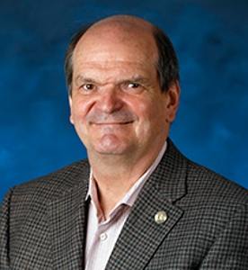 Krzysztof Palczewski合作论文数Department of Chemistry, School of Physical Sciences, University of California, Irvine;School of Medicine, University of California, Irvine11
 Robert K. Koenekoop合作论文数Department of Pediatric Surgery, Faculty of Medicine and Health Sciences, McGill University5
 Schuchard Ronald A合作论文数5
 Slawomir Filipek合作论文数Faculty of Chemistry, Warsaw University5
 Gislin Dagnelie合作论文数Department of Ophthalmology, School of Medicine, Johns Hopkins University4
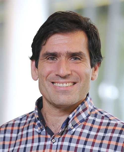 Dimitrios Fotiadis合作论文数Institute of Biochemistry and Molecular Medicine, Faculty of Medicine, University of Bern4L. Ingeborgh Van Den Born合作论文数4
 Yan Liang合作论文数3Christopher N. Singh合作论文数3
 Neil M. Bressler合作论文数Retinal Vascular Center, School of Medicine, Johns Hopkins University;Wilmer Eye Institute Research, School of Medicine, Johns Hopkins University3
 Philip J. Rosenfeld合作论文数Bascom Palmer Eye Institute, University of Miami3Akiko Maeda合作论文数3
 Thomas Aaberg合作论文数Michigan State University3
 Andreas Engel合作论文数The Center for Molecular Life Sciences, University of Basel;cryoWrite3Wenyu Sun合作论文数3
 Paul Sternberg合作论文数Department of Ophthalmology, Emory University3
 Ruifang Sui (睢瑞芳)合作论文数Peking Union Medical College Hospital3Robert H. Rosa合作论文数Cincinnati3Tadao Maeda合作论文数3
 Juliana Maria Ferraz Sallum合作论文数Instituto de Genética Ocular;Departamento de Oftalmologia, Universidade Federal de São Paulo3Ava K. Bittner合作论文数3Cs Barnes合作论文数2Troy A. Reaves合作论文数2
 Elise Heon合作论文数Department of Ophthalmology, Temerty Faculty of Medicine, University of Toronto;Department of Ophthalmology and Vision Sciences, Temerty Faculty of Medicine, University of Toronto;The Hospital for Sick Children2
 Janine Dale Mendola合作论文数Department of Ophthalmology, McGill University2
 Elias I. Traboulsi合作论文数Department of Pediatric Ophthalmology, Cleveland Clinic;Department of Graduate Medical Education, Cleveland Clinic;Center for Genetic Eye Diseases, Cole Eye Institute, Cleveland Clinic;Cleveland Clinic Lerner College of Medicine, Case Western Reserve University2
 Tony Moore合作论文数Institute of Ophthalmology, Faculty of Brain Sciences, University College London;European Vision Institute2
 Eberhart Zrenner合作论文数Werner Reichardt Centrum für Integrative Neurowissenschaften (CIN) Institute for Ophthalmic Research Centre for Ophthalmology, University of Tübingen2
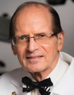 Gerald Fishman合作论文数Pangere Center for Inherited Retinal Diseases, Institute for Health Research and Policy, University of Illinois Chicago2
 David Birch合作论文数Rose-Silverthorne Retinal Degenerations Laboratory, Retina Foundation of the Southwest2
 Hendrik Scholl合作论文数Institute of Molecular and Clinical Ophthalmology Basel;Department of Ophthalmology, University of Basel;Department of Clinical Research, University of Basel;Department of Ophthalmology, University Hospital Basel;European Vision Institute2
 Michael D. Bennett (Michael Bennett)合作论文数Jodrell Laboratory2
 Astra Dinculescu合作论文数Department of Ophthalmology Research, College of Medicine, University of Florida2Michel Sickenberg合作论文数2Elliott M. Kanner合作论文数1g watson合作论文数1Annemarie Weisberger合作论文数1Lauren Van Der Kraak合作论文数1leah wood合作论文数1Ec Steele合作论文数1Scott Lawrence合作论文数1Ruben Stepanyan合作论文数1Patrick Leahy合作论文数1Philip D Kiser合作论文数1Zhu Li合作论文数1
 Peter A. Netland合作论文数Department of Ophthalmology, School of Medicine, University of Virginia1Todd Klesert合作论文数1Wen Yuquan合作论文数1
 David Mark Brown合作论文数Baylor College of Medicine;Brown Retina Institute, Retina Consultants of Texas;Greater Houston Retina Research Center;Blanton Eye Institute, Houston Methodist Hospital1




