C/EBPβ regulates oncogene-induced senescence (OIS) and the senescence-associated secretory phenotype (SASP) through activation by ERK1/2 and CK2. In tumor cells, C/EBPβ activity is suppressed by its 3'UTR via a mechanism termed 3'UTR regulation of protein activity (UPA), which spatially segregates CEBPB transcripts from kinase-rich perinuclear endosomes. Here, we identify kinase-proximal mRNA decay as the underlying mechanism. The mRNA decay factors UPF1 and STAU1/2 localize to perinuclear endosomes and promote degradation of CEBPB transcripts, thereby preventing C/EBPβ phosphorylation and activation. Disruption of this pathway restores C/EBPβ activity and induces senescence. In vivo, deletion of a G/U-rich regulatory element (GRE) in the 3'UTR impairs the progression of Kras-driven lung tumors and biases cells toward an AT2-like differentiation state with reduced EMT-associated transcriptional reprogramming. RAS-expressing GRE Δ/Δ fibroblasts show enhanced OIS that requires upregulation of the pro-senescent cytokine S100a9. These findings identify perinuclear mRNA decay as a mechanism suppressing C/EBPβ activity and senescence in cancer.
Lamin A/C (LMNA), a key component of the nuclear envelope, is essential for maintaining nuclear integrity and genome organization [W. Xie et al., Curr. Biol. 26, 2651-2658 (2016)]. While LMNA dysregulation has been implicated in genomic instability across cancer and aging, the underlying mechanisms remain poorly understood [S. Graziano et al., Nucleus 9, 258-275 (2018)]. Here, we define a mechanistic role for LMNA in preserving genome stability in small-cell lung cancer (SCLC), a malignancy marked by extreme genomic instability [N. Takahashi et al., Cancer Res. Commun. 2, 503-517 (2022)]. LMNA depletion promotes R-loop accumulation, transcription-replication conflicts, replication stress, DNA breaks, and micronuclei formation. Mechanistically, LMNA deficiency disrupts nuclear pore complex organization, specifically reducing phenylalanine-glycine (FG)-nucleoporin incorporation, resulting in impaired RNA export and nuclear retention of RNA. LMNA expression is repressed by EZH2 and reexpressed during SCLC differentiation from neuroendocrine (NE) to non-NE states, and low LMNA levels correlate with poor clinical outcomes. These findings establish LMNA as a key regulator of nuclear transport and genome integrity, linking nuclear architecture to SCLC progression and therapeutic vulnerability.
Lamin A/C (LMNA), a key component of the nuclear envelope, plays an essential role in maintaining nuclear integrity and regulating genome organization. While LMNA dysregulation has been linked to genomic instability in both cancer and aging, the specific mechanisms remain poorly understood. We investigated the role of LMNA in small cell lung cancer (SCLC), a highly lethal form of lung cancer characterized by neuroendocrine (NE) differentiation, low LMNA expression, and extreme genomic instability. We show that LMNA depletion leads to increased R-loop formation, transcription-replication conflicts, replication stress, DNA breaks, the formation of micronuclei and increases sensitivity to TOP1 and ATR inhibition. Mechanistically, LMNA loss compromises RNA export efficiency by disrupting the nuclear pore complex distribution causing clumping and reducing incorporation of phenylalanine-glycine-nucleoporins (FG-nucleoporins). We found that LMNA expression is dynamically regulated during SCLC differentiation from NE to a non-NE state by EZH2, a component of the polycomb repressive complex that epigenetically silences LMNA. Inhibition of EZH2 restored LMNA expression in SCLC cells, and functionally led to a reduction in R-loops. Low LMNA levels were associated with significantly reduced survival in SCLC patients implicating R-loops as potential detrimental factor in SCLC patients. To confirm this, we utilized multiplexed immunofluorescence and confirmed LMNA protein was inversely correlated with R-loops both within the same SCLC tumor and across SCLC tumors using an SCLC rapid autopsy sample alongside an SCLC tumor microarray. Interestingly, low expression of canonical R-loop suppressing genes (RNASEH1, BRD4, SETX and DHX9) also predicted poorer prognosis in SCLC patients, supporting R-loops as a detrimental factor for SCLC prognosis. Critically, we were able to replicate key findings linking low LMNA to decreased FG-nucleoporin incorporation and increased R-loops using patient derived young and old fibroblasts lines demonstrating generalizability of this finding across disease models. Our results reveal a novel mechanism by which LMNA safeguards genome integrity by repressing R-loops, with important implications for both cancer and aging. Christopher William Schultz, Sourav Saha, Anjali Dhall, Yang Zhang, Parth Desai, Lorinc Pongor, David A. Scheiblin, Valentin Magidson, Yilun Sun, Christophe Redon, Suresh Kumar, Manan Krishnamurthy, Henrique B. Dias, Vasilisa Askenova, Elizabeth Giordanio, Nobuyuki Takahashi, Michael Nirula, Yasuhiro Arakawa, Ukhyun Jo, Beverly A. Teacher, Mirit I. Aladjem, Stephen Lockett, Marry Dasso, Yves Pommier, Ajit K. Sharma, Anish Thomas. Lamin AC deficiency promotes genomic instability through increased R-loop accumulation [abstract]. In: Proceedings of the American Association for Cancer Research Annual Meeting 2025; Part 1 (Regular Abstracts); 2025 Apr 25-30; Chicago, IL. Philadelphia (PA): AACR; Cancer Res 2025;85(8_Suppl_1):Abstract nr 2835.
Lamin A/C (LMNA), a key component of the nuclear envelope, is essential for maintaining nuclear integrity and genome organization [1]. While LMNA dysregulation has been implicated in genomic instability across cancer and aging, the underlying mechanisms remain poorly understood [2]. Here, we investigate LMNA's role in small-cell lung cancer (SCLC), a highly aggressive malignancy characterized by extreme genomic instability [3, 4]. We demonstrate that LMNA depletion promotes R-loop accumulation, transcription-replication conflicts, replication stress, DNA breaks, and micronuclei formation. Mechanistically, LMNA loss disrupts nuclear pore complex distribution, reducing phenylalanine-glycine (FG)-nucleoporin incorporation and impairing RNA export efficiency. Furthermore, we show that LMNA expression is epigenetically repressed by EZH2 during SCLC differentiation from neuroendocrine (NE) to non-NE states. Clinically, low LMNA levels correlate with significantly worse survival in SCLC patients. These findings uncover a novel role for LMNA in safeguarding genome integrity and shaping tumor heterogeneity, with broad implications for cancer and aging.
C/EBPβ is a potent regulator of RAS-induced senescence (RIS) and the SASP. C/EBPβ is post- translationally activated in RIS cells by the effector kinases ERK1/2 and CK2, but in tumor cells activation is suppressed by the CEBPB 3’UTR. 3′UTR regulation of protein activity (UPA) requires a G/U-rich element (GRE) and its cognate binding protein, HuR. These components segregate CEBPB transcripts away from a perinuclear compartment harboring ERK1/2 and CK2, restricting C/EBPβ from its activating kinases. We report here that the mRNA decay proteins UPF1 and Staufen1/2 are essential UPA factors enriched within the perinuclear cytoplasm. STAU1/2 colocalize with CK2 on perinuclear signaling endosomes where they promote localized CEBPB mRNA decay. UPF1 or STAU1/2 depletion in tumor cells increased CEBPB transcripts adjacent to CK2 foci, coinciding with C/EBPβ activation and senescence. The GRE and an adjacent STAU binding site act to suppress C/EBPβ-mediated senescence, while a separate 3’UTR region inhibits SASP induction. Accordingly, KrasG12D-driven lung tumors in mice carrying a Cebpb GRE deletion rarely progressed to malignant adenocarcinomas. Our findings identify kinase-proximal mRNA decay as a novel mechanism to inhibit C/EBPβ activating modifications in tumor cells to facilitate senescence bypass.
Antitumor immune polarization is a key predictor of clinical outcomes to cancer therapy. An emerging concept influencing clinical outcome involves the spatial location of CD8+ T cells, within the tumor. Our earlier work demonstrated immunosuppressive effects of NOS2 and COX2 tumor expression. Here, we show that NOS2/COX2 levels influence both the polarization and spatial location of lymphoid cells including CD8+ T cells. Importantly, elevated tumor NOS2/COX2 correlated with exclusion of CD8+ T cells from the tumor epithelium. In contrast, tumors expressing low NOS2/COX2 had increased CD8+ T cell penetration into the tumor epithelium. Consistent with a causative relationship between these observations, pharmacological inhibition of COX2 with indomethacin dramatically reduced tumor growth of the 4T1 model of TNBC in both WT and Nos2- mice. This regimen led to complete tumor regression in ∼20–25% of tumor-bearing Nos2- mice, and these animals were resistant to tumor rechallenge. Th1 cytokines were elevated in the blood of treated mice and intratumoral CD4+ and CD8+ T cells were higher in mice that received indomethacin when compared to control untreated mice. Multiplex immunofluorescence imaging confirmed our phenotyping results and demonstrated that targeted Nos2/Cox2 blockade improved CD8+ T cell penetration into the 4T1 tumor core. These findings are consistent with our observations in low NOS2/COX2 expressing breast tumors proving that COX2 activity is responsible for limiting the spatial distribution of effector T cells in TNBC. Together these results suggest that clinically available NSAID’s may provide a cost-effective, novel immunotherapeutic approach for treatment of aggressive tumors including triple negative breast cancer.
Anti-tumor immune polarization is a key predictor of clinical outcomes to cancer therapy. An emerging concept influencing clinical outcome involves the spatial location of CD8 + T cells, within the tumor. Our earlier work demonstrated immunosuppressive effects of NOS2/ COX2 tumor expression. Here, we show that NOS2/COX2 levels influence the polarization and spatial location of lymphoid cells including CD8 + T cells. Importantly, elevated tumor NOS2/COX2 correlated with exclusion of CD8 + T cells from the tumor epithelium. In contrast, tumors expressing low NOS2/COX2 had increased CD8 + T cell penetration into the tumor epithelium. Consistent with a causative relationship between these observations, pharmacological inhibition of COX2 with indomethacin dramatically reduced tumor growth of the 4T1 model of TNBC in both WT and Nos2 -/- mice. This regimen led to complete tumor regression in ∼20% of tumor-bearing Nos2 -/- mice, and these animals were resistant to tumor rechallenge. Th1 cytokines were elevated in the blood of treated mice and intratumoral CD4 + and CD8 + T cells were higher in mice that received indomethacin when compared to control untreated mice. Multiplex immunofluorescence imaging confirmed our phenotyping results and demonstrated that targeted Nos2/Cox2 blockade improved CD8 + T cell penetration into the 4T1 tumor core. These findings are consistent with our observations in low NOS2/COX2 expressing breast tumors` proving that COX2 activity is responsible for limiting the spatial distribution of effector T cells in TNBC. Together these results suggest that clinically available NSAID’s may provide a cost-effective, novel immunotherapeutic approach for treatment of aggressive tumors including triple negative breast cancer.
The HIV-1 envelope glycoprotein (Env) contains a long cytoplasmic tail harboring highly conserved motifs that direct Env trafficking and incorporation into virions and promote efficient virus spread. The cellular trafficking factor Rab11a family interacting protein 1C (FIP1C) has been implicated in the directed trafficking of Env to sites of viral assembly. In this study, we confirm that small interfering RNA (siRNA)-mediated depletion of FIP1C in HeLa cells modestly reduces Env incorporation into virions. To determine whether FIP1C is required for Env incorporation and HIV-1 replication in physiologically relevant cells, CRISPR-Cas9 technology was used to knock out the expression of this protein in several human T-cell lines-Jurkat E6.1, SupT1, and H9-and in primary human CD4(+) T cells. FIP1C knockout caused modest reductions in Env incorporation in SupT1 cells but did not inhibit virus replication in SupT1 or Jurkat E6.1 T cells. In H9 cells, FIP1C knockout caused a cell density-dependent defect in virus replication. In primary CD4(+) T cells, FIP1C knockout had no effect on HIV-1 replication. Furthermore, human T-cell leukemia virus type 1 (HTLV-1)-transformed cell lines that are permissive for HIV-1 replication do not express FIP1C. Mutation of an aromatic motif in the Env cytoplasmic tail (Y795W) implicated in FIP1C-mediated Env incorporation impaired virus replication independently of FIP1C expression in SupT1, Jurkat E6.1, H9, and primary T cells. Together, these results indicate that while FIP1C may contribute to HIV-1 Env incorporation in some contexts, additional and potentially redundant host factors are likely required for Env incorporation and virus dissemination in T cells. IMPORTANCE The incorporation of the HIV-1 envelope (Env) glycoproteins, gp120 and gp41, into virus particles is critical for virus infectivity. gp41 contains a long cytoplasmic tail that has been proposed to interact with host cell factors, including the trafficking factor Rab11a family interacting protein 1C (FIP1C). To investigate the role of FIP1C in relevant cell types-human T-cell lines and primary CD4(+) T cells-we used CRISPR-Cas9 to knock out FIP1C expression and examined the effect on HIV-1 Env incorporation and virus replication. We observed that in two of the T-cell lines examined (Jurkat E6.1 and SupT1) and in primary CD4(+) T cells, FIP1C knockout did not disrupt HIV-1 replication, whereas FIP1C knockout reduced Env expression and delayed replication in H9 cells. The results indicate that while FIP1C may contribute to Env incorporation in some cell lines, it is not an essential factor for efficient HIV-1 replication in primary CD4(+) T cells.
Background There is a need to improve the treatment of prostate cancer (PCa) and reduce treatment side effects. Vascular-targeted photodynamic therapy (VTP) is a focal therapy for low-risk low-volume localised PCa, which rapidly disrupts targeted tumour vessels. There is interest in expanding the use of VTP to higher-risk disease. Tumour vasculature is characterised by vessel immaturity, increased permeability, aberrant branching and inefficient flow. FRT alters the tumour microenvironment and promotes transient ‘vascular normalisation’. We hypothesised that multimodality therapy combining fractionated radiotherapy (FRT) and VTP could improve PCa tumour control compared against monotherapy with FRT or VTP. Methods We investigated whether sequential delivery of FRT followed by VTP 7 days later improves flank TRAMP-C1 PCa tumour allograft control compared to monotherapy with FRT or VTP. Results FRT induced ‘vascular normalisation’ changes in PCa flank tumour allografts, improving vascular function as demonstrated using dynamic contrast-enhanced magnetic resonance imaging. FRT followed by VTP significantly delayed tumour growth in flank PCa allograft pre-clinical models, compared with monotherapy with FRT or VTP, and improved overall survival. Conclusion Combining FRT and VTP may be a promising multimodal approach in PCa therapy. This provides proof-of-concept for this multimodality treatment to inform early phase clinical trials.
The tumor microenvironment (TME) is multi-cellular, spatially heterogenous, and contains cell-generated gradients of soluble molecules. Current cell-based model systems lack this complexity or are difficult to interrogate microscopically. We present a 2D live-cell chamber that approximates the TME and demonstrate that breast cancer cells and macrophages generate hypoxic and nutrient gradients, self-organize, and have spatially varying phenotypes along the gradients, leading to new insights into tumorigenesis.
Abstract In breast cancer (BC), the presence of tumor infiltrating lymphocytes is associated with improved survival. A recent study showed that increased CD8 cells and Th17 cells are specifically associated with triple negative breast cancer (TNBC) patients, a highly aggressive subclass of breast cancer. However, they undergo functional reprogramming in the tumor micro-environment(TME) evident from decreased IFN-γ;; and granzyme B. These immune escape mechanisms contribute to inability of the immune system to control tumor progression. Thus modulation of TME is necessary to effectively target the tumor. Radiation therapy (RT) is commonly used in more than 60% of cancer patients including BC. Focal radiation limits systemic side effects commonly associated with chemotherapy and acts as immune modulator. Eventually the tumor comes out of the growth delay and tends to show more aggressive phenotype. We found that RT induced inflammation associated biomarkers nitric oxide synthase2 (NOS2) and cycloxygenase2 (COX2) in the TME, specifically in the tumor cells.We previously showed that co-expression of pro-inflammatory enzymes NOS2 and COX2is a powerful prognostic indicator of poor outcome (HR=21) among ER-patients which in turn drive major oncogenic pathways. Immunotherapy, on the other hand, is being used as a standard of care in lung cancer. However, currently there is no approved immunotherapy available for BC patients, early data from several ongoing clinical trials show activity in various subclasses of BC including TNBC. It is reported that PD-L1 is high in 20% of TNBCs and COX2 may be involved in its regulation in tumor-infiltrating myeloid cells. This led us to hypothesize that modulation of inflammation associated biomarkers in the TME would increase the efficacy of RT and immunotherapy by amplifying anti-tumor immunity. We investigated the effect of NOS2 or COX2 inhibition using commercially available inhibitor on radiation and αPD-L1 induced tumor growth delay and lung metastases in murine model of TNBC using 4T1 cell line implanted in flank of Balbc mice. Change in immune cell populations in the TME was investigated using confocal microscopy, CO-Detection by indEXing (CODEX) technology and flow-cytometry. We also measured the levels of inflammation associated cytokines in serum. We demonstrated that co-treatment with COX2 inhibitor led to tumor growth delay and reduced metastases compared to conventional therapy by changing the TME to support tumor clearance. Citation Format: Debashree Basudhar, Veena Somasundaram, David A. Scheiblin, Noemi Kedei, Robert Y. Cheng, Lisa A. Ridnour, Daniel W. McVicar, Stephen Lockett, David A. Wink. Regulation of cycloxygenase-2 in the tumor micro-environment improves radiation and immunotherapy [abstract]. In: Proceedings of the Annual Meeting of the American Association for Cancer Research 2020; 2020 Apr 27-28 and Jun 22-24. Philadelphia (PA): AACR; Cancer Res 2020;80(16 Suppl):Abstract nr 6198.
HIV-1 encodes an envelope glycoprotein complex (Env) containing a long cytoplasmic tail (CT) harboring trafficking motifs implicated in Env incorporation into virions. Although the requirement for the Env CT in viral transmission is known, the precise mechanism by which Env is incorporated into nascent virions and localizes to the virological synapse remains poorly defined. To further elucidate the mechanism of Env trafficking, we examined three HIV-1 strains: the lab-adapted clade B strain, NL4-3, and a transmitted/founder (T/F) clade C virus, K3016, and a T/F clade B virus, CH077. The HIV-1 Env CT contains two invariant trafficking motifs: tyrosine endocytosis motif, Y712SPL, and C-terminal dileucine motif, LL855. Virion Env incorporation analysis revealed that Y712SPL is necessary for efficient Env incorporation, while LL855 is dispensable. Spreading infection kinetics were analyzed in various T-cell lines and primary human PBMCs; the results indicated that both endocytic motifs contribute to efficient viral spread in culture. Analysis of Env localization to the T-cell uropod, the portion of the plasma membrane that forms a virological synapse with uninfected cells, was found to be dependent on the Env CT and the Y712SPL motif. Cell-to-cell and cell-free transmission assays using T cells infected with HIV-1 bearing Y712A or LL855AA Env CT mutations are ongoing to establish a role for these motifs in both modes of viral transmission. These studies will significantly enhance our understanding of Env trafficking and viral transmission, providing insights into viral Env–host interactions in physiologically relevant cells.
Background Radiotherapy enhances innate and adaptive anti-tumour immunity. It is unclear whether this effect may be harnessed by combining immunotherapy with radiotherapy fractions used to treat prostate cancer. We investigated tumour immune microenvironment responses of pre-clinical prostate cancer models to radiotherapy. Having defined this landscape, we tested whether radiotherapy-induced tumour growth delay could be enhanced with anti-PD-L1. Methods Hypofractionated radiotherapy was delivered to TRAMP-C1 and MyC-CaP flank allografts. Tumour growth delay, tumour immune microenvironment flow-cytometry, and immune gene expression were analysed. TRAMP-C1 allografts were then treated with 3 × 5 Gy ± anti-PD-L1. Results 3 × 5 Gy caused tumour growth delay in TRAMP-C1 and MyC-CaP. Tumour immune microenvironment changes in TRAMP-C1 at 7 days post-radiotherapy included increased tumour-associated macrophages and dendritic cells and upregulation of PD-1/PD-L1, CD8 + T-cell, dendritic cell, and regulatory T-cell genes. At tumour regrowth post-3 × 5 Gy the tumour immune microenvironment flow-cytometry was similar to control tumours, however CD8 + , natural killer and dendritic cell gene transcripts were reduced. PD-L1 inhibition plus 3 × 5 Gy in TRAMP-C1 did not enhance tumour growth delay versus monotherapy. Conclusion 3 × 5 Gy hypofractionated radiotherapy can result in tumour growth delay and immune cell changes in allograft prostate cancer models. Adjuncts beyond immunomodulation may be necessary to improve the radiotherapy-induced anti-tumour response.
Triple negative breast cancer (TNBC) is associated with lack of expression of human epidermal growth factor receptor 2 (HER2), estrogen receptor (ER) and progesterone receptor (PR), and do not respond to hormonal therapy. It is one of the most aggressive breast cancer phenotypes and remains a major health hazard among women with drug resistance being a limiting factor in treatment. Inflammation is a key driver of poor survival among TNBC patients through increase in metastasis and chemo-resistance. We recently demonstrated that co-expression of pro-inflammatory enzymes nitric oxide synthase2 (NOS2) and cycloxygenase2 (COX2) is a powerful prognostic marker of poor outcome (HR=21) among ER(-) patients where we showed that inflammatory loops involving these proteins globally drive major oncogenic pathways [1]. Apart from intramural signaling, the crosstalk of tumor cells with immune cells is a key driver of immuno-suppression. Tumor progression is associated with tumor infiltrating M2 macrophages and Th2 cells leading to immuno-suppression, aberrant activation of cytokines, chemokines and growth factors thus creating a conducive environment for tumor growth and metastasis. Our goal is to modulate the tumor micro-environment (TME) to increase efficacy of current radiation- and immunotherapy. Radiation therapy is a commonly used treatment option in different types of cancer including breast cancer. Focal radiation limits systemic side effects commonly associated with chemotherapy. It also activates the immune system. A key component of the immune system mediated tumor clearance is cytotoxic CD8 T cells. More recently a study found that increased CD8 cells and Th17 cells are specifically associated with TNBC patients [2]. However, they undergo functional reprogramming in the TME evident from decreased cytotoxic (IFN-γ) and proliferation marker (granzyme B). We used confocal microscopy and flow-cytometry techniques to investigate the role of NOS2 and COX2 in radiation induced tumor growth delay and metastasis. We also examined the ability of NOS2 and COX2 in regulation of the immune profile of the TME, thus emphasizing their importance in tumor growth and immune-surveillance. Lastly, we evaluated the role of COX2 and NOS2 inhibition using commercially available inhibitors on radiation induced tumor growth delay in murine models of ER- breast cancer. [1] Basudhar, D. et al, Proceedings of the National Academy of Sciences of the United States of America 2017, 114 (49), 13030-13035. [2] Gil Del Alcazar, et al., Cancer discovery 2017, 7 (10), 1098-1115. Note: This abstract was not presented at the meeting. Citation Format: Debashree Basudhar, Veena Somasundaram, David A. Scheiblin, Robert Y. Cheng, Stephen J. Lockett, David Wink, Lisa A. Ridnour. Role of NOS2-COX2 inhibition in radiation-induced tumor growth delay and immuno-modulation in the tumor micro-environment [abstract]. In: Proceedings of the American Association for Cancer Research Annual Meeting 2019; 2019 Mar 29-Apr 3; Atlanta, GA. Philadelphia (PA): AACR; Cancer Res 2019;79(13 Suppl):Abstract nr 1197.
The diffusion of metabolites in tissues produces gradients and heterogeneous microenvironments. As tumors grow, they eventually are no longer fully oxygenated by diffusion from normal vasculature. This leads to hypoxic regions within the tumor microenvironment that affect cellular processes and signaling pathways. Alterations in these pathways can lead to events such as angiogenesis and metastasis. We utilized a chamber system for cell culture that forms cell-generated hypoxic and metabolic gradients in two-dimensions by restricting the diffusive exchange of oxygen and metabolites to a monolayer of cells in a small volume – analogous to diffusion between a capillary and nearby tissue. Triple-negative mouse breast cancer cells (4T1s) were cultured in the chambers and live-cell imaging was used to quantify the evolution and extent of the oxygen concentration gradient. Cells migrated along the hypoxic gradient and formed a millimeter-scale disk of viable cells that was stable for at least one month. We quantified the spatial distribution of biomarkers such as HIF-1α, EMT markers, and proteins involved in inflammation by multiplexed, large-area immunofluorescence microscopy. The distribution of these markers correlated with the oxygen gradient. With this model, we studied changes in cellular signaling pathways as a function of cell position within the hypoxic gradient. Macrophages stimulated with lipopolysaccharide (LPS), an inflammatory cytokine, upregulated inducible nitric oxide synthase (iNOS), and nitric oxide (NO) production has been linked to decreased oxygen consumption in hypoxic environments in vitro. We investigated the interaction between iNOS in activated ANA-1 macrophages and hypoxia and demonstrated that treatment with LPS increased iNOS expression and altered the magnitude and spatial extent of hypoxic gradients. Funded by NCI Contract No. HHSN261200800001E.
The MT-4 human T-cell line expresses HTLV-1 Tax and is permissive for replication of an HIV-1 gp41 mutant lacking the cytoplasmic tail. MT-4 cells (lot 150048), distributed by the NIH AIDS Reagent Program (NIH-ARP), were found to be Tax deficient and unable to host replication of the gp41-truncated HIV-1 mutant. These findings, together with short tandem repeat profiling, established that lot 150048 are not bona fide MT-4 cells.
The role of nitric oxide (NO) in cancer progression has largely been studied in the context of tumor NOS2 expression. However, pro- versus anti-tumor signaling is also affected by tumor cell-macrophage interactions. While these cell-cell interactions are partly regulated by NO, the functional effects of NO flux on proinflammatory (M1) macrophages are unknown. Using a triple negative murine breast cancer model, we explored the potential role of macrophage Nos2 on 4T1 tumor progression. The effects of NO on macrophage phenotype were examined in bone marrow derived macrophages from wild type and Nos2-/- mice following in vitro stimulation with cytokine/LPS combinations to produce low, medium, and high NO flux. Remarkably, Nos2 induction was spatially distinct, where Nos2high cells expressed low cyclooxygenase-2 (Cox2) and vice versa. Importantly, in vitro M1 polarization with IFNγ+LPS induced high NO flux that was restricted to cells harboring depolarized mitochondria. This flux altered the magnitude and spatial extent of hypoxic gradients. Metabolic and single cell analyses demonstrated that single cell Nos2 induction limited the generation of hypoxic gradients in vitro, and Nos2-dependent and independent features may collaborate to regulate M1 functionality. It was found that Cox2 expression was important for Nos2high cells to maintain NO tolerance. Furthermore, Nos2 and Cox2 expression in 4T1 mouse tumors was spatially orthogonal forming distinct cellular neighborhoods. In summary, the location and type of Nos2high cells, NO flux, and the inflammatory status of other cells, such as Cox2high cells in the tumor niche contribute to Nos2 inflammatory mechanisms that promote disease progression of 4T1 tumors.
Abstract Tumor is often described as a wound that never heals. This leads to a chronic inflammatory tumor microenvironment characterized by infiltration of M2 macrophages and Th2 cells causing dysregulated release of multiple cytokines, chemokines and growth factors, thus creating a conducive environment for tumor growth and metastasis. In spite of significant progress in breast cancer treatment, metastatic breast cancer still remains a major health hazard with a high mortality rate among women. Moreover, there is cellular heterogeneity within and among different breast tumors, which poses a significant challenge in developing effective therapeutics, thus making it important to understand subtype-specific mechanisms. Our laboratory and other groups have previously shown that inducible nitric oxide synthase (NOS2), an enzyme involved in production and regulation of endogenous nitric oxide (NO), is a predictor of poor survival among highly metastatic ER-negative (ER-) breast cancer patients. Another proinflammatory enzyme, cyclooxygenase-2 (COX2,) responsible for conversion of arachidonic acid to prostaglandin E2 (PGE2), is also highly expressed in breast cancer and is detectable in ductal carcinoma in situ, invasive breast carcinoma, and metastatic lesions. We investigated the role of inflammation associated enzymes, NOS2 and COX2, and established that their simultaneous elevated expression significantly reduced patient survival (33%) when compared to greater than 95% survival of ER- patients with low NOS2/COX2 tumor expression. We further investigated their tumor subtype specific novel signaling mechanism in vitro and showed TNFα and/or endoplasmic reticulum stress as key players. Proinflammatory cytokines present in tumor microenvironment play a key role in regulation of this pathway and effectiveness of chemotherapeutics. Moreover, the ability of NOS2 and COX2 to regulate different cytokines in the tumor microenvironment further emphasizes the importance of their crosstalk in tumor progression, metastasis and ability of cancer cells to escape immune surveillance. Last, we demonstrated that simultaneous inhibition of COX2 and NOS2 using commercially available inhibitors significantly reduced tumor growth in murine models of ER- breast cancer, thus suggesting the beneficial effects of dual NOS2/COX2 therapy. Citation Format: Debashree Basudhar, Sharon Glynn, Madison Greer, Veena Somasundaram, Jae H. No, David A. Scheiblin, Pablo Garrido, William F. Heinz, Aideen E. Ryan, Jonathan M. Weiss, Robert Y. Cheng, Lisa A. Ridnour, Stephen J. Lockett, Daniel W. McVicar, Stefan Ambs, David A. Wink. Role of NOS2-COX2 crosstalk in tumor microenvironment of estrogen receptor-negative breast cancer and its therapeutic implications [abstract]. In: Proceedings of the American Association for Cancer Research Annual Meeting 2018; 2018 Apr 14-18; Chicago, IL. Philadelphia (PA): AACR; Cancer Res 2018;78(13 Suppl):Abstract nr 3789.
 Stephen J. Lockett合作论文数14
 David A. Wink Jr合作论文数Cancer Innovation Laboratory, Center for Cancer Research, National Cancer Institute, National Institutes of Health13
 Veena Somasundaram合作论文数11
 William F. Heinz合作论文数Department of Physiology, Johns Hopkins University School of Medicine9
 Robert Cheng合作论文数9Lisa A Ridnour合作论文数National Cancer Institute, National Institutes of Health9
 Melinda K. Duncan合作论文数Department of Biological Sciences;University of Delaware;Department of Biological Sciences, University of Delaware8
 Daniel W. Mcvicar合作论文数Medical College of Virginia, Department of Anatomy Virginia Commonwealth University8
 Debashree Basudhar合作论文数NCI, NIH7Kirk J. Czymmek合作论文数Department of Biological Sciences, University of Delaware7
 Salil Lachke合作论文数Center for Bioinformatics and Computational Biology, University of Delaware/Department of Biological Sciences, University of Delaware5
 Noemi Kedei合作论文数4
 Stefan Ambs合作论文数National Cancer Institute, National Institutes of Health4
 Richard Bryant合作论文数Nuffield Department of Surgical Sciences, University of Oxford;Cancer Research UK4
 Sharon A Glynn合作论文数National Institutes of Health Laboratory of Human Carcinogenesis, National Cancer Institute4Christine Dang合作论文数3
 Eric O. Freed合作论文数Center for Cancer Research, National Cancer Institute, NIH3
 Erika Mariana Palmieri合作论文数Department of Biosciences, Biotechnologies and Biopharmaceutics, University of Bari3
 Kakrana Atul合作论文数University of Delaware, University of Delaware3Vladimir Simirskii合作论文数3Smriti A. Agrawal合作论文数2Madison Greer合作论文数2Mk Duncan合作论文数2Abigail J Walke合作论文数2Adelaide L Wink合作论文数2Fahmy Mamuya合作论文数2
 Dibyangana Bhattacharyya合作论文数2Andrew C. Warner合作论文数2Lwar Naing合作论文数2
 Pablo Garrido合作论文数2
 Milind Pore合作论文数Imaging Mass Cytometry Laboratory, Frederick National Laboratory for Cancer Research2Hanna Sjoberg合作论文数Univ Liverpool Liverpool Sch Trop Med, Dept Parasitol, Pembroke Pl, Liverpool L3 5QA, Merseyside, England2Weiss Jonathan M合作论文数2
 Melissa Victoria Fernandez合作论文数Cancer Institute, New York University Langone Medical Center;Cancer Institute, New York University Langone Medical Center2No Jae Hong合作论文数2Dorsey Tiffany H合作论文数2
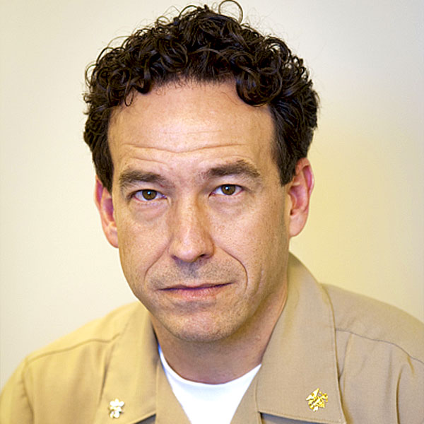 Stephen M. Hewitt合作论文数Laboratory of Pathology, Center for Cancer Research, National Cancer Institute, National Institutes of Health2
 Alastair Lamb合作论文数Medical Sciences Division, Nuffield Department of Surgical Sciences, University of Oxford2Lipkowitz Stanley合作论文数2
 Abhyudai Singh合作论文数Center for Applications of Mathematics in Medicine, University of Delaware/Center for Bioinformatics and Computational Biology, University of Delaware2
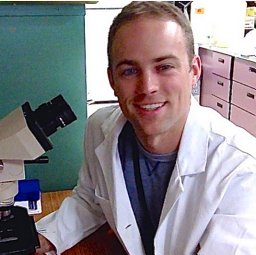 Edmondson Elijah F合作论文数Environmental and Radiological Health Sciences Department, Colorado State University2
 Ian G. Mills合作论文数Nuffield Department of Surgical Sciences, University of Oxford2Siddam Archana D合作论文数2Roop Victoria H合作论文数2
 Yiannis Philippou合作论文数2
 Ruth Muschel合作论文数Department of Oncology, Medical Sciences Division, University of Oxford;Oxford Institute for Radiation Oncology, University of Oxford;St Hilda's College, University of Oxford2Sherimay D. Ablan合作论文数2
 Freddie Hamdy合作论文数Nuffield Department of Surgical Sciences, University of Oxford2David C. Beebe合作论文数2




