The application of cryo-correlative light and cryo-electron microscopy (cryo-CLEM) gives us a way to locate structures of interest in the electron microscope. In brief, the structures of interest are fluorescently tagged, and images from the cryo-fluorescent microscope (cryo-FM) maps are superimposed on those from the cryo-electron microscope (cryo-EM). By enhancing cryo-FM to include single-molecule localization microscopy (SMLM), we can achieve much better localization. The introduction of cryo-SMLM increased the yield of photons from fluorophores, which can benefit localization efforts. Dahlberg and Moerner (2021, Annual Review of Physical Chemistry, 72, 253-278) have a recent broad and elegant review of super-resolution cryo-CLEM. This paper focuses on cryo(F)PALM/STORM for the cryo-electron tomography community. I explore the current challenges to increase the accuracy of localization by SMLM and the mapping of those positions onto cryo-EM images and maps. There is much to consider: we need to know if the excitation of fluorophores damages the structures we seek to visualize. We need to determine if higher numerical aperture (NA) objectives, which add complexity to image analysis but increase resolution and the efficiency of photon collection, are better than lower NA objectives, which pose fewer problems. We need to figure out the best way to determine the axial position of fluorophores. We need to have better ways of aligning maps determined by FM with those determined by EM. We need to improve the instrumentation to be easier to use, more accurate, and ice-contamination free. The bottom line is that we have more work to do.
Superresolution microscopy has fundamentally altered our ability to resolve subcellular proteins, but improving on these techniques to study dense structures composed of single-molecule-sized elements has been a challenge. One possible approach to enhance superresolution precision is to use cryogenic fluorescent imaging, reported to reduce fluorescent protein bleaching rates, thereby increasing the precision of superresolution imaging. Here, we describe an approach to cryogenic photoactivated localization microscopy (cPALM) that permits the use of a room-temperature high-numerical-aperture objective lens to image frozen samples in their native state. We find that cPALM increases photon yields and show that this approach can be used to enhance the effective resolution of two photoactivatable/switchable fluorophore-labeled structures in the same frozen sample. This higher resolution, two-color extension of the cPALM technique will expand the accessibility of this approach to a range of laboratories interested in more precise reconstructions of complex subcellular targets.
Prior to the development of 3D reconstruction, images were interpreted in terms of models made from simple units like ping-pong balls. Generally, people eye-balled the images and with other knowledge about its structure, such as the number of subunits, proposed models to account for the images. How was one to know if the models were correct and to what degree they faithfully represented the true structure? The analysis of electron micrographs of negatively stained viral structures led to the answers and 3D reconstruction.
Thermodynamic Molecular Switch in the Hydrophobic Interaction of 35 Dipeptide Pairs Paul W. Chun University of Florida, PO Box 100245, Gainesville, Florida 32610-0245 Applying the Planck-Benzinger methodology, the sequencespecific hydrophobic interactions of 35 dipeptide pairs were examined over a temperature range of 273-333 K. The results imply that the negative Gibbs free energy minimum at a well-defined stable temperature, , has its origin in the sequence-specific hydrophobic interactions, which are highly dependent on details of molecular structure. Each case confirms the existence of a thermodynamic molecular switch wherein a change of sign in DCpo(T) leads to true negative minimum in the Gibbs free energy change of reaction and a maximum in the related equilibrium constant. All interacting biological systems examined using the PlanckBenzinger methodology have shown such a thermodynamic switch at the molecular level, suggesting its existence may be universal.
Villin is an F-actin nucleating, crosslinking, severing, and capping protein within the gelsolin superfamily. We have used electron tomography of 2D arrays of villin-crosslinked F-actin to generate 3D images revealing villin's crosslinking structure. In these polar arrays, neighboring filaments are spaced 125.9 +/- 7.1 angstrom apart, offset axially by 17 angstrom, with one villin crosslink per actin crossover. More than 6500 subvolumes containing a single villin crosslink and the neighboring actin filaments were aligned and classified to produce 3D subvolume averages. Placement of a complete villin homology model into the average density reveals that full-length villin binds to different sites on F-actin from those used by other actin-binding proteins and villin's close homolog gelsolin.
If Fexp(ialpha) are the set of structure factors for a structure f, the amplitudes can be converted to those of an uncorrelated structure g (amplitude swapping) by multiplying each F by the positive number G/F. Correspondingly, the image f is convoluted with k, the Fourier transform of G/F; k has a large peak at the origin, so that f * k approximately f. For swapped phases, the image f is convoluted with l, the Fourier transform of exp(iDeltaalpha), where Deltaalpha, the phase difference between F and G, is a random variable; l does not have a large peak at the origin, so that f * l does not resemble f. The paper provides quantitative descriptions of these arguments.
Objectives. To develop a computerized algorithm to quantify fetal heart rate (FHR) variability and compare it to perinatologists' interpretation of FHR variability.Methods. FHR variability was calculated using data from 30 women who had a fetal scalp electrode placed for a clinical indication, and compared to the assessment of FHR variability from four perinatologists who interpreted paper tracings of the same data. Inter-rater reliability was calculated and receiver-operator curve analysis was done.Results. Correlation between the computer algorithm's assessment of variability and the perinatologists' assessment (0.27-0.68) was similar to the inter-rater reliability between perinatologists (0.33-0.72).Conclusions. A computer-based algorithm can assess FHR variability as well as expert clinicians.
Abstract The single-particle approach to solving macromolecular structure (see Frank, 1996, 2006) is based on the premise that the specimen is in the form of many isolated particles that are randomly oriented, collectively presenting a large range of views. It is further assumed, at least initially, that through application of biochemical methods of isolation and purfication, the specimen is homogeneous; that is, only macromolecules of one kind are present. Finally, if the specimen is prepared in such a way that anisotropic compression is avoided, then all the molecules on the electron microscopic grid can be assumed to have identical structure, and hence the geometrical relationships among the molecules in three-dimensional (3-D) space can be modeled as a set of rigid body movements. However, even if this assumption of structural identity is violated, due to some native structural variability, we can still use the single-particle approach for solving the structure at low resolution, and treat the structural variability as a high-resolution perturbation, a topic to be discussed later on.
Extended abstract of a paper presented at Microscopy and Microanalysis 2007 in Ft. Lauderdale, Florida, USA, August 5 – August 9, 2007
Abstract This chapter gives an overview of how x-ray crystallographyis used to determine the structure of proteins and other biological macromolecules at high resolution. Our brief review of x-ray crystallography is designed to identify the most important mathematical concepts that electron crystallography has in common with x-ray crystallography. Thus, this chapter is meant to be a summary of concepts that the reader should be familiar with in order to work independently and proficiently in the field of electron crystallography. Our review is not intended to teach these concepts in full detail; that would be the subject of an entire book of its own.
Localization of proteins in cells or complexes using electron microscopy has mainly relied upon the use of heavy metal clusters, which can be difficult to direct to sites of interest. For this reason, we would like to develop a clonable tag analogous to the clonable fluorescent tags common to light microscopy. Instead of fluorescing, such a tag would initiate formation of a heavy metal cluster. To test the feasibility of such a tag, we exploited the metal-binding protein, metallothionein (MT). We created a chimeric protein by fusing one or two copies of the MT gene to the gene for maltose binding protein. These chimeric proteins bound many gold atoms, with a conservative value of 16 gold atoms per copy of metallothionein. Visualization of gold-labeled fusion proteins by scanning electron microscopy required one copy of metallothionein while transmission electron microscopy required two copies. Images of frozen-hydrated samples of simple complexes made with anti-MBP antibodies hint at the usefulness of this method.
Abstract A number of idealized assumptions formed the basis of the majority of what was said about electron microscopy of biological macromolecules in the previous chapters. The most important of these assumptions were: (1) elastically scattered electrons undergo no more than one scattering event, (2) curvature of the Ewald sphere can be neglected, and (3) inelastic scattering can be ignored. While these represent satisfactory approximations for suitably thin specimens, each one becomes progressively less good as the specimen thickness increases.
Abstract Proteins can self-assemble into highly symmetrical particles that are biologically active and functional. Many viral proteins assemble into icosahedral shells containing a viral genome to become an infectious particle (Casjens, 1997). The icosahedral shell serves as a protective layer for the viral genome and is also involved in various biological processes during the viral infection and replication cycle. The outer shell can interact with immunoresponsive molecules, cellular receptors, and cytoskeletal proteins. In addition, some large enzyme complexes exist as icosahedral particles (Ladenstein et al., 1988; Zhou et al., 2001b; Milne et al., 2002). Structural studies of icosahe-dral particles thus are highly relevant to understanding their assembly principles and functional mechanisms.
Extended abstract of a paper presented at Microscopy and Microanalysis 2007 in Ft. Lauderdale, Florida, USA, August 5 – August 9, 2007
Abstract Helical structures are essential for life. They connect and integrate function between distantly separated structures within a cell. Actin and microtubules are examples of helical structures that are part of the cell’s cytoskeleton and that define cell shape. They also act as the railroad tracks along which molecular motors tow subcellular material between locations within the cell. Helical structures also occur in pathological states such as the paired helical filaments associated with Alzheimer’s disease or fibers of hemoglobin in sickle cell anemia. Helical structures can serve mechanical roles; for example, the bacterial flagellar filament converts torque into thrust to propel the bacterium. In addition to naturally occurring helical structures, some molecules can be induced to form helical structures after biochemical isolation, as in the case of the nicotinic acetylcholine receptor.
Abstract A substantial amount of image processing is required to extract the information contained in high-resolution electron micrographs of biological macromolecules. Descriptions of commonly used software packages for processing electron microscope images have been published in the Journal of Structural Biology, volume 116, issue 1 (1996). In this chapter we focus on the methodology for extracting structural information from images of thin, two-dimensional crystals.
Abstract This chapter covers many of the practical details that are associated with digitizing electron diffraction patterns and processing the data. The subsequent use of the diffraction intensities to compute density maps is covered at length in chapter 11. Before getting started with the practical aspects of data processing, however, section 9.2 gives a general overview of how diffraction data are used in the context of electron crystallography.
Abstract The repetition of a particular structure, or unit cell, in a pattern generated by some form of translational symmetry, is a unique, defining characteristic of crystals. While crystals may also have additional types of symmetry, such as that corresponding to rotation about a certain axis, the periodic repetition of a given structural motif over an extended region of space is something that uniquely defines what is meant by a crystal.
 Lewis G. Tilney合作论文数University of Pennsylvania25
 David Gene Morgan合作论文数14
 Kenneth H. Downing合作论文数Molecular Biophysics and Integrated Bioimaging Division, Lawrence Berkeley National Laboratory13
 Tanvir R. Shaikh合作论文数Wadsworth Center, New York State Department of Health13Nr Francis合作论文数12Katsumi Imada合作论文数12
 Edward H. Egelman合作论文数School of Medicine, University of Virginia11
 Joachim Frank (阿希姆·弗兰克)合作论文数Howard Hughes Medical Institute, Department of Biochemistry and Molecular Biophysics, College of Physicians and Surgeons, Columbia University;Department of Biological Sciences, Columbia University11
 Wah Chiu合作论文数Departments of Bioengineering, School of Engineering, Stanford University;Department of Microbiology and Immunology, School of Medicine, Stanford University;SLAC National Accelerator Laboratory11
 Keiichi Namba合作论文数11Shlomo Trachtenberg合作论文数10
 Dorit Hanein合作论文数Department of Chemistry & Biochemistry, University of California, Santa Barbara10
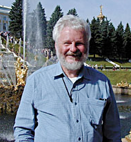 Robert M. Macnab合作论文数Department of Biophysics and Biochemistry, Yale University9H. Matsunami合作论文数8N Francis合作论文数6D Thomas合作论文数6
 Samatey Fadel A合作论文数JST, ICORP6
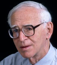 Aaron Klug合作论文数Laboratory of Molecular Biology, Medical Research Council6Noreen R. Francis合作论文数6M. S. Tilney合作论文数6Dennis R. Thomas合作论文数5Ge Sosinsky合作论文数5Ouyang G合作论文数5Terence Wagenknecht合作论文数5
 Paul Thomas Matsudaira合作论文数Department of Biological Sciences, National University of Singapore5
 Lucy Shapiro合作论文数Department of Developmental Biology, School of Medicine, Stanford University5James Z. Chen合作论文数4S Nagashima合作论文数4T. Ruiz合作论文数4Fa Samatey合作论文数3
 David L. Stokes合作论文数Laboratory of David Stokes, School of Medicine, New York University;Skirball Institute for Biomolecular Medicine, Department of Cell Biology, Grossman School of Medicine, New York University3
 Kenneth A. Taylor合作论文数Department of Biological Science, Biological Science Faculty, Florida State University3Kathleen M. Trybus合作论文数3
 Robert M. Glaeser合作论文数University of California, Berkeley;Lawrence Berkeley National Laboratory3Susan Lowey合作论文数3
 Koji Yonekura合作论文数Protonic NanoMachine Project;Advanced Technology Research Laboratories;Matsushita Electric Industrial Co . , Ltd3
 Akio Kitao合作论文数Department of Chemistry Faculty of Science, Kyoto University3
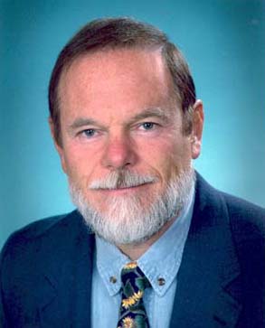 James F. Hainfeld合作论文数Nanoprobes, Inc./Department of Biomedical Engineering, State University of New York at Stony Brook3
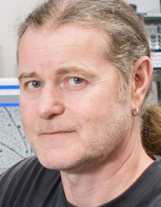 Niels Volkmann合作论文数Department of Electrical and Computer Engineering, College of Engineering, UC Santa Barbara;Institut Pasteur3Adré J. Du Plessis合作论文数3
 Donald L. D. Caspar (Donald Caspar)合作论文数Institute of Molecular Biophysics, Department of Biological Science, Florida State University3Adam Driks合作论文数3Adam Wolfberg合作论文数Tufts Medical Center, Boston, MA3S. Maki合作论文数3
 Zeeshan Syed合作论文数3David Acker合作论文数2Christopher P. Mercogliano合作论文数2Js Wall合作论文数2Le Rost合作论文数2




