BACKGROUND:Skin quality is an aggregate term encompassing a broad range of skin attributes that convey its overall look, feel, and health. OBJECTIVE:To assess consistency in definitions and treatment outcomes for various skin quality attributes. MATERIALS AND METHODS:We searched PubMed, MEDLINE, and Embase to perform 2 systematic reviews. The initial search identified published articles (January 1, 2000-October 13, 2021) and abstracts (January 1, 2019-October 13, 2021) that used an objective or subjective instrument to assess skin quality at ≥2 timepoints. A supplemental search of meta-analyses published from 2010 to May 2024 was conducted to evaluate treatment outcomes for each skin quality attribute. RESULTS:The initial search included 903 studies comprising 4,668 individual observations. Subjective end points comprised 87% of observations. Substantial inconsistencies were identified in the definition of individual skin quality attributes across studies, with multiple definitions in use in the literature for each attribute (range, 7-17) and no clear consensus. CONCLUSION:This extensive review of the current state of the aesthetic medicine literature revealed an absence of consensus definitions for skin quality attributes, which poses a barrier to comparative evaluation of treatment options and could affect physician recommendations, potentially with downstream effects on patient outcomes.
BACKGROUNDEnergy-based interventions improve signs of skin aging by inducing controlled thermal injury and activating regenerative wound healing pathways.OBJECTIVEThis study investigates the underlying mechanism of acute and long-term extracellular matrix (ECM) remodeling induced by Synchronous Parallel Ultrasound technology leading to skin regeneration.METHODS/MATERIALSAn in vivo porcine model was used to investigate the acute thermal injury. In vivo human skin biopsies were obtained from subjects at baseline and at 1, 4, 7, and 10 months post-treatment. Histological evaluation was performed using Hematoxylin and Eosin, Masson's Trichome, Verhoeff-Van Gieson, Unna Taenzer, and Alcian Blue stains to assess changes in ECM protein expression.RESULTSThermal injury was restricted to the dermal layer. Histological analysis revealed a gradual increase in connective tissue collagen and elastin with unique reorganization and parallel realignment of the dermal fibers. Increased expression of glycosaminoglycans (GAGs) was also observed, indicating sustained fibroblast activity and matrix regeneration across all time points.CONCLUSIONThese findings provide histological evidence of continuing and long-lasting dermal remodeling after Synchronous Parallel Ultrasound treatment, characterized by neocollagenesis, neoelastogenesis, and GAGs production. The study supports the efficacy of Synchronous Parallel Ultrasound in the long-term induction of skin regeneration.
Objectives:Evaluate the effects of a new antioxidant containing topical allyl pyrroloquinoline quinone (TAP) on expression of key markers and assess the efficacy and tolerability in subjects with photodamaged skin.Methods:Donor skin tissue was irradiated prior to and following application of study products (TAP; a leading antioxidant cream [L-VC]). Expression of markers related to epidermal homeostasis and oxidative stress were assessed at 48 hours and compared to untreated, irradiated control (n=3 each). Evaluation of lines/wrinkles, skin texture, skin tone, dullness, and erythema from baseline occurred over 12 weeks in subjects with mild-to-moderate photodamaged skin. Histological evaluation occurred at Weeks 6 and 12 (n=4).Results:Following application of TAP, significant expression of markers related to epidermal homeostasis and repair, recycling and removal, and oxidative stress were demonstrated, compared to control (p<0.05). Reduced expression of collagen degrading enzymes, compared to control, were observed (p<0.05). Application of L-VC demonstrated nonsignificant expression of markers versus control. In 40 subjects evaluated over 12 weeks, significant mean improvements from baseline were observed at Week 4 in skin texture and dullness (both p<0.0001) and skin tone and lines/wrinkles (both p=0.01). The study product was highly tolerable. Histologic evaluation demonstrated reductions in solar elastosis from baseline at Weeks 6 (33%, p=0.01) and 12 (60%, p=0.002).Conclusion:An antioxidant containing TAP addresses internal and external manifestations of photoaging. TAP demonstrated significant expression of key markers associated with epidermal homeostasis and counteracting oxidative stress. Significant, early improvements in the appearance of photodamaged skin and histological improvements in solar elastosis were observed.
The growing interest in improving the quality of body (as distinct from facial) skin may be in part attributable to the expanding use of noninvasive body contouring procedures. In this review, we describe a new framework characterizing the factors that define skin quality (including visual, textural, and biomechanical attributes) that provides a foundation for improved assessment of skin quality and its response to treatment. We then highlight critical biological pathways responsible for body skin restoration and maintenance that have been identified during the development of restorative topical products. Each of these pathways, including extracellular matrix support, suppression of lipogenesis, and enhancement of cellular/macromolecular recycling and clearance, lymphatic drainage, and lipolysis, is a potential target of 1 or more bioactive substances. A survey of available topical products marketed for skin quality improvement suggests that none target more than 2 of these pathways (including extracellular matrix support, lipolysis, and autophagy, a component of cellular recycling), leaving abundant opportunity for development of new topical formulations that target all or most of the critical pathways. Such formulations may provide improved outcomes when used as standalone products for general skin quality improvement and rejuvenation, in addition to their potential for post-procedure use, and also for pre-procedure skin conditioning.
Living better longer? Optimizing healthspan? Enhancing natural beauty? These are worthy goals and things which we all might aspire to both as physicians and scientists but also for all peoples and cultures. This special supplement highlights the 2020 Science of Aging Symposium, which was held virtually due to the COVID-19 pandemic. The pandemic has highlighted and emphasized the importance of both the expanding our understanding of the science of aging to preservation of aging concepts Although these goals are aspirational, they are becoming reality. The Science of Aging Symposium exists to bring together world leading experts in various facets of aging research who might not ordinarily meet and exchange their ideas. The symposium provides a forum for rapid dissemination and sharing of emerging new scientific discoveries and a platform to teach and advance and accelerate research into the science of aging. It also helps to facilitate translating these aspirational goals into reality and to transform healthcare for our patients in our lifetimes. As physicians, we are called to serve our patients as healers, consolers, and teachers. What better way than to have a forum to rapidly exchange ideas and how to implement new scientific discoveries into our patient’s care. We know that lifestyle choices and environmental conditions impact health span and lifespan more than genetics. This presents a special opportunity and also highlights the importance for physicians and healthcare providers to stay informed and educated about newly emerging discoveries. This supplement chronicles the 2020 Science of Aging Symposium, which included topics such as the fundamentals of living longer better and how to extend health span through innovative gene therapies. New discoveries regarding skin pigmentation dyschromia were presented and some remarkable 3D visual presentations were seen as we learned about new artificial intelligence and machine learning advances in visualizing, measuring, and understanding human facial and body skin as well as how computer vision-based systems can affect patient self-perception. On the therapeutic side of aesthetic medicine, new options for skin care using serotherapeutic agents were reviewed as well as how regenerative medicine can be used for applications such as hair restoration and some striking research advances were presented regarding producing plant-derived recombinant human collagen from tobacco plants for aesthetic medicine applications. The presenting scientists generously shared their time and their research during the symposium and their work is represented in this supplement. We thank them for their time and also encourage them as they continue their exciting research. The overriding goal and long-term benefits of this tremendous global multidisciplinary effort is to spark new ideas and innovations to bring this “science of aging” into the clinical realm and reality of everyone on the earth. They also join a growing community of leading scientists from around the globe who are part of the Science of Aging faculty who have dedicated their careers to aging related research. Past, present, and future faculty communicate, collaborate, and educate both each other and the research and medical communities in the world at large to help elevate and accelerate the science of aging. We thank all of our speakers past and present who continue to be a part of our growing group as we reach across diverse specialties to share knowledge. We know that you will enjoy reading this special supplement (and viewing the remarkable videos) and that each of you will learn something new which impacts your research, your patients and your own lives as we each strive to “live better longer”! We would like to thank all the authors who took time out of their busy careers to prepare these outstanding papers. Special thanks to Dr. Stephanie Manson Brown and Dr. Darin Messina who co-founded the Science of Aging Symposium and to the other core symposium team members Chinmay Kanuga and Dr. Artem Kutikov. The staff at Plastic and Reconstructive Surgery were tireless in their efforts and provided superb support at all levels. Thank you to all who worked behind the scenes as well and we appreciate everyone’s commitment to this special supplement.
Antioxidants (AOX) are free radical scavengers both occurring naturally in the skin and supplemented topically or orally to mitigate potentially damaging effects of oxidative stress on keratinocytes and dermal collagen. Used frequently in alternative medicine, AOX have been incorporated into cosmeceuticals in more recent decades as a way to protect and defend the epidermis and dermis against oxidative damage by ultraviolet radiation, and more recently as a possible defense against visible light, high-energy visible light, infrared radiation, and air pollution. Common topical AOX include vitamin C, vitamin E, polyphenols, and flavonoids. Most are harvested and extracted from botanicals (e.g., green tea, red grapes, dark red berries, milk thistle, etc.) for use in both oral and topical preparations. Combinations of multiple AOX are most common in cosmeceuticals, as several AOX have synergistic effects. For example, vitamin C naturally recycles vitamin E to its active form, allowing for more rapid neutralization of both oxygen-based and peroxyl radicals. Topical AOX augment sunscreens, repair damage from oxidative stress, and have become an integral part of the cosmeceutical armamentarium.
1Department of Dermatology and Skin Science, University of British Columbia, Vancouver, British Columbia, Canada 2Humphrey Cosmetic Dermatology, Vancouver, British Columbia, Canada 3Institute of Aging Research, Virginia Beach, Virginia, USA 4Department of Biological Sciences, Old Dominion University, Hampton, Virginia, USA 5Skin of Color Research Institute, Hampton University, Hampton, Virginia, USA 6School of Science, Hampton University, Hampton, Virginia, USA
Tropospheric ozone (O3) is a source of oxidative stress. This study examined the ability of a topical antioxidant (WEL-DS) to inhibit O3-mediated damage in a human epidermal skin model. Four groups of tissues (N = 24) were compared: Group 1 (control) were untreated and unexposed; Group 2 were untreated and exposed to O3 (0.4 ppm, 4 h); Group 3 were pretreated with WEL-DS and unexposed; Group 4 were pretreated with WEL-DS and exposed to O3 (0.4 ppm, 4 h). Pretreated tissues were topically treated with 20 uL of WEL-DS and incubated for up to 20 h at 37 °C [humidified, 5% carbon dioxide (CO2)]. After 24 h, tissues were re-treated with WEL-DS and exposed to O3. Tissues were evaluated for Reactive Oxygen Species (ROS), hydrogen peroxide (H2O2), 4-hydroxynonenal (4-HNE) protein adducts, NF-κB p65 response and histology. In O3-exposed groups, WEL-DS significantly inhibited ROS formation vs. untreated tissues (p < 0.05). Pretreatment with WEL-DS inhibited H2O2 production vs. untreated tissues (p < 0.05), and decreased NF-κB p65 transcription factor signal. Oxidative stress induction in O3-exposed tissues was confirmed by increased levels of 4-HNE protein adducts (marker of lipid peroxidation); WEL-DS application reduced this effect. WEL-DS inhibited damage in tissues exposed to O3 with no significant changes in epidermal structure. A comprehensive topical antioxidant significantly diminished O3-induced oxidative damage in a human epidermal skin model.
Objective: This study evaluated the effects of twice-daily application of a new rebalancing moisture treatment (TRMT) cream that aims to optimize skin moisturization and enhance skin barrier by supporting skin’s natural lipid bilayer and enhancing NMFs and HA in subjects with moderate to severe photodamaged skin.
Objectives To evaluate skin barrier and hydration effects of a new rebalancing moisture treatment (TRMT) and to assess efficacy and tolerability in subjects with photodamaged skin. Methods In an epidermal skin model, tissues (n = 5/group) were topically treated with 25 mu L of TRMT, 25 mu L of a market-leading moisturizer (MLM), or untreated for 60 minutes. Hydration was measured at 0, 15, and 30 minutes. Tissues were harvested for gene expression analysis of markers associated with skin barrier and hydration: Claudin (CLD), Aquaporin (AQP), Hyaluronic Acid Syntheses (HAS), and Hyaluronidase (HYAL). A clinical study evaluated twice-daily application of TRMT, assessing changes in fine lines/wrinkles, brightness, texture, erythema, and tolerability from baseline through week 8. Hydration was measured using electrical impedance. Results TRMT and MLM demonstrated significant increases in hydration vs untreated tissue at each timepoint (P < .005), with greater hydration effects observed for TRMT vs MLM. TRMT-treated tissues demonstrated greater expression of CLD, AQP, and HA, and reduced expression of HYAL vs untreated and MLM-treated tissues. Twice-daily application of TRMT demonstrated significant improvements at 2 weeks in fine lines/wrinkles (P < .001), brightness (P < .0001), texture (P < .0004), and hydration (P < .004). At 8 weeks, statistically significant improvements were achieved in all categories. Conclusion In an epidermal skin model, TRMT demonstrated significant increases in hydration, greater hydration effects, and expression of key markers associated with skin barrier and hydration vs a MLM. Twice-daily application of TRMT was well tolerated and resulted in early, significant improvements in hydration and visible improvements in skin brightness, texture, fine lines/wrinkles, and erythema at 8 weeks.
Objectives: Investigators sought to evaluate the antioxidant capacity of a comprehensive topical antioxidant (WEL-DS), its ability to protect skin against the oxidizing effects of UVA/UVB radiation, and to assess the effectiveness and tolerability of WEL-DS for visible improvements in facial photodamage. Study Designs:In-vitro testing utilized a hydrogen peroxide assay to detect activity in human skin explants following application with WEL-DS, a leading antioxidant serum (L-AOX), and a saline control. Clinical studies included a minimal erythema dose (MED) trial in female subjects, aged 35 to 60 years. Skin was initially irradiated to determine each subject's MED. WEL-DS was applied for four days to one site on the lower back of subjects; the other site remained untreated. Both sites were irradiated with 1X, 2X and 3X each subject's MED, digital images were obtained, and punch biopsies were collected from the 3X MED irradiated areas for histological analysis. A second clinical study evaluated efficacy and tolerability of twice daily application of WEL-DS in female subjects, aged 25 to 65 years with mild-to-moderate photodamage. Changes in fine lines/ wrinkles, dyschromia, erythema, skin tone, pores, and tolerability were assessed at baseline and Weeks 4, 8, and 12. A subset of subjects were evaluated through Week 16. Results: Skin treated with WEL-DS neutralized up to 53 percent more oxidative stress relative to L-AOX. WEL-DS-treated skin demonstrated significantly less UV-induced erythema at 1X, 2X, and 3X MED and demonstrated cellular protective effects versus untreated irradiated skin (N=5). WEL-DS demonstrated average improvements from baseline of 37 percent, fine lines/ wrinkles; 17 percent, skin tone; 13 percent, dyschromia; 18 percent, erythema; and four percent, pores (N=21; Week 12). Continued improvements were demonstrated in all parameters in an extension study (n=14; week 16). WEL-DS was well-tolerated. Conclusion: These studies demonstrate WEL-DS's innate ability to quench free radicals, protect skin from the oxidizing effects of UV radiation, and reduce the visible effects of facial photodamage.
wide variety of hypopigmented scars, and striae alba had mean final pigment correction rates of approximately 100% by colorimetric analysis after 9 treatments of 308-nm excimer laser biweekly. However, maintenance treatment every 1 to 4 months is suggested because the pigment gradually declined toward baseline values during a 6month follow-up. Although the authors performed 308-nm excimer light only once weekly for 6 times in our patient, she had a good response and rapid repigmentation within 3 months, and no recurrence of hypopigmentation was found at a 10-month follow-up. A possible hypothesis is that she had no genetic predisposition for vitiligo, and she had a darker Fitzpatrick skin type. Patients with Fitzpatrick skin Type III and above generally respond better to an excimer laser than those with lower skin types. To the best of our knowledge, this is the first case of corticosteroid-induced perilymphatic linear hypopigmentation treated with 308-nm excimer light.
Background: Anecdotal reports indicate the use of microfocused ultrasound with visualization (MFU-V) improves facial redness.Objective: The purpose of this pilot study was to assess the safety and effectiveness of MFU-V for improving the signs and symptomsof erythematotelangiectatic rosacea.Methods & Materials: Healthy adults with a clinical diagnosis of erythematotelangiectatic rosacea were enrolled (N=91). Eligiblesubjects had baseline Clinician Erythema Assessment (CEA) scores ≥3 and Patient Self-Assessment (PSA) of erythema scores ≥2.Subjects were randomized to receive one or two low-density MFU-V treatments or one or two high-density MFU-V treatments. Subjectswere evaluated at 90, 180, and 365 days after treatment. The primary effectiveness endpoint was treatment success, defined asa 1-point change in CEA scores at 90 days post-treatment.Results: Across groups, 75 to 91.3% of subjects achieved treatment success at 90 days post-treatment. Notable adverse events includebruising (44%), tenderness/soreness (43%), and redness (35%). Treatment results were sustained, lasting up to 1 year. Subjectsatisfaction was high based on self-assessment questionnaires.Conclusion: The results of this study demonstrated that a single, high-density MFU-V treatment may be effective for treating erythematotelangiectaticrosacea. Based on these results, a large, randomized controlled study of single, high-density MFU-V treatment forerythematotelangiectatic rosacea is warranted.J Drugs Dermatol. 2019;18(6):522-531.
Background: Pigmentation disorders are therapeutically challenging to treat, requiring complicated regimens. Objectives: Alternatives to hydroquinone (HQ) are desired. We evaluated the efficacy and tolerability of a non-HQ multi-action skin tone corrector (ETCS) developed to inhibit melanin production and improve skin quality. Design and Methods: Twice-daily use of ETCS and ETCS + AHA-Ret, a retinoid-based alpha hydroxy acid cream, was evaluated in subjects with mild to severe dyschromia. Digital images were obtained at baseline, 4, 8, and 12 weeks and included assessment of dyschromia, erythema, fine lines/wrinkles, pores, texture, and global improvement. Melanin Index (MI) measurements were obtained at baseline, 4, 8, and 12 weeks. Subject self-assessments were obtained over the course of the study. Adverse Events (AEs) were collected throughout the study. An extension study evaluated use over 16-weeks. Results: Significant mean reductions from baseline occurred in dyschromia for ETCS (n=42) and ETCS + AHA-Ret (n=10) over 12 weeks (P<0.0001, each). Significant mean reductions from baseline in MI were achieved in both groups at every timepoint (ETCS: P<0.0001; ETCS + AHA-Ret: P<0.02, 4 weeks; P<0.0001, 8 and 12 weeks). Substantial improvements were demonstrated in global improvement, fine lines/wrinkles, erythema, pores, and texture at 12 weeks. Reductions from baseline occurred in dyschromia and MI (P<0.0001, each) at 16 weeks. High levels of subject satisfaction were reported with nearly all subjects reporting reduced appearance of uneven skin tone/discoloration and lightened darker patches, and improvement in overall skin tone. Mild, transient AEs were reported with no discontinuations due to an AE. Conclusions: Twice daily use of ETCS led to early, significant reductions in dyschromia and melanin index. Combination use with a retinoid-based, AHA cream in the evening demonstrated enhanced reductions. ETCS effectively reduced hyperpigmentation, improved overall skin appearance, and was highly tolerable. J Drugs Dermatol. 2019;18(7):642-648.
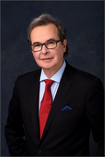 Robert Arnold Weiss合作论文数Maryland Laser, Skin & Vein Institute;School of Medicine, University of Maryland17
 Roy G. Geronemus合作论文数Ronald O. Perelman Department of Dermatology, Grossman School of Medicine, New York University;Langone Hospitals, New York University12
 Diane Nelson合作论文数10Mitchell S. Wortzman合作论文数10Chris Mazur合作论文数8
 Margaret A Weiss合作论文数7
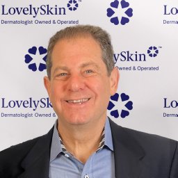 Joel Schlessinger合作论文数Advanced Skin Research Center5
 Paul Graham合作论文数Edinburgh Parallel Computing Centre, The University of Edinburgh, James Clerk Maxwell Building, Mayfield Road, Edinburgh EH9 3JZ, U.K.
5Christopher Mazur合作论文数4
Paul Graham合作论文数Edinburgh Parallel Computing Centre, The University of Edinburgh, James Clerk Maxwell Building, Mayfield Road, Edinburgh EH9 3JZ, U.K.
5Christopher Mazur合作论文数4 Jeffrey Dover合作论文数4Mark Lupin合作论文数3
 Kayi Monica Li合作论文数3Rosalyn George合作论文数3Anne Mahoney合作论文数3Christian Halvorson合作论文数3
 Sabrina G Fabi合作论文数3
 Derek Jones合作论文数Brain Research Imaging Centre, School of Psychology, Cardiff University3Deland Maitland M合作论文数3Dennis DaSilva合作论文数2
 Lisa Dispensa合作论文数2Jay E Birnbaum合作论文数2Doug Canfield合作论文数2Sarah Hsu合作论文数2
 Rahul C Mehta合作论文数2Kerry Shaughnessy合作论文数2Abigail Waldman合作论文数2Jeremy A. Brauer合作论文数2Karen Beasley合作论文数2Sean Doherty合作论文数2Laura Harrington合作论文数2Tsing Cheng合作论文数Allergan Aesthet2
 Mitchel P. Goldman合作论文数Cosmetic Laser Dermatology2
 Shannon Humphrey合作论文数2
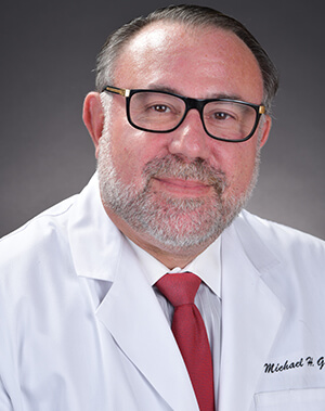 Michael H. Gold合作论文数School of Nursing, Vanderbilt University ;Gold Skin Care Center;Advanced Aesthetics Medical Spa;The Laser & Rejuvenation Center;Tennessee Clinical Research Center2Alam Murad合作论文数2Le Moigne Anne合作论文数2Amato David合作论文数2Jiang Lily I合作论文数2Heinz Moritz合作论文数2Schlöbe Andrea合作论文数2Ogilvie Patricia合作论文数2
 Emily Poon合作论文数2Patricia K. Farris合作论文数2Thomas J. Stephens合作论文数2Suzanne L. Kilmer合作论文数2
 David J. Goldberg合作论文数Skin Laser & Surgery Specialists of New York|Mount Sinai School of Medicine2Lisa Donofrio合作论文数2
 Alessandra Pecorelli合作论文数Plants for Human Health Institute, N.C. State University2Thomas E. Rohrer合作论文数2




