Background/Objectives: Internal tremor (IT) is often reported by patients with post-acute sequelae of SARS-CoV-2, also known as Long COVID, as a distressing and disabling symptom. Similarly, physicians are typically perplexed by the nature and etiology of IT and find it extremely challenging to manage. Methods: We describe a patient with Long COVID who experienced IT as part of post-COVID postural orthostatic tachycardia syndrome (POTS) and small fiber neuropathy (SFN) and review the limited literature available on this topic. Results: Our patient’s IT improved significantly after intravenous saline infusions, but there was no effect on IT with oral hydration, increased oral sodium chloride intake, neuropathic pain medications, muscle relaxants, or medications used for the treatment of POTS. Conclusions: Based on this case, our clinical experience, and the limited literature available to date, we believe IT is a manifestation of POTS and SFN, which may be driven by hypovolemia, cerebral hypoperfusion, sympathetic overactivity, neuropathic pain, and mast cell hyperactivation. Subjective description, objective findings, and diagnostic and therapeutic considerations in patients with IT and Long COVID are discussed.
The goal of this review is to guide the physician in ordering laboratory tests for a patient with a neuropathic disorder after the patient has been placed in one of the eleven neuropathic patterns (See this issue: Barohn et al.Pattern Recognition to Neuropathy and Neuronopathy, Pages 4-27, 2023).Once the patient has been placed into one of these eleven patterns, the physician is well on their way to a correct diagnosis for the neuropathic disorder that will ultimately lead to a targeted approach, including treatment.The neuropathic pattern will determine what laboratory tests should be ordered to confirm the diagnosis.
Background: Quantitative sensory testing (QST) is a subjective but reliable and quantifiable method to detect patient thresholds to different sensory stimuli. QST is used to measure small- and large-fiber nerve function and can be used in conjunction with other diagnostic modalities in the evaluation of peripheral neuropathy (PN). The utility of QST to distinguish among different types of PN, however, has not been explored. The objective of the study was to evaluate if different patterns of QST abnormalities could distinguish between PN types. Methods: This single-center retrospective cohort study evaluated the frequency of QST abnormalities to vibratory, cold and heat detection thresholds in a large population of PN cases evaluated at the University of Texas Southwestern Medical Center peripheral neuropathy clinic between 1995-2000. PN was categorized by etiology. Results: A total of 559 QST studies were performed in this study. The average age of patients (n=557) was 60 years with a male-to-female ratio of 1:1. The most common diagnosis was cryptogenic sensory polyneuropathy (CSPN, n=294), followed by Charcot–Marie–Tooth disease (n=84)). Meta-regression of vibration and cold indicate that the expected proportion of abnormal responses is less for the vibration test (p = 0.0002), relative to the cold test. However, no differences were observed between diagnoses. Conclusions: Though abnormal QST thresholds were seen in most patients with PN, patterns of QST abnormalities do not distinguish between different types of PN. The routine clinical utility of QST is likely limited.
Background: A database which documents the diagnosis of neuromuscular patients is useful for determining the types of patients referred to academic centers and for identifying participants for clinical trials and other studies. The ICD-9 or ICD-10 numeric systems are insufficiently detailed for this purpose. Objective: To develop a database for neuromuscular diagnoses Methods: We developed a detailed diagnostic coding system for neuromuscular diseases called DEMAND: Database Evaluation for Muscle and Nerve Diseases that has been adopted by neuromuscular clinics at University of Texas Health Science Center San Antonio (UTHSCSA), Ohio State University (OSU), University of Kansas Medical Center (KUMC), and University of Texas Southwestern (UTSW). At the initial visit, patients are assigned a diagnostic code which can be revised later if appropriate. Fields include patient’s name, date of birth, and diagnostic code. The neuromuscular database consisted of 457 codes. Each code has a prefix (MUS or PNS) followed by a three-digit number. Depending on whether muscle or nerve is primarily involved, there are eight broad groups: motor neuron disease (MUS codes 100-139); neuromuscular junction disorders (MUS 200-217); acquired and hereditary myopathies (MUS 300-600s); acquired and hereditary polyneuropathies (PNS 100-400); mononeuropathies (PNS 500s); plexopathies (PNS 600s); radiculopathies (PNS 700s); and mononeuritis multiplex (PNS 800s). Results: During a period of 10 years, 17,163 of patients were entered (1,752 at UTHSCSA, 1,840 at OSU, 3,699 at KUMC, 9,872 at UTSW). The number of patients in several broad categories are: 3,080 motor neuron disease; 1,575 neuromuscular junction disease; 1,851 muscular dystrophies; 633 inflammatory myopathies; 1,090 hereditary neuropathies; 1,001 immune-mediated polyneuropathies; 620 metabolic/toxic polyneuropathies; 535 mononeuropathies; 296 plexopathies; and 769 radiculopathies. Conclusion: A detailed diagnostic neuromuscular database can be utilized at multiple academic centers. The database should be simple without too many fields to complete, to ensure compliance during busy clinic operations. This database has been very useful in identifying groups of patients for retrospective, observational studies and for prospective treatment studies including trials for Amyotrophic Lateral Sclerosis (ALS), Muscular Dystrophies (MD), Myasthenia Gravis (MG), and retrospective studies of Primary Lateral Sclerosis (PLS), chronic inflammatory demyelinating neuropathy (CIDP), etc.
BACKGROUND AND PURPOSE:Data on maintenance therapy with subcutaneous immunoglobulin (SCIg) in myasthenia gravis (MG) are limited. We report on transitioning acetylcholine receptor (AChR) antibody-positive (Ab+) MG patients on stable intravenous immunoglobulin (IVIg) regimens as part of routine clinical care to SCIg 1:1.2.METHODS:This multicenter North American open-label prospective investigator-initiated study had two components: the IVIg Stabilization Period (ISP) enrolling patients already on IVIg as part of routine clinical care (Weeks -10 to -1), followed by transition of stable MG subjects to SCIg in the Experimental Treatment Period (ETP; Weeks 0 to 12). We hypothesized that >65% of patients entering the ETP would have a stable Quantitative Myasthenia Gravis (QMG) score from Week 0 to Week 12. Secondary outcome measures included other efficacy measures, safety, tolerability, IgG levels, and treatment satisfaction.RESULTS:We recruited 23 patients in the ISP, and 22 entered the ETP. A total of 12 subjects (54.5%) were female, and 18 (81.8%) were White, with mean age 51.4 ± 17 years. We obtained Week 12 ETP QMG data on 19 of 22; one subject withdrew from ETP owing to clinical deterioration, and two subjects withdrew due to dislike of needles. On primary analysis, 19 of 22 participants (86.4%, 95% confidence interval = 0.72-1.00) were treatment successes using last observation carried forward (p = 0.018). Secondary efficacy measures supported MG stability. SCIg was safe and well tolerated, and IgG levels were stable. Treatment satisfaction was comparable between ISP and ETP.CONCLUSIONS:MG patients on IVIg as part of their routine clinical care remained stable on monthly IVIg dosage, and most maintained similar disease stability on SCIg.
Intravitreal anti-vascular endothelial growth factor (VEGF) injections are commonly used to treat eyes with macular edema secondary to hemiretinal vein occlusion (HRVO) or central retinal vein occlusion (CRVO). Information on whether differences exist in outcomes after anti-VEGF therapy can help guide treatment for each of the different disease types.To compare baseline characteristics, treatment burden, and outcomes of macular edema treatment in participants with HRVO with those of participants with CRVO.This post hoc outcome analysis from the Study of Comparative Treatments for Retinal Vein Occlusion 2 randomized clinical trial included 362 participants with macular edema caused by HRVO or CRVO treated at 66 US sites. Randomization began in September 2014, and the last month 24 follow-up visit occurred in February 2018. Data were analyzed from April 2020 to May 2021.Eyes were initially randomized to 6 monthly intravitreal injections of aflibercept or bevacizumab and were treated according to protocol between months 6 to 12 depending on 6-month outcome. After month 12, patients were treated per investigator discretion and observed through month 60.Mean visual acuity letter score (VALS).Of 362 included patients, 157 (43.4%) were female, and the mean (SD) age was 68.9 (12.0) years. Outcome data were analyzed up to month 24 owing to substantial missing data at later visits. A significantly greater proportion of participants with HRVO than those with CRVO were Black (37% vs 11%). Treatment rates between months 12 to 23 were 0.36 (95% CI, 0.32-0.40) injections per month for patients with CRVO and 0.28 (95% CI, 0.19-0.36) for patients with HRVO (P = .11). The mean VALS from months 1 to 24 of an HRVO study eye exceeded that of a CRVO study eye by 5.5 (95% CI, 1.5-9.5; P = .01), consistent with the magnitude of the VALS difference between eyes with CRVO and HRVO at baseline. Eyes with CRVO presented at baseline with more macular edema than eyes with HRVO (difference in central subfield thickness [CST], 86 μm; 95% CI, 48-124; P < .001), with no difference in CST between the groups throughout months 1 to 24.Black race was more prevalent among participants with HRVO than CRVO, and no differences were observed in the frequency of treatments for macular edema between eyes with CRVO and HRVO. Although eyes with CRVO presented with worse visual acuity and more macular edema on average than did eyes with HRVO, the magnitude of VALS improvement, central retinal thickness in response to anti-VEGF therapy, and treatment burden were similar between the groups.
Small fiber neuropathy has a broad array of presentations. Length-dependent symptoms and findings present little diagnostic difficulty, but non-length-dependent or multifocal symptoms can be challenging. Intraepidermal nerve fiber density (IENFD) testing in apparent fibromyalgia warrants further study, but skin biopsy testing of this patient population is reasonable. Avoidance of IENFD testing in situations where diagnosis of neuropathy is already clear or where neuropathy is not the cause of symptoms helps to prevent incorrect conclusions. Careful history and physical examination plus pretest probability are important factors to consider when assessing the results of an IENFD test report.
INTRODUCTION:Causes of small-fiber peripheral neuropathies (SFN) are often undefined. In this study we investigated associations of serum autoantibodies, immunoglobulin G (IgG) vs fibroblast growth factor receptor-3 (FGFR-3), and immunoglobulin M (IgM) vs trisulfated heparan disaccharide (TS-HDS) in cryptogenic SFN.METHODS:One hundred fifty-five patients with biopsy-proven SFN and no identified cause for their neuropathy were blindly tested for serum IgM vs TS-HDS and IgG vs FGFR-3.RESULTS:Forty-eight percent of SFN patients had serum antibodies, 37% with IgM vs TS-HDS and 15% with IgG vs FGFR-3. TS-HDS antibodies were more frequent in SFN patients than in controls (P = .0012). Both antibodies were more common in females, and with non-length-dependent nerve pathology. Nintey-two percent of patients with acute-onset SFN had serum IgM vs TS-HDS.DISCUSSION:Autoantibodies directed against TS-HDS and FGFR-3 suggest an immune disorder in otherwise idiopathic SFN. Serum IgM vs TS-HDS may be a marker for SFN with an acute onset.
Neurodegenerative diseases are estimated to afflict hundreds of thousands of Americans with vastly more worldwide. The etiologies of amyotrophic lateral sclerosis (ALS) and multiple sclerosis (MS) have yet to be established. Previous studies have suggested an association of these diseases with viruses, bacteria, and eukaryotic microbes, no new therapies have been forthcoming. High-throughput DNA sequencing has enabled the comprehensive analysis of microbial DNA profiles in diseased populations. To date, no amplicon-based next-generation DNA sequencing prokaryotic and eukaryotic community profiling studies have been completed for these diseases. Analysis of peripheral blood samples from control participants as well as ALS and MS participants was used to characterize the hematologic population of microbial DNA. Categorical and multivariate analysis with control for multiple comparisons and aged matched controls revealed differences in microbial DNA contribution in ALS patients compared to others. Notably, sequences that belonging to Ochrophyta were enriched in ALS patient samples. Mechanisms underlying this association, the role of microbial DNA sequences, and the development or progression of ALS may become a fertile subject of inquiry.
Objective: Objectives: To evaluate the efficacy, safety and tolerability of subcutaneous immunoglobulin (SCIg) in the treatment in myasthenia gravis (MG) patients who are on IVIg as part of routine clinicalcare. Background: Background: IVIg has been demonstrated to improve the Quantitative MG (QMG) score as compared to placebo and in another study to be efficacious in comparison to plasma exchange. In routine care, SCIg might be easier to administer than IVIg. Design/Methods: Methods: This multi-center North American open label prospective investigator-initiated study had 2 components: IVIg Screening Phase (ISP; Weeks −10 to −1) followed by Experimental Treatment Phase (ETP; Weeks 0 to 12). We hypothesized that more than 65% of the patients entering the ETP would have stable QMG score (primary outcome) at Week 12. We recruited 23 patients in the ISP and 22 entered the ETP. 12/22(54.5%) were females and 18(78%) were white, with mean age 51.4 years±17. We had complete ETP QMG data on 19/22; one subject withdrew from ISP owing to worsened condition and two subjects withdrew before Week 4 (needle dislike). The per protocol primary statistical analysis was conducted for n=22 subjects using a one-sided z-test of proportions at the 5% significance level. Sensitivity analyses were conducted using a cohort of n=22 subjects using ‘worst-case’ imputation scenario as well as post hoc analysis. Results: Findings: On primary analysis, 19/22 (86.4%; 95% CI: 0.72–1.00)were treatment “successes” (p=0.018). Sensitivity analysis using the ‘worst-case’ imputation resulted in 17/22 (77.3%;0.60–0.95) declared as treatment success (p=0.114). Post hoc analysis of the primary outcome confirmed treatment success in 17/20 (85%;0.69–1.00) (p=0.0304). There was no difference in the secondary outcome measures though MG composite was better at Week 12. SCIg was safe and well tolerated in this population. Conclusions: Conclusion: Most MG patients who were doing well on IVIg maintained disease stability for another 12 weeks once transitioned to SCIg. Disclosure: Dr. Dimachkie has received personal compensation for consulting, serving on a scientific advisory board, speaking, or other activities with Alnylam, Audentes, Biomarin, Catalyst, CSL-Behring, Genzyme, Mallinckrodt, Momenta, Novartis, NuFactor, Octapharma, RMS Medical, Sanofi, Shire and Terumo. Dr. Dimachkie has received research support from Alexion, Alnylam, Amicus, Biomarin, Bristol-Myers Squibb, Catalyst, CSL-Behring, FDA/OPD, GlaxoSmithKline, Genentech, Grifols, MDA, NIH, Novartis, Genzyme, Octapharma, UCB Biopharma, Viromed and TMA. Dr. Bril has received personal compensation for consulting, serving on a scientific advisory board, speaking, or other activities with CSL Behring, UCB, Alnylam, Alexion, Grifols, Octapharma, Shire, Pfizer and Bionevia. Dr. Bril has received research support from CSL Behring, UCB, Alnylam, Alexion, Grifols, Octapharma, Shire, and Bionevia. Dr. Levine has received personal compensation for consulting, serving on a scientific advisory board, speaking, or other activities with Griffons, Alexion, Nufactor, Diplomat, Shire, and Corinthian Reference Labs. Dr. Levine holds stock and/or stock options in Corinthian Reference Labs. Dr. Levine has received research support from Alnylam Pharmaceuticals and Malinckrodt. Dr. Trivedi has received personal compensation for consulting, serving on a scientific advisory board, speaking, or other activities with Sanofi. Dr. Trivedi has received research support from Sanofi, CSL Behring. Dr. Silvestri has received personal compensation for consulting, serving on a scientific advisory board, speaking, or other activities with Alexion Pharmaceuticals and OptionCare. Dr. Phadnis has nothing to disclose. Dr. Saperstein has nothing to disclose. Dr. Nations has received research support from Catalyst Pharmaceutical. Dr. Katzberg has received personal compensation for consulting, serving on a scientific advisory board, speaking, or other activities with CSL Behring, Amazentis, Flexpharma, Momenta, Octapharma, Terumo, Grifols, and Sanofi Genzyme. Dr. Katzberg has received research support from CSL Behring, Octapharma and Grifols. Dr. Wolfe has received personal compensation for consulting, serving on a scientific advisory board, speaking, or other activities with Grifols, Shire, and Alexion. Dr. Wolfe has received research support from ArgenX, Ra, and Immunovant. Dr. Herbelin has nothing to disclose. Dr. Higgs has nothing to disclose. Dr. Heim has nothing to disclose. Dr. McVey has nothing to disclose. Dr. Rico has nothing to disclose. Dr. Statland has received personal compensation for consulting, serving on a scientific advisory board, speaking, or other activities with Acceleron Pharma, Inc., Expansion Therapeutics, , Fulcrum Therapeutics, Genea Biocells, and Strongbridge Biopharma. Dr. Statland has received research support from FSH Society, Muscular Dystrophy Association, and the National Institutes of Health. Dr. Barohn has received personal compensation for consulting, serving on a scientific advisory board, speaking, or other activities with NuFACTOR Specialty Pharmacy. Dr. Barohn has received research support from Alexion, BioMarin, Cytokinetics, Eli Lilly, Ionis, Neuraltus, Novartis, PTC Therapeutics, Sanofi/Genzyme, Sarepta, Teva. Dr. Barohn has received personal compensation for consulting, serving on a scientific advisory board, speaking, or other activities with NuFACTOR Specialty Pharmacy. Dr. Barohn has received research support from Alexion, BioMarin, Cytokinetics, Eli Lilly, Ionis, Neuraltus, Novartis, PTC Therapeutics, Sanofi/Genzyme, Sarepta, Teva. Dr. Pasnoor has received personal compensation for consulting, serving on a scientific advisory board, speaking, or other activities with TerumoBCT, Alexion Pharmaceuticals and Momenta Pharmaceuticals.
Sporadic inclusion body myositis (IBM) is the most common idiopathic inflammatory myopathy occurring in patients over the age of 45 years. IBM muscle displays both inflammatory and degenerative features, yet immune suppression has proven to be ineffective. Modulating the cytoprotective "heat shock response" (HSR) represents a therapeutic strategy targeting both inflammation and degeneration. Arimoclomol is an orally administered pharmacological agent that can up-regulate the HSR by amplifying heat shock protein expression. In a pilot Phase 2a study Arimoclomol was safe and well tolerated and demonstrated a preliminary signal for potential therapeutic benefit in patients with IBM. These data supported further research of Arimoclomol in IBM. To evaluate the efficacy in a phase 2/3 randomized controlled trial of Arimoclomol in IBM and to gather data on safety and tolerability. We present herein the study protocol of NCT02753530. In this double-blind, placebo-controlled, multicenter, international study, 150 subjects are being randomizing 1:1 to receive orally three times a day either Arimoclomol 400 mg or matching placebo capsules for 20 months. The primary outcome measure is the IBM functional rating scale (IBMFRS) at Month 20. Secondary outcome measures include other efficacy measures, namely manual muscle testing (MMT), dynamometry of the quadriceps and grip, Health Assessment Questionnaire (HAQ-DI) and Short-Form 36 (SF-36). Safety and tolerability will also be evaluated. MRI of the lower extremities is an exploratory outcome measure. This is an ongoing study. We anticipate the last subject will be enrolled by April of 2019. We present herein this pivotal study design and anticipate being able to share results in 2021.
To assess efficacy, safety and tolerability of subcutaneous immunoglobulin (SCIg) in the treatment in myasthenia gravis (MG) patients who are on IVIg as part of routine clinical care. IVIg has been demonstrated to improve the MG status as compared to placebo and in another study to be equally efficacious to plasma exchange. In routine care, SCIg might be preferred to administer over IVIg. This multi-center North American open label prospective investigator-initiated study had 2 components: IVIg Screening Phase (ISP; Weeks -10 to -1) followed by Experimental Treatment Phase (ETP; Weeks 0 to 12). We hypothesized that more than 65% of the patients entering the ETP would have a stable a QMG score (primary outcome) at Week 12. We recruited 23 patients in the ISP and 22 entered the ETP. 12/22 (54.5%) were females and 18 cases were white; mean age 51.4 ± 17 years. We had complete ETP QMG data on 19/22; one subject withdrew from ISP owing to worsened condition, and two subjects who disliked needles withdrew before Week 4. The per protocol primary statistical analysis was conducted for n=22 subjects using a one-sided z-test of proportions at the 5% significance level. Sensitivity analyses were conducted using a cohort of n=22 subjects using 'worst-case' imputation scenario as well as post hoc analysis. On primary analysis, 19/22 (86.4%; 95% CI:0.72–1.00) were treatment "successes" (p=0.018). Sensitivity analysis using the 'worst-case' imputation resulted in 17/22 (77.3%;0.60–0.95) declared as treatment success (p=0.114). Post hoc analysis of the primary outcome confirmed treatment success in 17/20 (85%;0.69–1.00) (p=0.0304). There was no difference in the secondary outcome measures although MG composite performed better at Week 12. SCIg was safe and well tolerated in this population. Most MG patients who were doing well on IVIg maintained disease stability for another 12 weeks once transitioned to 1:1.2 SCIg.
ImportanceTwo-year outcomes are reported comparing eyes originally assigned to aflibercept or bevacizumab to assess the need for continued anti-vascular endothelial growth factor (VEGF) therapy for macular edema due to central retinal vein occlusion (CRVO) or hemiretinal vein occlusion (HRVO) from participants in the Study of Comparative Treatments for Retinal Vein Occlusion 2 (SCORE2) trial.ObjectiveTo investigate outcomes 1 year after cessation of the SCORE2 treatment schedule.Design, Setting, and ParticipantsIn this secondary analysis of the SCORE2 randomized clinical trial, follow-up included 117 participants originally randomized to aflibercept and 119 participants originally randomized to bevacizumab between September 17, 2014, and November 18, 2015. Data for the analyses were frozen on September 13, 2018.InterventionsSCORE2 participants completed the treatment protocol at month 12, were subsequently treated at investigator discretion, and underwent assessment at month 24.Main Outcomes and MeasuresVisual acuity letter score (VALS) and central subfield thickness (CST) on spectral-domain optical coherence tomography.ResultsAmong 362 participants randomized to aflibercept or bevacizumab, 65.2% (236 of 362) completed a protocol visit at month 24 (mean [SD] age, 68.5 (12.0) years; 53.8% male). The mean (SD) VALS improved from baseline to 12 months by 21.6 (14.5) in the aflibercept group compared with 21.9 (16.6) in the bevacizumab group (difference, -0.3; 99% CI, -5.6 to 4.9), then worsened from those values by a mean (SD) VALS of 7.6 (17.5) in the aflibercept group and 7.5 (14.5) in the bevacizumab group (difference, -0.1; 99% CI, -5.6 to 5.3) at month 24. The mean (SD) CST improved from baseline to 12 months by 394 (231) μm in the aflibercept group compared with 420 (274) μm in the bevacizumab group (difference, 26 μm; 99% CI, -62 to 114 μm), then worsened from those values by a mean (SD) of 58 (192) μm in the aflibercept group compared with 48 (186) μm in the bevacizumab group (difference, 10 μm; 99% CI, -58 to 78 μm) at month 24.Conclusions and RelevanceNo differences in VALS or CST outcomes at month 24 were identified when participants originally assigned to aflibercept were compared with those assigned to bevacizumab. Caution in interpretation is needed because of loss to follow-up. In both groups, VALS and CST improved through month 12 and then worsened somewhat during the second year, when treatment was at investigator discretion. This analysis suggests that CRVO and HRVO warrant close monitoring and treatment as needed over at least 2 years to optimize outcomes in eyes treated with anti-VEGF therapy.Trial RegistrationClinicalTrials.gov identifier: NCT01969708.
 Levine Todd D合作论文数60
 Richard J. Barohn合作论文数Department of Neurology, the University of Kansas Medical Center47Mazen M. Dimachkie合作论文数University of Texas–Houston Medical School37
 Tahseen Mozaffar合作论文数College of Medicine, University of California36
 Gil I Wolfe合作论文数Department of Neurology, University of Texas Southwestern Medical Center26Robert G. Miller合作论文数21
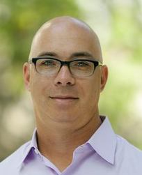 Jonathan N. Katz合作论文数Division of the Humanities and Social Sciences, California Institute of Technology21Laura Herbelin合作论文数University of Kansas21
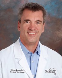 Robert Bowser合作论文数Department of Pathology, University of Pittsburgh Medical School21Wang Yunxia合作论文数20John T. Kissel合作论文数20
 Hiroshi Mitsumoto合作论文数Vagelos College of Physicians & Surgeons, Columbia University19
 Dan Moore合作论文数Department of Geography, Faculty of Arts, The University of British Columbia19
 Michael D Weiss合作论文数19Jonathan Goldstein合作论文数18George Sachs合作论文数18
 Pamela P. Kittrell Msn合作论文数18Rup Tandan合作论文数18
 Michael Rivner合作论文数Department of Neurology, Medical College of Georgia18Noah Lechtzin合作论文数17Melanie L Leitner合作论文数17
 Richard Stanley Bedlack合作论文数Department of Neurology, School of Medicine, Duke University17Polak Meraida合作论文数17
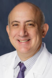 Jonathan D Licht合作论文数University of Florida Health Cancer Center;Department of Medicine, College of Medicine, University of Florida;University of Florida Health/Shands Hospital17
 Orla Hardiman合作论文数Trinity College Institute of Neuroscience, Trinity College Dublin, The University of Dublin;RCSI Clinical Research Centre, Royal College of Surgeons in Ireland17
 Jeffrey Rothstein合作论文数The Solomon H. Snyder Department of Neurology, School of Medicine, Johns Hopkins University;Institute for Clinical and Translational Research, Johns Hopkins University17Dan Pastula合作论文数17Muddasir Quereshi合作论文数17Ashok Verma合作论文数17Daniel MacGowan合作论文数17Leo McClusky合作论文数17
 Jeremy M. Shefner合作论文数Department of Neurology, Barrow Neurological Institute;Gregory W. Fulton ALS & Neuromuscular Disease Center, Barrow Neurological Institute17Gary L. Pattee合作论文数17
 Carlayne E Jackson合作论文数17Stacy A. Rudnicki合作论文数17
 Cathy Lomen-Hoerth合作论文数School of Medicine, University of California, San Francisco;Weill Institute for Neurosciences, University of California, San Francisco17
 Khema Sharma合作论文数17
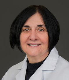 Terry Heiman-Patterson合作论文数Lewis Katz School of Medicine, Temple University17
 Stephen J. Kolb合作论文数Howard Hughes Medical Institute, University of Pennsylvania17
 Eric Sorenson合作论文数16Bertorini Tulio合作论文数16
 Oskarsson Björn合作论文数Department of Neurology, University of California Davis Medical Center16Mamede De Carvalho合作论文数16Dallas Forshew合作论文数16Kevin B. Boylan合作论文数16Caress James B合作论文数15Jim Wymer合作论文数15Bonnie Gerecke合作论文数15Megan Grosso合作论文数15




