e16337 Background: Appendiceal adenocarcinomas (AAs) are a rare and heterogeneous mix of tumors for which few preclinical models exist for drug discovery. These tumors are commonly treated with chemotherapy according to best practices developed for colorectal cancer, despite clear evidence that the two cancers are distinctly different in their clinical behavior and molecular profiles. In this study, we created functional tumoroid models (MicroOrganoSpheres or MOS) of peritoneal carcinomatosis from appendix cancer and mesothelioma and used these MOS to test the efficacy of different chemotherapy treatments. Methods: After obtaining informed consent, fresh tumor samples were obtained from 5 patients with peritoneal carcinomatosis undergoing surgical resection at The University of Texas MD Anderson Cancer Center and 5 samples were obtained from previously established PDX models. Tissue was digested using xMRS solution and MOS culture was established. H&E was performed on tissue and paired MOS. Results: Five MOS models were successfully established, including four AA and one primary peritoneal mesothelioma (to our knowledge the first time this has been done from a peritoneal mesothelioma). Four (80%) of the 5 PDX models samples successfully generated MOS. Three out of five from the fresh samples generated p0 MOS, but only one continued to grow (20%). The MOS successfully established from fresh biopsy was a moderately differentiated mucinous adenocarcinoma with GNAS R201C and KRAS G12R mutation, to our knowledge the first time a Patient Derived Organoid (PDO) model of this disease has been created. Identified failure modes were delayed processing times (>48 hours after extraction in 3 out of 5 clinical samples) and low tumor content in samples. Histologically, the MOS recapitulated several important characteristics of the original patients’ tumors, including degree of differentiation, abundance of mucin, and presence of signet ring features. Response of AA MOS models to chemotherapeutic agents and regimes has been evaluated in one model thus far. AAPDX-13, derived from a low-grade mucinous adenocarcinoma, was tested against common CRC treatments: 5-FU (pIC 50 5.90), Oxaliplatin (pIC 50 4.51), SN-38 (pIC 50 5.16). Relative to the mean from multiple CRC MOS, this was less sensitive to 5FU and more sensitive to irinotecan. Conclusions: Novel MOS models for AA and peritoneal mesothelioma were successfully established. MOS recapitulated key histologic features of AA and mesothelioma. Data acquired from this MOS system can aid in identifying appropriate chemotherapy treatments in select patients where there are no existing preclinical disease models.
In vitro tissue models hold great promise for modeling diseases and drug responses. Here, we used emulsion microfluidics to form micro-organospheres (MOSs), which are droplet-encapsulated miniature three-dimensional (3D) tissue models that can be established rapidly from patient tissues or cells. MOSs retain key biological features and responses to chemo-, targeted, and radiation therapies compared with organoids. The small size and large surface-to-volume ratio of MOSs enable various applications including quantitative assessment of nutrient dependence, pathogen-host interaction for anti-viral drug screening, and a rapid potency assay for chimeric antigen receptor (CAR)-T therapy. An automated MOS imaging pipeline combined with machine learning overcomes plating variation, distinguishes tu-morspheres from stroma, differentiates cytostatic versus cytotoxic drug effects, and captures resistant clones and heterogeneity in drug response. This pipeline is capable of robust assessments of drug response at individual-tumorsphere resolution and provides a rapid and high-throughput therapeutic profiling platform for precision medicine.
Patient-derived xenografts (PDXs) and patient-derived organoids (PDOs) have been shown to model clinical response to cancer therapy. However, it remains challenging to use these models to guide timely clinical decisions for cancer patients. Here, we used droplet emulsion microfluidics with temperature control and dead volume minimization to rapidly generate thousands of micro-organospheres (MOSs) from low-volume patient tissues, which serve as an ideal patient-derived model for clinical precision oncology. A clinical study of recently diagnosed metastatic colorectal cancer (CRC) patients using an MOS-based precision oncology pipeline reliably assessed tumor drug response within 14 days, a timeline suitable for guiding treatment decisions in the clinic. Furthermore, MOSs capture original stromal cells and allow T cell penetration, providing a clinical assay for testing immuno-oncology (IO) therapies such as PD-1 blockade, bispecific antibodies, and T cell therapies on patient tumors.
e12628 Background: Patient-derived breast cancer (BC) organoids are valuable preclinical models to study patient drug responses, demonstrating good correlations with patients’ clinical outcomes. However, establishment and expansion of such organoids from patient tumors for drug screening is currently a time-consuming and labor-intensive process. A more rapid and high-throughput method will enable broader utility in diagnostics and drug development. Methods: An automated, rapid and scalable microfluidic platform was used to process and develop BC micro-organospheres. Drug sensitivities studies on BC micro-organospheres were performed on day 3 and day 6 using 10-FDA approved drugs, including palbociclib, adriamycin, 5-FU, gemcitabine, methotrexate, everolimus, paclitaxel, docetaxel, ixabepilone, and vinblastine. The responses of micro-organospheres and organoids to the drugs were assessed by CellTiter 3D Glo assay on day 6 after the drug treatment. The growth and establishment of the micro-organospheres by imaging. The drug sensitivity and resistance of the micro-organospheres were analyzed by calculating the percentage cell viability and normalized growth rate inhibition (GRI) and compared to organoids. Results: We successfully established micro-organospheres from eight patient-derived BC organoids with a 100% success rate. The micro-organospheres preserved similar cell morphologies to the bulk organoids. 7/8 micro-organosphere models had similar drug response patterns to organoids between day 3 and day 6 as evident by the GRI heatmap. Specifically, we treated matching micro-organospheres and conventional organoids from two patients with 10 frontline BC chemotherapy drugs, and both showed similar response patterns with GRI heatmap. For the other 6 patient-derived models, the responses of micro-organospheres to docetaxel and everolimus also matched the historical drug responses of in bulk organoid culture with similar GRI heatmap. Conclusions: We have now shown the feasibility of establishing micro-organospheres as a rapid, scalable, and cost-effective platform to study patient-derived BC drug response. This technology has the potential to be used for both diagnostics to guide patient treatment and as a screening platform for new BC drug discovery.
149 Background: Patient-derived organoids (PDO) have been shown to have a high degree of similarity to the original patient tumors. PDO have also been used to perform high throughput drug screens and shown to correlate with patient response to therapy. Unfortunately, PDO require too much tissue, take too long to establish and are too inefficient and costly for adoption into the clinic. The ideal assay for clinical use would be one that could be performed in less than14 days from a core biopsy to minimize delay in therapy. We have now circumvented these barriers by leveraging recent technological advances in emulsion microfluidics and droplet generators to develop MicroOrganoSpheres (MOS) that can be established and used to predict drug sensitivity within 14 days of obtaining a biopsy. Methods: 18-gauge core biopsy specimens from patients with colorectal cancer liver metastasis who subsequently received an oxaliplatin based therapy were first obtained. Biopsy specimens were minced, enzymatically digested and mixed with components necessary to generate MOS. The mixture was then processed through a custom fabricated flow-focusing droplet microfluidic chip in our MOS Generator Device to generate MOS. After culturing for 8-10 days, MOS were then used to perform drug screen with oxaliplatin. Results: A total of twelve CRC biopsies from liver metastasis were obtained and processed to generate MOS with a success rate of 12/12 (100%). Furthermore, drug screens with oxaliplatin were performed on all twelve samples with an average time to drug screen of 10.1 days. We next wanted to determine if there was a correlation between MOS drug sensitivity and patient clinical outcome (ie. time on treatment). For the first eight patients, MOS was used to predict sensitivity to oxaliplatin and using a drug sensitivity cut-off of 1 uM, four patients were predicted to be sensitive to oxaliplatin and four patients were predicted to be resistant. Three of the four patients predicted to be sensitive to oxaliplatin continue to be on treatment (> 6 months), whereas 3 of the four patients predicted to be resistant to oxaliplatin progressed on oxaliplatin based therapy within 8 weeks (sensitivity = 80%, specificity = 100%, positive predictive value = 100%, negative predictive value = 75%). Conclusions: MOS can be generated from core biopsies and correlates to time on treatment. Although further studies will need to be conducted, the ability to generate MOS and perform a drug screen in < 14 days will allow for the development of a precision oncology platform that can be rapidly used in the clinic to guide therapy.
Blood-based methods using cell-free DNA (cfDNA) are under development as an alternative to existing screening tests. However, early-stage detection of cancer using tumor-derived cfDNA has proven challenging because of the small proportion of cfDNA derived from tumor tissue in early-stage disease. A machine learning approach to discover signatures in cfDNA, potentially reflective of both tumor and non-tumor contributions, may represent a promising direction for the early detection of cancer. Whole-genome sequencing was performed on cfDNA extracted from plasma samples (N = 546 colorectal cancer and 271 non-cancer controls). Reads aligning to protein-coding gene bodies were extracted, and read counts were normalized. cfDNA tumor fraction was estimated using IchorCNA. Machine learning models were trained using k-fold cross-validation and confounder-based cross-validations to assess generalization performance. In a colorectal cancer cohort heavily weighted towards early-stage cancer (80% stage I/II), we achieved a mean AUC of 0.92 (95% CI 0.91–0.93) with a mean sensitivity of 85% (95% CI 83–86%) at 85% specificity. Sensitivity generally increased with tumor stage and increasing tumor fraction. Stratification by age, sequencing batch, and institution demonstrated the impact of these confounders and provided a more accurate assessment of generalization performance. A machine learning approach using cfDNA achieved high sensitivity and specificity in a large, predominantly early-stage, colorectal cancer cohort. The possibility of systematic technical and institution-specific biases warrants similar confounder analyses in other studies. Prospective validation of this machine learning method and evaluation of a multi-analyte approach are underway.
Abstract Introduction: Blood-based tests hold great promise as cancer diagnostics but until now have largely been restricted to the analysis of a single class of molecules (eg, circulating tumor DNA, platelet mRNA, circulating proteins). The ability to analyze multiple analytes simultaneously from the same biological sample may increase the sensitivity and specificity of such tests by exploiting independent information between signals. Here, we describe an experimental and analytical system that we developed and implemented for the integrated analysis of multiple analytes from a single blood sample, which revealed examples of both correlations and orthogonality among individual analytes. Methods: De-identified blood samples were obtained from healthy individuals, as well as individuals with pre-malignant conditions and stage I-IV colorectal cancer (CRC). After plasma separation, multiple types of analytes were assayed: cell-free DNA (cfDNA) content was assessed by low-coverage whole-genome sequencing (lcWGS) and whole-genome bisulfite sequencing (WGBS), cell-free microRNA (cf-miRNA) was assessed by small-RNA sequencing, and levels of circulating proteins were measured by quantitative immunoassay. Results: lcWGS of plasma cfDNA was able to identify CRC samples with high tumor fraction (>20%) on the basis of copy number variation (CNV) across the genome. High tumor fractions, while more frequent in late-stage cancer samples, were observed in some stage I and II patients. Aberrant signals in each of the three other analytes—cf-miRNA profiles discordant with those in healthy controls, genome-wide hypomethylation at LINE1 (long interspersed nuclear element 1) CpG loci, and elevated levels of circulating carcinoembryonic antigen (CEA) and cytokeratin fragment 21-1 (CYFRA 21-1) proteins—were also observed in cancer patients. Strikingly, aberrant profiles across analytes were indicative of high tumor fraction (as estimated from cfDNA CNV), rather than cancer stage. Conclusion: Our data suggest that tumor fraction is correlated with cancer stage but has a large potential range, even in early stage samples. Previous literature on blood-based screens for detection of cancer has displayed discordance in the claimed ability of different single analytes to detect early stage cancer. Tumor fraction may be able to explain the historical disagreement, as we found that aberrant profiles among cf-miRNA, cfDNA methylation, and circulating protein levels were more strongly associated with high tumor fraction than with late stage. These findings suggest that some positive “early stage” detection results may in fact be “high tumor fraction” detection results. Our results further demonstrate that assaying multiple analytes from a single sample may enable the development of classifiers that are reliable at low tumor fraction and for detecting pre-malignant or early-stage disease. Citation Format: Daniel Delubac, Eric Ariazi, Jonathan Berliner, Adam Drake, John Dulin, Riley Ennis, Erik Gafni, Kate Niehaus, Gabriel Otte, Jennifer Pecson, Girish Putcha, Corey Schaninger, Aarushi Sharma, Mike Singer, Abraham Tzou, Jill Waters, David Weinberg, Brandon White, Imran S. Haque. Multi-analyte profiling reveals relationships among circulating biomarkers in colorectal cancer [abstract]. In: Proceedings of the American Association for Cancer Research Annual Meeting 2018; 2018 Apr 14-18; Chicago, IL. Philadelphia (PA): AACR; Cancer Res 2018;78(13 Suppl):Abstract nr 2227.
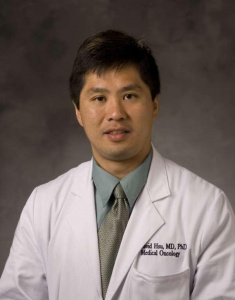 Shiao-Wen David Hsu合作论文数Department of Medicine, Duke University School of Medicine4
 Carolyn Hsu合作论文数3Daniel Nelson合作论文数3Abraham Tzou合作论文数2Jennifer Pecson合作论文数2Gabriel Otte合作论文数2Gabrielle Rupprecht合作论文数2Riley Ennis合作论文数2Athena Yeung合作论文数2
 Marcos Negrete合作论文数2
 Aarushi Sharma合作论文数Regenerat Engn Lab, Indian Inst Technol2Naveen Natesh合作论文数Dept Biomed Engn, Duke Univ2Erik Gafni合作论文数Freenome2Girish Putcha合作论文数2Shree Bose合作论文数Department of Pharmacology and Cancer Biology, Duke University School of Medicine2
 Rosemary Millen合作论文数Sir Peter MacCallum Dept Oncol, Univ Melbourne2Grecia O Rivera合作论文数2
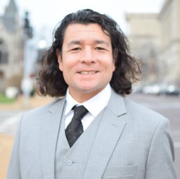 John Paul Shen合作论文数Univ Texas MD Anderson Canc Ctr, Dept Gastrointestinal Med Oncol, Houston, TX 77030 USA2
 Shengli Ding合作论文数Department of Biomedical Engineering, Pratt School of Engineering, Duke University2
 Imran S. Haque合作论文数Kimia Therapeutics2Smith Amber R合作论文数2
 Brandon White合作论文数2
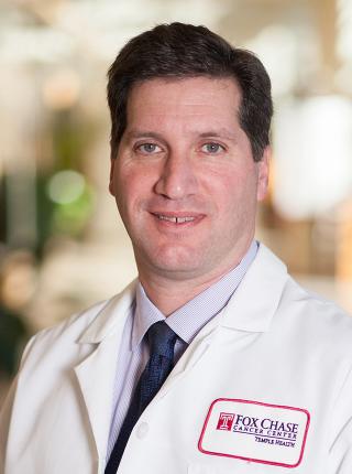 David S. Weinberg合作论文数Fox Chase Cancer Center2
 Hans Clevers合作论文数Utrecht University;Hubrecht Institute2
 Eric A. Ariazi合作论文数2Ravian L. van Ineveld合作论文数1Zhaohui Wang合作论文数1Mahmoud Yousef合作论文数1
 John Alden St John合作论文数1Marvin Bertin合作论文数1Signe Fransen合作论文数1Kassandra Spiller合作论文数1Jill Waters合作论文数1Tomer Rotstein合作论文数1Estelle Marrer-Berger合作论文数1
 Abdelrahman Yousef合作论文数1
 Matteo Giovanni Boretto合作论文数1
 Ajay Kannan合作论文数Google DeepMind1Loren Hansen合作论文数1Adam Drake合作论文数1Jessica DeVilla合作论文数1Corey Schaninger合作论文数1Catherina Tang合作论文数1Haipei Yao合作论文数1
 Nicholas S Giroux合作论文数1Emilio Cortes-Sanchez合作论文数Dept Oncol Sci, Univ Utah1




