Perinatally acquired HIV (PHIV) and antiretroviral therapy (ART) can alter innate immune cells, (monocytes and natural killer [NK] cells) which are important in the pathogenesis of cardiovascular disease (CVD). We compare cardiovascular biomarkers and immune signatures between adolescents with PHIV (APHIV) on suppressive ART and HIV-unexposed, adolescents without HIV in Uganda. Carotid intima-media thickness (IMT) is increased in APHIV, suggesting a higher CVD risk. Flow cytometry analysis reveals greater activation, memory, and migratory capabilities of NK cells, and increased pro-inflammatory intermediate monocytes in APHIV, and these observations are supported by transcriptomics. Many of these innate immune cell subsets are associated with carotid IMT. Plasma oxidized-LDL (Ox-LDL) is significantly lower among APHIV, and negatively correlates with pro-inflammatory, memory-like NK subsets. We demonstrate increased uptake of Ox-LDL by macrophages in the presence of activated, memory-like NK cells in vitro, suggesting a possible mechanism for greater CVD risk in APHIV. Collectively, our data demonstrate associations between dysregulated NK cell signatures and increased CVD risk among APHIV.
Natural killer (NK) cell-driven effector mechanisms, such as antibody-dependent cell-mediated cytotoxicity, emerged as a secondary correlate of protection in the RV144 HIV vaccine clinical trial, the only vaccine thus far demonstrating some efficacy in human trials. Therefore, leveraging NK cells with enhanced cytotoxic effector responses may bolster vaccine-induced protection against HIV. Here, we investigated the effect of orally administering indole-3-carbinol (I3C), an aryl hydrocarbon receptor (AHR) agonist, as an adjuvant to an RV144-like vaccine platform in a mouse model. We demonstrate the expansion of KLRG1-expressing NK cells induced by the vaccine together with I3C. This NK cell subset exhibited enhanced vaccine antigen-specific cytotoxic memory-like features. Our study underscores the potential of incorporating I3C as an oral adjuvant to HIV vaccine platforms to enhance antigen-specific cytotoxicity of NK cells against HIV-infected cells. This approach may contribute to enhancing the protective efficacy of HIV preventive vaccines against HIV acquisition.
Abstract Bacillus Calmette-Guérin (BCG) vaccine is the only approved vaccine against tuberculosis (TB). BCG has been shown to elicit nonspecific protection as well as detrimental effects during diseases unrelated to TB. We investigate the impact of BCG on modulating NK cell responses and its effect on altering susceptibility to HIV acquisition. The C57BL/6 mouse model was used to compare splenic NK cell responses of control (n=4) and BCG vaccinated mice (n=4) following subsequent exposure to Mycobacterium bovis (Mtb) and HIV Gag peptide antigens. Statistical comparisons were made using Wilcoxon ranked sum test. The data acquired though flowcytometry suggested that mature, BCG trained memory-like (KLRG1+ Ly49H+) NK cells showed increased proinflammatory cytokine responses and polyfunctionality upon re-exposure to Mtb antigens. However, when these trained cells were exposed to HIV Gag antigen, we observed a significant reduction in cytokine production and polyfunctionality. Specifically, these BCG trained NK cells showed marked decrease in IFNγ, TNFα, IL1β and Granzyme B production alone or in combination (p<0.05). Furthermore, negative correlations between NK polyfunctionality and mature BCG trained NK cells were observed, following HIV Gag peptide exposure. This suggests that NK cells are impaired in their antiviral effects during HIV exposure, soon after BCG vaccination. This may suggest higher risk of HIV acquisition in BCG vaccinated neonates that are frequently exposed to HIV.
BACKGROUND:Evidence suggests that COVID-19 predisposes to cardiovascular diseases (CVDs). While monocytes/macrophages play a central role in the immunopathogenesis of atherosclerosis, less is known about their immunopathogenic mechanisms that lead to CVDs during COVID-19. Natural killer (NK) cells, which play an intermediary role during pathologies like atherosclerosis, are dysregulated during COVID-19. Here, we sought to investigate altered immune cells and their associations with CVD risk during severe COVID-19.METHODS:We measured plasma biomarkers of CVDs and determined phenotypes of circulating immune subsets using spectral flow cytometry. We compared these between patients with severe COVID-19 (severe, n=31), those who recovered from severe COVID-19 (recovered, n=29), and SARS-CoV-2-uninfected controls (controls, n=17). In vivo observations were supported using in vitro assays to highlight possible mechanistic links between dysregulated immune subsets and biomarkers during and after COVID-19. We performed multidimensional analyses of published single-cell transcriptome data of monocytes and NK cells during severe COVID-19 to substantiate in vivo findings.RESULTS:During severe COVID-19, we observed alterations in cardiometabolic biomarkers including oxidized-low-density lipoprotein, which showed decreased levels in severe and recovered groups. Severe patients exhibited dysregulated monocyte subsets, including increased frequencies of proinflammatory intermediate monocytes (also observed in the recovered) and decreased nonclassical monocytes. All identified NK-cell subsets in the severe COVID-19 group displayed increased expression of activation and tissue-resident markers, such as CD69 (cluster of differentiation 69). We observed significant correlations between altered immune subsets and plasma oxidized-low-density lipoprotein levels. In vitro assays revealed increased uptake of oxidized-low-density lipoprotein into monocyte-derived macrophages in the presence of NK cells activated by plasma of patients with severe COVID-19. Transcriptome analyses confirmed enriched proinflammatory responses and lipid dysregulation associated with epigenetic modifications in monocytes and NK cells during severe COVID-19.CONCLUSIONS:Our study provides new insights into the involvement of monocytes and NK cells in the increased CVD risk observed during and after COVID-19.
Purpose of review Despite decades of insights about the role of natural killer (NK) cells in HIV infection, their persistent dysregulation despite antiretroviral therapy (ART) and its pathological consequences have been incompletely delineated. In this review, we highlight recent findings on the immunophenotypic and functional alterations of NK cells during virally suppressed HIV infection and explore their potential impact on promoting non-AIDS related comorbidities among people living with HIV (PLWH). Recent findings Of note are the apparent persistent activated profiles of NK cells and pathophysiological events such as endoplasmic reticulum (ER) stress in potentially driving NK cell derived inflammation and tissue destruction. Additionally, recent interest in trained immunity is discussed as a potential mediator of ongoing NK cell dysregulation, contributing to comorbidities such as cardiovascular disease and neurocognitive disorders, both with an inflammatory etiology. Summary Clinical and mechanistic evidence suggests persistent activation and dysregulation of the innate immune system are major drivers of non-AIDS comorbidities during virally suppressed HIV infection. Delineating the mechanistic role of specific components of innate immunity such as NK cells in inducing these pathologies will lead to the identification of novel therapeutic/prophylactic strategies to improve the overall health of PLWH.
Bacille-Calmette-Guerin (BCG) is the only approved vaccine against Mycobacterium tuberculosis (MTB), offering protection not only against tuberculosis (TB) but also non-related infections. 'Trained immunity' of innate immune cells is considered one of the mechanisms of this broad protection derived through BCG. Here, we investigated the effect of BCG on Natural Killer (NK) cells, a key innate immune cell type, and their subsequent responses to mycobacterial and HIV antigens. We found that BCG-induced KLRG1+ NK cells exhibit significantly higher production of IFN gamma, compared to KLRG1- cells, indicating their memory-like responses upon exposure to these antigens (p < 0.05). These findings may be important in regions of high burden of HIV and TB where BCG is routinely administered.
HIV-infected patients are at higher risk of developing oral mucosal infection and Epstein–Barr virus (EBV)-associated B cell malignancies. However, the potential role of oral immunity in the pathogenesis of oral lesions is unknown. Tonsils are oral-pharyngeal mucosal-associated lymphoid tissues that play an important role in oral mucosal immunity. In this study, we investigated the changes of innate and adaptive immune cells in macaque tonsils during chronic SIV infection. We found significantly higher frequencies of classical monocytes, CD3+CD56+ (NKT-like) cells, CD3+CD4+CD8+ (DP), and CD161+ CD4 T cells in tonsils from chronic infected compared to naïve animals. On the contrary, intermediate monocytes and CD3+CD4-CD8- (DN) cells were lower in chronic SIV-infected macaques. We further confirmed a recently described small B-cell subset, NKB cells, were higher during chronic infection. Furthermore, both adaptive and innate cells showed significantly higher TNF-α and cytotoxic marker CD107a, while IL-22 production was significantly reduced in innate and adaptive immune cells in chronic SIV-infected animals. A dramatic reduction of IFN-γ production by innate immune cells might indicate enhanced susceptibility to EBV infection and potential transformation of B cells in the tonsils. In summary, our observation shows that the SIV-associated immune responses are distinct in the tonsils compared to other mucosal tissues. Our data extends our understanding of the oral innate immune system during SIV infection and could aid future studies in evaluating the role of tonsillar immune cells during HIV-associated oral mucosal infections.
Abstract Vα24-invariant natural killer T cells (NKT) possess innate antitumor properties that can be exploited for cancer immunotherapy. We have shown previously that the CD62L+ central memory-like subset of these cells drives the in vivo antitumor activity of NKTs, but molecular mediators of NKT central memory differentiation remain unknown. Here, we demonstrate that relative to CD62L– cells, CD62L+ NKTs express a higher level of the gene encoding the Wnt/β-catenin transcription factor lymphoid enhancer binding factor 1 (LEF1) and maintain active Wnt/β-catenin signaling. CRISPR/Cas9-mediated LEF1 knockout reduced CD62L+ frequency after antigenic stimulation, whereas Wnt/β-catenin activator Wnt3a ligand increased CD62L+ frequency. LEF1 overexpression promoted NKT expansion and limited exhaustion following serial tumor challenge and was sufficient to induce a central memory–like transcriptional program in NKTs. In mice, NKTs expressing a GD2-specific chimeric-antigen receptor (CAR) with LEF1 demonstrated superior control of neuroblastoma xenograft tumors compared with control CAR-NKTs. These results identify LEF1 as a transcriptional activator of the NKT central memory program and advance development of NKT cell–based immunotherapy. See related Spotlight by Van Kaer, p. 144
Vα24-invariant natural killer T cells (NKTs) have anti-tumor properties that can be enhanced by chimeric antigen receptors (CARs). Here we report updated interim results from the first-in-human phase 1 evaluation of autologous NKTs co-expressing a GD2-specific CAR with interleukin 15 (IL15) (GD2-CAR.15) in 12 children with neuroblastoma (NB). The primary objectives were safety and determination of maximum tolerated dose (MTD). The anti-tumor activity of GD2-CAR.15 NKTs was assessed as a secondary objective. Immune response evaluation was an additional objective. No dose-limiting toxicities occurred; one patient experienced grade 2 cytokine release syndrome that was resolved by tocilizumab. The MTD was not reached. The objective response rate was 25% (3/12), including two partial responses and one complete response. The frequency of CD62L+NKTs in products correlated with CAR-NKT expansion in patients and was higher in responders (n = 5; objective response or stable disease with reduction in tumor burden) than non-responders (n = 7). BTG1 (BTG anti-proliferation factor 1) expression was upregulated in peripheral GD2-CAR.15 NKTs and is a key driver of hyporesponsiveness in exhausted NKT and T cells. GD2-CAR.15 NKTs with BTG1 knockdown eliminated metastatic NB in a mouse model. We conclude that GD2-CAR.15 NKTs are safe and can mediate objective responses in patients with NB. Additionally, their anti-tumor activity may be enhanced by targeting BTG1. ClinicalTrials.gov registration: NCT03294954 .
Human immunodeficiency virus (HIV) is associated with persistent immune activation and dysfunction in people with HIV despite treatment with antiretroviral therapy (ART). Modulation of the immune system may be driven by: low-level HIV replication, co-pathogens, gut dysbiosis /translocation, altered lipid profiles, and ART toxicities. In addition, perinatally acquired HIV (PHIV) and lifelong ART may alter the development and function of the immune system. Our preliminary data and published literature suggest reprogramming innate immune cells may accelerate aging and increase the risk for future end-organ complications, including cardiovascular disease (CVD). The exact mechanisms, however, are currently unknown. Natural killer (NK) cells are a highly heterogeneous cell population with divergent functions. They play a critical role in HIV transmission and disease progression in adults. Recent studies suggest the important role of NK cells in CVDs; however, little is known about NK cells and their role in HIV-associated cardiovascular risk in PHIV adolescents. Here, we investigated NK cell subsets and their potential role in atherogenesis in PHIV adolescents compared to HIV-negative adolescents in Uganda. Our data suggest, for the first time, that activated NK subsets in PHIV adolescents may contribute to atherogenesis by promoting plasma oxidized low-density lipoprotein (Ox-LDL) uptake by vascular macrophages.
Clinical data demonstrate an increased predisposition to cardiovascular disease (CVD) following severe COVID-19 infection. This may be driven by a dysregulated immune response associated with severe disease. Monocytes and vascular tissue resident macrophages play a critical role in atherosclerosis, the main pathology leading to ischemic CVD. Natural killer (NK) cells are a heterogenous group of cells that are critical during viral pathogenesis and are known to be dysregulated during severe COVID-19 infection. Their role in atherosclerotic cardiovascular disease has recently been described. However, the contribution of their altered phenotypes to atherogenesis following severe COVID-19 infection is unknown. We demonstrate for the first time that during and after severe COVID-19, circulating proinflammatory monocytes and activated NK cells act synergistically to increase uptake of oxidized low-density lipoprotein (Ox-LDL) into vascular tissue with subsequent foam cell generation leading to atherogenesis despite recovery from acute infection. Our data provide new insights, revealing the roles of monocytes/macrophages, and NK cells in COVID-19-related atherogenesis.
COVID-19, the disease caused by SARS-CoV-2, has led to a global public health emergency. Severity of disease course may be related to a dysregulated immune response and pre-existing health conditions. Recent studies have demonstrated that SARS-CoV-2 infection may directly or indirectly lead to an increase in cardiometabolic complications in patients with pre-existing type-2 diabetes mellites (T2DM) when compare to non-DM patients. A lack of mechanistic and systematic studies on how SARS-CoV-2 infection related immune responses may contribute to increase risk of cardiometabolic complications in pre-existing T2DM patients, hinder early risk identification and therapeutic interventions. Thus, in this study we investigate the biomarkers of cardiometabolic risk in non-DM and T2DM, severe COVID-19 patients admitted to the Intensive care unit. Using high-dimensional flowcytometry and immune biomarker assays, we investigated functional and phenotypic changes in immune subsets in whole blood and plasma biomarkers of cardiovascular disease in healthy donors (n=17), T2DM severe-COVID-19 patients (n=10), non-diabetic severe-COVID-19 patients (n=10) admitted to the OSU medical center's intensive care unit. We found neutrophils and Intermediate monocytes (ITM) were significantly higher in the T2DM group compared to non T2DM patients. However, activated (HLA-DR+) NKT-like cells and NKG2A+ CD56 Dim CD16+ NK cells, were significantly lower in the T2DM-COVID-19+ group. Interestingly, LBP, FABP4, sCD14, IL-1b, RANTES and MIP-1a were significantly higher in the COVID-19 T2DM patients. In this study, we identify core immune signatures that may predict increased cardiovascular disease risk in T2DM patients who had severe COVID-19. Supported by PI's startup funding
Covid-19, the disease caused by SARS-CoV-2 infection, has resulted in millions of deaths and led to a global public health emergency. SARS-CoV-2 infected patients exhibit a wide variety of phasic clinical manifestations ranging from asymptomatic to severe complications and death. SARS-CoV-2 infection can lead to excessive immune activation, inflammation and multi-organ damage. Clinical data showed that COVID-19 may promote the development of cardiovascular disorders (CVDs). Immune activation, thrombosis, cytokine storm, and altered adhesion molecule expression on leukocyte populations have been proposed as possible mechanisms that trigger COVID-19 associated CVDs. A lack of systematic studies on how SARS-CoV-2 infection triggered immune responses that may lead to CVDs, hinder early risk identification and therapeutic interventions. In this study, by using deep immune profiling and extensive cytokine and chemokine profiling, we explore potential mechanisms of developing CVDs in severe COVID-19 patients (ICU) (n=20) as well as patients recovered from COVID-19 (RD) (n=30). We identify core immune signatures in ICU patients and RD compared to healthy controls (n=17) that may predict potential cardiovascular risk. We found that significantly elevated eosinophils and neutrophils and increased circulating levels of tissue factor, fatty acid binding protein 4 and, LPS binding protein in ICU patients suggested increased immune activation and thrombotic risk. Interestingly, we found significant elevation of several immune parameters (TIMP-1, TIMP-2, Monocytes) that were associated with cardiometabolic risk, in RD group. Thus, our data suggest a possible mechanistic link between severe COVID-19 and cardiometabolic risk.
Background: Severe Acute Respiratory Syndrome Coronavirus 2 (SARS-CoV-2) is a highly pathogenic corona virus which causes COVID-19 and resulted in millions of deaths and led to a global public health emergency. SARS-CoV-2 infected patients exhibit a wide variety of clinical manifestations ranging from asymptomatic to severe complications and death. SARS-CoV-2 infection can lead to excessive immune activation, inflammation and multi-organ damage. Clinical data showed that COVID-19 may promote the development of cardiovascular disorders (CVDs). Immune activation, thrombosis, cytokine storm, and altered adhesion molecule expression on leukocyte populations, have been proposed as possible mechanisms that trigger COVID-19 associated CVDs. A lack of systematic studies on how SARS-CoV-2 infection triggered immune responses that may lead to CVDs, hinder early risk identification and therapeutic interventions. Methods: In this study, by using deep immune cell profiling (high dimensional flowcytometry) in fresh whole blood and extensive plasma cytokine and chemokine profiling, we explore potential mechanisms that could lead to CVDs in severe COVID-19 patients that did not have previous known CVDs (ICU) (n=20) as well as patients recovered from COVID-19 (RD) (n=30) compared to healthy donors (n=17). To identify the major statistically significant immune signatures that predict CVD risk in ICU patients and RD, we performed parametric (ANOVA) and non-parametric (Kruskal-Wallis) statistical tests with Dunn's and Tukey's post hoc tests. Integrative correlation and network analysis were performed by computing Spearman's coefficients. Correlations with r > 0.3 , r <-0.3 and P < 0.01 were considered significant. Results: We found that significantly elevated eosinophils, neutrophils and increased circulating levels of tissue factor, fatty acid binding protein 4 and, LPS binding protein in ICU patients suggested increased immune activation and thrombotic risk. Interestingly, we found significant elevation of several immune parameters (TIMP-1, TIMP-2, M-CSF, Monocytes) that were associated with cardiometabolic risk, even 3-4 months after the recovery of initial COVID-19 infection in RD. Furthermore, we found unique relationship with cytokine and cellular responses in ICU and RD groups compare with HD. Conclusion: Our data strongly suggest a possible mechanistic link between SARS-CoV-2 induced dysregulated immune responses and increased cardiometabolic risk in severe COVID-19 patients.
Severe Acute Respiratory Syndrome Coronavirus 2 (SARS□CoV□ 2) is a highly pathogenic corona virus which causes COVID-19 and resulted in millions of deaths and led to a global public health emergency. An effective and appropriate immune response is essential to control and eliminate viral infections, however, dysregulated immune responses may lead to immunopathology in viral infections. Clinical data showed that COVID-19 may promote the development of cardiovascular disorders (CVDs). However, exact mechanism associated with CVDs in COVID-19 patients is currently unknown. Monocytes are one of the major immune cell subsets that contribute to host defense against many infections but also associated with immunopathology and development of CVDs. Thus, in this study we investigated the role of monocyte subsets in COVID-19 associated CVDs. By using high dimensional flowcytometry, immune biomarker assays and multi variant data analysis in healthy donors (n=17), severe-COVID-19 patients (n=20) (ICU) and COVID-19 recovered individuals (n=30), we found elevated intermediate monocytes and Monocyte chemoattractant protein-1, one of the most important chemokines that regulates migration and infiltration of monocytes to the subendothelial space, where they may become foam cells. Spearman correlation network analysis showed that strong correlation between increase intermediate monocytes in the whole blood and plasma cardimetabolic biomarkers such as fatty acid binding protein 4, C-reactive protein, soluble CD14 and LPS binding protein in ICU patients. Our data show for the first time that the possible role of monocytes in the development of CVDs in ICU patients.
The new pandemic virus SARS-CoV-2 emerged in China and spread around the world in <3 months, infecting millions of people, and causing countries to shut down public life and businesses. Nearly all nations were unprepared for this pandemic with healthcare systems stretched to their limits due to the lack of an effective vaccine and treatment. Infection with SARS-CoV-2 can lead to Coronavirus disease 2019 (COVID-19). COVID-19 is respiratory disease that can result in a cytokine storm with stark differences in morbidity and mortality between younger and older patient populations. Details regarding mechanisms of viral entry via the respiratory system and immune system correlates of protection or pathogenesis have not been fully elucidated. Here, we provide an overview of the innate immune responses in the lung to the coronaviruses MERS-CoV, SARS-CoV, and SARS-CoV-2. This review provides insight into key innate immune mechanisms that will aid in the development of therapeutics and preventive vaccines for SARS-CoV-2 infection.
Abstract Adoptive cellular therapy (ACT) has dramatically changed the landscape of immunotherapy; however, only a small proportion of solid tumor patients have benefited from these advances due to i) heterogeneity of tumor antigen expression, ii) tumor escape (e.g., only one target is addressed), or iii) off-target toxicities (e.g., expression of targets on normal tissues). ACTolog® concept, utilizing antigen specific T-cells (IMA101), identified by the Immatics’ proprietary XPRESIDENT® technology, is intended to overcome these limitations by introducing multiple novel tumor targets. ACTolog® is a personalized ACT approach in which autologous T-cell products are manufactured against the most relevant tumor target peptides for individual patients whose tumors are positive against a predefined target warehouse. Target positive tumors are identified by qPCR. Expression levels predictive for antigen presentation are determined by mass spectrometry. Autologous T-cells against ACTolog targets are in vitro primed in the presence of IL-21 followed by HLA tetramer-guided cell sorting and expansion prior to infusion. IMA101-101 is a first-in-human clinical trial in HLA-A*02:01 positive patients with relapsed or refractory solid tumors using the multitargeted ACTolog® approach in which up to four products with different tumor target-specificities are manufactured and infused for each patient. We developed two flow cytometric phenotyping assays that allow us to determine the frequency of target-specific cells in the final product and persisting cells in the blood as well as to deeply characterize the memory marker expression (CD45RA, CCR7, CD27, CD28, CD45RO, CD62L, CD57, CD127) and immune checkpoint expression (CD137, LAG-3, PD-1, TIGIT, TIM-3) of target-specific cells. Product characterization and initial persistence data of the three first treated patients revealed a high prevalence of persisting target-specific cells in the blood until 2 months after infusion as well as a favorable phenotype of target-specific cells. At the conference 6 months’ data for one patient will be available and presented. Citation Format: Regina Mendrzyk, Alexander Ulges, Thorsten Demberg, Geoffrey Stephens, Carsten Reinhardt, Steffen Walter, Dominik Maurer. Cellular immunomonitoring for personalized adoptive cellular therapy trial ACTolog® (IMA101-101) [abstract]. In: Proceedings of the Fourth CRI-CIMT-EATI-AACR International Cancer Immunotherapy Conference: Translating Science into Survival; Sept 30-Oct 3, 2018; New York, NY. Philadelphia (PA): AACR; Cancer Immunol Res 2019;7(2 Suppl):Abstract nr A015.
 Marjorie Robert-Guroff合作论文数Center for Cancer Research, National Cancer Institute42
 David Venzon合作论文数Center for Cancer Research, National Cancer Institute26
 Diego A Vargas-Inchaustegui合作论文数18
 Namal Liyanage合作论文数College of Veterinary Medicine, The Ohio State University;The Ohio State University Wexner Medical Center14Thomas Musich合作论文数13Venkatramanan Mohanram合作论文数11
 Manuja Gunasena合作论文数10
 Peng Xiao合作论文数National Cancer Institute, National Cancer Institute10Dhanuja Kasturiratna合作论文数9Vs Kalyanaraman合作论文数9Katherine M McKinnon合作论文数9Tuero Iskra合作论文数9Ranajit Pal合作论文数Advanced BioScience Laboratories, Inc.9
 Jean L. Patterson合作论文数Department of Virology and Immunology, Southwest Foundation for Biomedical Research9
 Egidio Brocca-Cofano合作论文数Department of Histology, Microbiology and Medical Biotechnology, University of Padova8Nicholas T Funderburg合作论文数8Mario Alles合作论文数7Janet Dipasquale合作论文数7
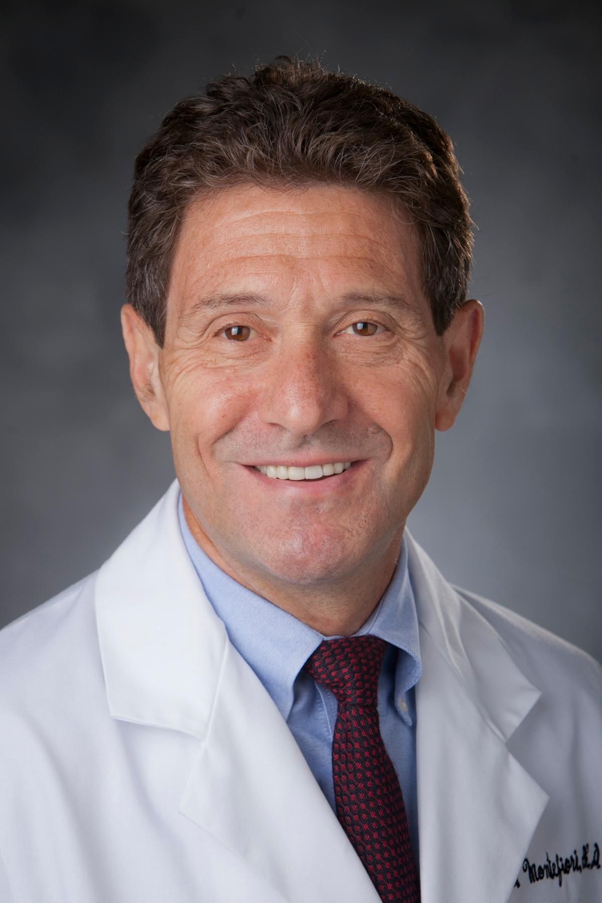 David Charles Montefiori合作论文数Laboratory for HIV and COVID-19 Vaccine Research and Development, Department of Surgery, Division of Surgical Sciences, Duke University School of Medicine;Duke Human Vaccine Institute, Duke University7
 Eun Mi Lee合作论文数Department of Internal Medicine;College of Medicine;Korea University;College of Medicine, Korea University7Irene Kalisz合作论文数7Will Mulhern合作论文数6Krishanthi Weragalaarachchi合作论文数6Aaren Kettelhut合作论文数6Yasasvi Wijewantha合作论文数6Bowman Emily R合作论文数6Bednash Joseph S合作论文数6
 Anna Vilgelm合作论文数Institute of Molecular Biology, Russian Academy Sciences6Janelle Gabriel合作论文数Div Med Lab Sci, Ohio State Univ5
 Linjie Guo合作论文数5
 Amrendra Kumar合作论文数Molecular Immunogenetics laboratory Institute of Genomics and Integrative Biology Mall Road Delhi 110007 India5Ruth H. Florese合作论文数5
 Genoveffa Franchini合作论文数Center for Cancer Research, National Cancer Institute, National Institutes of Health5Leia K Miller-Novak合作论文数4Michael Wood合作论文数4
 Gabriel A Barragan合作论文数4
 Erica J. Di Pierro合作论文数Center for Advanced Innate Cell Therapy, Baylor College of Medicine4Chunchao Zhang合作论文数Dept Pediat, Baylor Coll Med4Pannu Sonal合作论文数4
 Xin Xu合作论文数4Courtney Amy N合作论文数Department of Pediatrics., Texas Children's Cancer Center4
 Tian Gengwen合作论文数4
 Jay A. Berzofsky合作论文数Center for Cancer Research, National Cancer Institute4
 George N. Pavlakis合作论文数Center for Cancer Research, National Cancer Institute, National Institutes of Health4
 Aurelio Cafaro合作论文数4Leonid S. Metelitsa合作论文数4Barbara Ensoli合作论文数4Kay Larsen合作论文数4susan whoriskey合作论文数3




